In Vivo Electroporation: A Site-Specific Method to Transfect Zebrafish

Published: April 30, 2023

Abstract

Source: Durovic, T., et al. Electroporation Method for In Vivo Delivery of Plasmid DNA in the Adult Zebrafish Telencephalon. J. Vis. Exp. (2019).

This video describes in vivo electroporation, which is a technique for DNA delivery into ependymoglial cells in adult zebrafish.

Protocol

1. Electroporation

  1. Remove the fish from the injection set-up while still holding it in the sponge.
  2. Immerse the inner side of the tip of the electrodes in the ultrasound gel.
  3. Cover the fish telencephalon with a small amount of ultrasound gel.
  4. Position the fish head between the electrodes, placing the positive electrode at the ventral side of the fish’s head and the negative electrode on the dorsal side (Figure 1C), while still holding the fish’s body in the sponge. This sets the direction of the flow of the current necessary to electroporate ependymoglia positioned at subventricular zone.
  5. Press the electrodes gently and precisely against the telencephalon (Figure 1 C). Administer the current with the foot pedal. Hold the electrodes in place until all five pulses are finished.

Representative Results

Figure 1
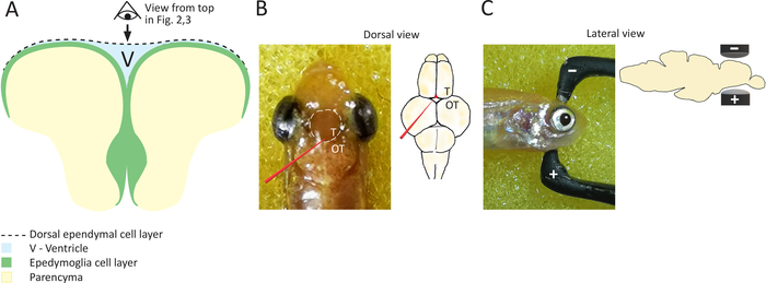
Figure 1: Schematic representation of coronal section of the everted zebrafish telencephalon. (A) Scheme of a coronal section of zebrafish telencephalon, highlighting the position of ependymoglial cells, which are lining ventricular surface and building the ventral ventricular wall. Dorsal ependymal layer is bridging the two hemispheres and covering the ventricle (V), located in between two cell layers: ependymoglial and ependymal. (B) On the left, a photograph of the zebrafish head taken from above, highlighting the position of telencephalon with a white dashed line. A glass capillary is depicted in red, along with the target site for capillary insertion. Pictured on the right is a schematic of zebrafish brain showing the position of plasmid injection in red. It should be noted that the glass capillary does not touch the telencephalon and that the plasmid is injected just above the telencephalon into the ventricle. T = telencephalon, OT = optic tectum. (C) Depicted on the left is a photograph of the zebrafish head (side view), and on the right is a depiction of the head (side view) showing the position of electrodes in order to target the telencephalon.

Materials

Ultrasound gel SignaGel, Parker laboratories INC 15-60 Electrode Gel
BTX Tweezertrodes Electrodes Platinum Tweezertrode, BTX Harvard Apparatus 45-0486 1mm diameter
Electroporation device BTX ECM830 Square Wave Electroporation System, BTX Harvard Apparatus 45-0662
check_url/pt/20193?article_type=t

Play Video

Citar este artigo
In Vivo Electroporation: A Site-Specific Method to Transfect Zebrafish. J. Vis. Exp. (Pending Publication), e20193, doi: (2023).

View Video