Summary

FISH for Pre-implantation Genetic Diagnosis

Published: February 23, 2011
doi:

Summary

This article describes the selection of suitable probes for single-cell FISH, spreading techniques for blastomere nuclei, and in situ hybridization and signal scoring, applied to pre-implantation genetic diagnosis (PGD) in a clinical setting.

Abstract

Pre-implantation genetic diagnosis (PGD) is an established alternative to pre-natal diagnosis, and involves selecting pre-implantation embryos from a cohort generated by assisted reproduction technology (ART). This selection may be required because of familial monogenic disease (e.g. cystic fibrosis), or because one partner carries a chromosome rearrangement (e.g. a two-way reciprocal translocation). PGD is available for couples who have had previous affected children, and/or in the case of chromosome rearrangements, recurrent miscarriages, or infertility. Oocytes aspirated following ovarian stimulation are fertilized by in vitro immersion in semen (IVF) or by intracytoplasmic injection of an individual spermatozoon (ICSI). Pre-implantation cleavage-stage embryos are biopsied, usually by the removal of a single cell on day 3 post-fertilization, and the biopsied cell is tested to establish the genetic status of the embryo. Fluorescence in situ hybridization (FISH) on the fixed nuclei of biopsied cells with target-specific DNA probes is the technique of choice to detect chromosome imbalance associated with chromosome rearrangements, and to select female embryos in families with X-linked disease for which there is no mutation-specific test. FISH has also been used to screen embryos for spontaneous chromosome aneuploidy (also known as PGS or PGD-AS) in order to try and improve the efficiency of assisted reproduction; however, the predictive value of this test using the spreading and FISH technique described here is likely to be unacceptably low in most people’s hands and it is not recommended for routine clinical use. We describe the selection of suitable probes for single-cell FISH, spreading techniques for blastomere nuclei, and in situ hybridization and signal scoring, applied to PGD in a clinical setting.

Protocol

1. Lysing and Spreading a Nucleus from a Biopsied Blastomere

  1. Cell lysis buffer for spreading cells (0.2% Tween20 in 0.01 M HCl, pH 2.0) should be prepared 24 hours in advance and stored at -20°C. Prepare 100 mL and filter 20 mL into a 30 mL sterile universal container using a sterile 20 mL syringe and syringe filter. Dispense 1 mL aliquots into 10-15 1.7 mL sterile microcentrifuge tubes, close and label the tubes prior to freezing. It is recommended to have two different batches is use, the new batch and the previous batch, which has been tested and can be used if the new batch is unsatisfactory.
  2. Defrost the lysis buffer at room temperature 30 minutes before the biopsy procedure. For practical purposes the working temperature will be between room temperature and hand temperature.
  3. Score a small circle (approximately 5 mm diameter) on the underside of an amine-coated slide (e.g. Genetix) using a diamond pen and pre-label the slide with the case number, unique slide number, and biopsy date. Use a separate slide for each blastomere in numerical order, and label with the embryo number. Slides should be labeled with a hard pencil such as 4H, and “blotted” with a latex glove to remove any graphite dust.
  4. Place a small volume of lysis buffer within the circle.
  5. Transfer the biopsied blastomere into the lysis buffer. If necessary add lysis buffer within the circle until the cell begins to lyse; the cell should lyse completely and the cytoplasm disperse before the buffer dries.
  6. Observe the nucleus to ensure that it remains within the circle and is not lost; if the cell does not have a nucleus or has multiple nuclei, biopsy another cell.
  7. Leave the slide to air-dry at room temperature.

2. In Situ Hybridization of a Single Blastomere Nucleus

  1. Prepare an ethanol series (70, 90 and 100%) made up in sterile distilled water.
  2. Turn on and set the hot block (e.g. Hybaid Omnislide or Vysis Hybrite) to 75°C.
  3. Defrost probes, vortex, and centrifuge. The reagents should be witnessed and verified to be correct before making up the probe mixture. Pipette volumes as specified by the manufacturer to make up the probe mixture in a 0.65 mL sterile microcentrifuge tube and vortex and centrifuge before use. The total volume of probe mixture should be sufficient to allow 2 μL per nucleus to be tested and rounded up to the nearest 10 μL to allow a safety margin.
  4. Pre-wash the slides in coplin jars using phosphate-buffered saline (PBS) (pH 7.0: 0.14 M NaCl, 3 mM KCl, 10 mM Na2HPO4, 2 mM KH2PO4) for 5 min at room temperature.
  5. Rinse the slides twice in sterile distilled water.
  6. Dehydrate the slides with the ethanol series (70, 90, and 100%) for 2 min each at room temperature and air-dry. Ensure slides are fully immersed and if any graphite dust floats to the surface, soak up with a clean tissue.
  7. Record the position of the nucleus within the circle by visualizing with a phase contrast microscope.
  8. Dehydrate with 100% ethanol for 2 min at room temperature and air-dry.
  9. Apply 2 μL of probe mixture, and cover with a 9 x 9 mm coverslip (one quarter of an 18 x 18 mm no.1 coverslip).
  10. Seal the edges of the coverslip with rubber cement (e.g. Cow Gum; Cow Proofing).
  11. Codenature the slides on a hot block at 75°C for 5 min, and then hybridize the slides overnight (16-20 h) in a humidified chamber at 37°C. Probe mixes that consist entirely of centromere probes (i.e. for sex-linked cases) will give a satisfactory result after 60 min of hybridization.
  12. Prepare a water bath with sufficient coplin jars and heat to 71°C.
  13. Prepare a 0.4x standard saline citrate (SSC) stringent wash solution (pH 7.0 at 71°C, 0.06 M NaCl, 6 mM C6H5Na3O7.2H2O) and heat in the water bath.
  14. Dispense 50 mL of stringent wash per coplin jar required and check that the temperature is 71°C immediately prior to use using a clean thermometer.
  15. Carefully remove the rubber cement from each slide and rinse off the coverslip using 4x SSC/0.05% Tween20 (pH 7.0) at room temperature.
  16. Wash the slides in the 0.4x SSC stringent wash at 71°C for 5 min. Wash no more than 6 slides per coplin jar.
  17. Wash the slides in 4x SSC/0.05% Tween20 at room temperature for 2 min.
  18. If the probe mix contains indirectly labelled probe(s), drain the slides of excess liquid and apply 20 μL of fluorescently conjugated antibody under a 20 x 20 mm square of Parafilm.
  19. Incubate in a humidified chamber at 37°C for 15 min.
  20. Remove the Parafilm and wash once in 4x SSC/0.05% Tween20 at room temperature for 2 min.
  21. Wash twice for 2 min in PBS at room temperature and drain the slides.
  22. Apply 6 μL of DAPI/Vectashield (160 ng of 4′,6-diamidino-2-phenylindole dihydrochloride in 1 mL of Vectashield mounting medium, Vector Laboratories) to a 22 x 22 mm no. 0 coverslip and invert the slide over the coverslip.
  23. Blot and seal the edges of the coverslip with clear nail varnish.

3. Analysis using a Fluorescence Microscope Suitably Equipped with Appropriate Filters for the Probes Used.

  1. Score signals by direct visualization using a fluorescence microscope and single band-pass filters for each fluorochrome in the assay. Each nucleus should be scored by two analysts. A general guideline should lead to scoring of a single signal where two closely spaced signals are less than one domain (signal-width) apart; however, judgment based on experience needs to be exercised to interpret signals of varying size, intensity, and separation.
  2. Use imaging software (e.g. Isis, MetaSystems, Altlussheim, Germany; CytoVision, Genetix) to capture an image of the nucleus for confirmation of the visual diagnosis, and for image archiving as part of a laboratory quality assurance plan.

4. Representative Results:

Metaphase and interphase nuclei from cultured peripheral blood lymphocytes should be examined to confirm that the selected probes are specific for the translocation chromosomes, informative for the breakpoints (the subtelomere probes should hybridize only to the translocated segments and the centromere probe(s) to the centric segment(s)) and the signals in interphase nuclei should be bright and discrete. Scoring the number of signals for each probe in 100 interphase nuclei from each partner is recommended to assess the efficiency of the assay. In this case 2 signals were scored in 95%-99% of nuclei for each probe. Figure 1 shows a metaphase and interphase nucleus from a preparation for a reciprocal translocation between the short arms of chromosomes 5 and 9.

Signals in interphase nuclei from embryo blastomeres should be bright and discrete and scored using separate band pass filters for the colors used. Figure 2 shows a blastomere nucleus with a normal signal pattern (2 copies for all the loci tested) consistent with a normal or balanced chromosome complement for chromosome 5 and 9, and a nucleus with an abnormal signal pattern consistent with an unbalanced product of the translocation that has monosomy (one copy) for the translocated segment of chromosome 5 and trisomy (3 copies) for the translocated segment of chromosome 9.

Figure 1
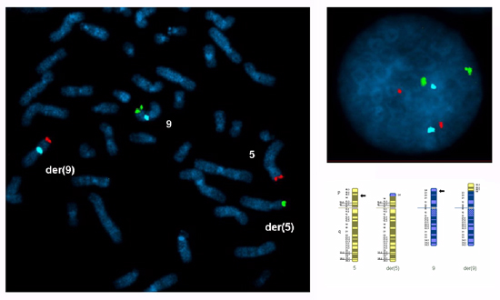
Figure 1. A metaphase spread and an interphase nucleus prepared from cultured peripheral blood lymphocytes from a carrier of a reciprocal translocation between the short arms of chromosomes 5 and 9 with breakpoints at 5p14.3 and 9p24.1: 46,XY,t(5;9)(p14.3;p24.1).ish t(5;9)(5ptel48-,9ptel30+;9ptel30-,5ptel48+,9cen+). FISH probes were selected for both translocated segments (5p14.3→5pter, red Cytocell subtelomere5ptel48 TexasRed ; 9p24.1→9pter, green Cytocell subtelomere 9ptel30 FITC) and the centric segment of chromosome 9 (9p24.1→9qter, blue Abbott CEP 9 alpha satellite SpectrumAqua).

Figure 2
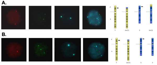
Figure 2. Signals in interphase blastomere nuclei from day-3 embryos captured using a different filter for each fluorochrome and merged to form a composite image. (A) Two signals for each probe indicating two copies of each locus, which is consistent with a normal or balanced complement for the translocation chromosomes. (B) One red signal, three green signals and two blue signals indicating one copy of the translocated segment of chromosome 5, three copies of the translocated segment of chromosome 9 and two copies of the centric segment of chromosome 9, which is consistent with adjacent-1 segregation of the translocation resulting in an unbalanced product with monosomy for 5p14.3→5pter and trisomy for 9p24.1→9pter.

Discussion

The application of fluorescence in situ hybridization (FISH) to a single embryo cell (blastomere) presents special challenges both in practicalities and in interpretation of the signal pattern. The biopsied cell needs to be spread within a pre-defined area on the slide in order to facilitate its localization following FISH; extreme care needs to be taken in ensuring that the cell is lysed, that the cytoplasm has been dispersed, and that the nucleus is visible and intact; and, as the diagnosis depends on the results from this single cell, stringent scoring and interpretation guidelines should be applied. However, in experienced hands, FISH is a robust technique for pre-implantation genetic diagnosis (PGD) in clinical practice. The principle of PGD by FISH is that target-specific DNA probes labelled with different fluorochromes or haptens can be used to detect the copy number of specific loci, and thereby to detect chromosome imbalance associated with meiotic segregation of chromosome rearrangements (1), including Robertsonian translocations, reciprocal translocations, inversions, and complex rearrangements (2). FISH can also be used to select female embryos in families with X-linked disease, for which there is no mutation-specific test (3, 4). More controversially, FISH has also been used to screen for sporadic chromosome aneuploidy in order to try and improve the efficiency of assisted reproduction (5, 6); however, the predictive value of this test using FSIH is likely to be unacceptably low in most people’s hands and it is not recommended for routine clinical use (7). This article concentrates on the technical aspects and the limitations of FISH applied to clinical single-cell diagnosis.

Methods of spreading and fixing single blastomeres include methanol/acetic acid (5, 8), Tween/HCl (9), and a combination of Tween/HCl and methanol/acetic acid (10). Variations include hypotonic treatment of cells prior to spreading and/or pepsin and paraformaldehyde treatment after fixation. The method should be appropriately validated for the laboratory (14). The Tween/HCl method is described in detail in this article. The Tween/HCl method is technically simple and highly reproducible in different laboratories. This method can be used to prepare single nuclei for the FISH diagnosis of sex determination and chromosome rearrangements with acceptable diagnostic accuracy (7).

Probe mixes can combine directly labelled and indirectly labelled probes, and probes from different manufacturers. Probes for known polymorphic chromosome regions (11, 12), or those known to cross-hybridize significantly with other chromosomes (13) should be avoided, although can be used if shown to be specific and suitable for PGD by prior testing on both reproductive partners. Available fluorochromes/haptens and strategies for discriminating probes include the following: Texas Red (TR), fluorescein isothiocyanate (FITC), SpectrumGreen (Vysis), SpectrumOrange (Vysis), SpectrumAqua, biotinylated probes detected with TR-avidin, FITC-avidin, or Cy-5 streptavidin (visualized using a FarRed filter), a mix of red and green probes to produce a yellow signal, a second round of hybridization and a third color created by sequential capturing of SpectrumOrange using a TexasRed and a SpectrumGold filter).

A probe set containing three probes, specific for the centromere regions of the X and Y chromosomes and one autosome, is recommended for sex determination (14); the autosomal probe is used to establish ploidy and thereby to differentiate between trisomy X (2n, 47,XXX) and triploidy (3n, 69,XXX), and between tetrasomy X (48,XXXX) and tetraploidy (4n, 92,XXXX). A typical probe set applied in this setting is the Abbott AneuVysion mix containing alpha-satellite X, Y, and 18; this probe set has been demonstrated to have a very low polymorphism rate, and therefore pre-treatment work-up of the reproductive partners is not required (14).

The probe mix for any specific rearrangement should: Ideally contain probes at least sufficient to detect all the expected products of the rearrangement with chromosome imbalance. If this is not possible, probe mixes where the undetected unbalanced products have been assessed to be non-viable in a recognizable pregnancy and are likely to have very low prevalence may be acceptable (14). Be tested on cultured lymphocyte metaphases from both reproductive partners. At least ten metaphase spreads should be examined for probe specificity, polymorphisms and cross-hybridization, and, for the chromosome rearrangement carrier, to ensure that the probes hybridize as expected to the different segments of the rearrangement. In addition, at least 100 interphase nuclei from these preparations should be scored to assess signal specificity, brightness, and discreteness (14).

Commercial PGS probe sets are available (e.g. Abbott MultiVysion PB or PGT), targeting the chromosomes most frequently found to be aneuploid in products of conception, and comprising a single probe per chromosome targeted. Typically the nucleus may have a second hybridization with probes for additional chromosomes providing an assay for chromosomes 13, 15, 16, 18, 21, 22, and XY (14). The predictive value of this test using the spreading and FISH technique described here is likely to be unacceptably low in most people’s hands and it is not recommended for routine clinical use (7, 15).

Techniques such as comparative genomic hybridization (CGH) applied to single cells are able to test for the copy number of every chromosome (Wilton et al. 2001), and the use of single nucleotide polymorphisms (SNP) arrays and quantitative analysis (Wells et al. 2008) or “karyomapping” genotyping (Handyside et al. 2009) are promising alternative techniques to detect chromosome aneuploidy. However, the resolution limit and accuracy for segment imbalance remain uncertain and it is likely that FISH will continue to be the technique of choice for chromosome rearrangements involving small segments.

Declarações

The authors have nothing to disclose.

Materials

Material Name Tipo Company Catalogue Number Comment
Hydrochloric Acid, 2.5L   VWR 101254H  
Sodium Chloride, 5Kg   VWR 443827W  
Potassium Chloride, 250g   VWR 26764.232  
di-Sodium Hydrogen Orthophosphate Anhydrous, 500g   VWR 102494C  
Potassium Dihydrogen Orthophosphate, 500g   VWR 10203 4B  
Sodium Hydroxide Pellets, 500g   VWR 28245.265  
Tween20, 500mL   VWR 663684B  
Ethanol, 2.5L   VWR 8.18760.2500 Duty Paid
Tri-Sodium Citrate, 5Kg   VWR 27833.363  
Cow Gum, 250mL   Cow Proofings Ltd. No longer available Old stocks can still be found in art shops
DAPI/Antifade ES, 0.125μg/mL, 500ul   Cytocell DES500L 150μl vial rec’d with each Cytocell TEL Probe
Nail Varnish, 12mL   Boots 10088316001  
Amine-coated Slides, 25   Genetix K2615  
DAPI, 0.1mL   Sigma-Aldrich D-9542  
Vectashield, 10mL   Vector Laboratories H1000  
Texas-Red-avidin, 0.001mL   Vector Laboratories A-2016  
FITC-avidin, 0.001mL   Vector Laboratories A-2011  
Cy-5 Streptavidin, 0.001mL   GE Healthcare PA45001  
Microcentrifuge Tubes, 1.75mL, box of 500   Thistle Scientific AX-MCT-175-C  
Microcentrifuge Tubes, 0.6mL, box of 1000   Thistle Scientific AX-MCT-060-C-CS  
30ml Universal Containers, box of 400   Sterilin 128B Available from NHS Supply Chain, Cat No. KCP053
Hot Block        
Spirit Burner
Spirit Burner Socket
Spirit Burner Wick
  VWR
VWR
VWR
451-0107
451-3112
451-3111
 
Plugged Glass Pipettes   Fisher Scientific PMK-300-052R  
Dissecting Microscope   Wild Heerbrugg    
Tissues, 100boxes   NHS SUPPLY CHAIN MJT058  
Metal Culture Trays        
Plastic Melamine Trays   VWR 682-009  
Oven        
Mouth Pipette Mouthpiece   Scientific Laboratory Supplies HAE3700  
Tubing        
Syringe Filter, 0.2μm pore size, 25mm diameter   Fisher Scientific FDM-340-010U  
Diamond Pen   VWR 818-0021  
Hot Block for Slides   Thermo Hybaid HBOSBB  
Hybridization Chamber        
Tissue Roll   NHS SUPPLY CHAIN MJT004  
Schieferdecker Jar   VWR 631-9313  
Coplin Jar   VWR 631-9331  
Forceps, Fine   VWR 232-2123  
Forceps, Blunt   VWR 232-2113  
1000mL Beaker   VWR 213-1642  
Phase Contrast Microscope   Nikon E200  
Fibre-free Blotting Cards, box of 100   Fisher Scientific (Raymond Lamb) SDE1  
Blue-Frosted Microscope Slides, box of 100   Scientific Laboratory Supplies MIC3022  
Coverslips, 18x18mm, No. 1, box of 200   Scientific Laboratory Supplies MIC3110  
Coverslips, 22x22mm, No. 0, box of 200   Scientific Laboratory Supplies MIC3104  
Coverslips, 22x50mm, No. 0, box of 100   Scientific Laboratory Supplies MIC3206  
1mL Syringe, box of 100   NHS Supply Chain FWC045  
1mL Pipette Tip, 10racks of 1000   Thistle Scientific AX-T-1000-C-L-R  
Incubator   LEEC    
Waterbath   Grant W22  
Alcohol Thermometer       Various sources available
pH Meter   Jenway 3305  
pH Electrode   VWR 662-1759 BNC Plug to fit Jenway pH meter 3305
Heated Stirrer   Bibby HC502  
1mL Pasteur Pipettes, box of 500   Scientific Laboratory Supplies PIP4202  
Parafilm, 50mm x 75m   VWR 291-1214  

Referências

  1. Scriven, P. N., Handyside, A. H., Ogilvie, C. M. Chromosome translocations: segregation modes and strategies for preimplantation genetic diagnosis. Prenat. Diagn. 18, 1437-1449 (1998).
  2. Mackie Ogilvie, C., Scriven, P. N., Harper, J. C. Preimplantation genetic diagnosis for chromosome rearrangements. Preimplantation Genetic Diagnosis. , 194-201 (2009).
  3. Harper, J. C., Coonen, E., Ramaekers, F. C. S., Delhanty, J. D. A., Handyside, A. H., Winston, R. M. L., Hopman, A. H. N. Identification of the sex of human preimplantation embryos in two hours using an improved spreading method and fluorescent in situ hybridisation using directly labelled probes. Hum. Reprod. 9, 721-724 (1994).
  4. Mackie Ogilvie, C., Scriven, P. N., Harper, J. C. Preimplantation genetic diagnosis for sex-linked diseases and sex-selection for non-medical reasons. Preimplantation Genetic Diagnosis. , 230-235 (2009).
  5. Munné, S., Lee, A., Rosenwaks, Z., Grifo, J., Cohen, J. Diagnosis of major chromosome aneuploidies in human preimplantation embryos. Hum. Reprod. 8, 2185-2192 (1993).
  6. Mackie Ogilvie, C., Scriven, P. N., Harper, J. C. Preimplantation genetic diagnosis for infertility (preimplantation genetic screening). Preimplantation Genetic Diagnosis. , 203-229 (2009).
  7. Scriven, P. N., Bossuyt, P. M. Diagnostic accuracy: theoretical models for preimplantation genetic testing of a single nucleus using the fluorescence in situ hybridization technique. Hum Reprod. Cytogenetics. 5, 394-400 (2010).
  8. Coonen, E., Dumoulin, J. C., Ramaekers, F. C., Hopman, A. H. Optimal preparation of preimplantation embryo interphase nuclei for analysis by fluorescence in-situ hybridization. Hum. Reprod. 9, 533-537 (1994).
  9. Dozortsev, D. I., McGinnis, K. T. An improved fixation technique for fluorescence in situ hybridization for preimplantation genetic diagnosis. Fertil. Steril. 76, 186-188 (2001).
  10. Hsu, L. Y., Benn, P. A., Tannenbaum, H. L., Perlis, T., Carlson, A. D. Chromosomal polymorphisms of 1, 9, 16, and Y in 4 major ethnic groups: a large prenatal study. Am. J. Med. Genet. 26, 95-101 (1987).
  11. Shim, S. H., Pan, A., Huang, X. L., Tonk, V. S., Varma, S. K., Milunsky, J. M., Wyandt, H. E. FISH variants with D15Z1. J. Assoc. Genet. Technol. 29, 146-151 (2003).
  12. Knight, S. J., Flint, J. Perfect endings: a review of subtelomeric probes and their use in clinical diagnosis. J. Med. Genet. 37, 401-409 (2000).
  13. Thornhill, A. R., de Die-Smulders, C. E., Geraedts, J. P., Harper, J. C., Harton, G. L., Lavery, S. A., Moutou, C., Robinson, M. D., Schmutzler, A. G., Scriven, P. N., Sermon, K. D., Wilton, L. ESHRE PGD Consortium (PGS) ESHRE PGD Consortium ‘Best practice guidelines for clinical preimplantation genetic diagnosis (PGD) and preimplantation genetic screening (PGS)’. Hum. Reprod. 20, 35-48 (2005).
  14. Munné, S., Wells, D., Cohen, J. Technology requirements for preimplantation genetic diagnosis to improve assisted reproduction outcomes. Fertil. Steril. 94, 408-430 (2010).
  15. Wilton, L., Williamson, R., McBain, J., Edgar, D., Voullaire, L. Birth of a healthy infant after preimplantation confirmation of euploidy by comparative genomic hybridization. N. Engl. J. Med. 345, 1537-1541 (2001).
  16. Wells, D., Alfarawati, S., Fragouli, E. Use of comprehensive chromosomal screening for embryo assessment: microarrays and CGH. Mol. Hum. Reprod. 14, 703-710 (2008).
  17. Handyside, A. H., Harton, G. L., Mariani, B., Thornhill, A. R., Affara, N., Shaw, M. A., Griffin, D. K. Karyomapping, a universal method for genome wide analysis of genetic disease based on mapping crossovers between parental haplotypes. J. Med. Genet. 47, 651-658 (2010).
check_url/pt/2570?article_type=t

Play Video

Citar este artigo
Scriven, P. N., Kirby, T. L., Ogilvie, C. M. FISH for Pre-implantation Genetic Diagnosis. J. Vis. Exp. (48), e2570, doi:10.3791/2570 (2011).

View Video