Insect hemocytes carry out many important functions, both immune and non-immune, throughout all stages of insect development. Our present knowledge of hemocyte types and function comes from studies on insect genetic models. Here, we present a method for extracting, quantifying and visualizing hemocytes from wild caterpillars.
Insect hemocytes (equivalent to mammalian white blood cells) play an important role in several physiological processes throughout an insect’s life cycle 1. In larval stages of insects belonging to the orders of Lepidoptera (moths and butterflies) and Diptera (true flies), hemocytes are formed from the lymph gland (a specialized hematopoietic organ) or embryonic cells and can be carried through to the adult stage. Embryonic hemocytes are involved in cell migration during development and chemotaxis regulation during inflammation. They also take part in cell apoptosis and are essential for embryogenesis 2. Hemocytes mediate the cellular arm of the insect innate immune response that includes several functions, such as cell spreading, cell aggregation, formation of nodules, phagocytosis and encapsulation of foreign invaders 3. They are also responsible for orchestrating specific insect humoral defenses during infection, such as the production of antimicrobial peptides and other effector molecules 4, 5. Hemocyte morphology and function have mainly been studied in genetic or physiological insect models, including the fruit fly, Drosophila melanogaster 6, 7, the mosquitoes Aedes aegypti and Anopheles gambiae 8, 9 and the tobacco hornworm, Manduca sexta 10, 11. However, little information currently exists about the diversity, classification, morphology and function of hemocytes in non-model insect species, especially those collected from the wild 12.
Here we describe a simple and efficient protocol for extracting hemocytes from wild caterpillars. We use penultimate instar Lithacodes fasciola (yellow-shouldered slug moth) (Figure 1) and Euclea delphinii (spiny oak slug) caterpillars (Lepidoptera: Limacodidae) and show that sufficient volumes of hemolymph (insect blood) can be isolated and hemocyte numbers counted from individual larvae. This method can be used to efficiently study hemocyte types in these species as well as in other related lepidopteran caterpillars harvested from the field, or it can be readily combined with immunological assays designed to investigate hemocyte function following infection with microbial or parasitic organisms 13.
1. Material Preparation
2. Injection of Caterpillars with Collection Solution
3. Extraction of Hemocytes
4. Quantification of Hemocytes
Total volume of each square of the hemocytometer grid: (0.04 mm2 area) × (0.1 mm depth) = 0.004 mm3 × 15 squares = 0.06 mm3 = the total volume of all 15 counted squares.
(Sum of the total number of hemocytes in the 15 squares counted / the total volume of all 15 counted squares) x 2 dilution factor = the number of hemocytes/mm3 x 1,000 = the number of hemocytes/ml of hemolymph.
The protocol described herein allows the collection of a minimum volume of 10-20 μl of insect hemolymph from individual caterpillars. Hemocytes collected using this method are free of cell clumping, melanization defects, tissue debris or other contaminants. Therefore hemocytes can be readily observed and counted under the microscope, and several insects can be observed within a few hosur. We observed that the majority of hemocytes in our samples consisted of plasmatocytes 3 (hemocytes spreading asymmetrically) (Figure 4). We are currently characterizing the different hemocyte types found in these caterpillars in detail (Stoepler et al., unpublished data). The method described here provides a quick and accurate means for counting total hemocyte numbers for individual caterpillars collected from field experiments.
Although hemocyte density can vary greatly between and within insect species 1, we found that hemocyte numbers for L. fasciola caterpillars range from 1.10 × 10⁶ – 8.23 × 10⁶ cells/ml of hemolymph (mean: 3.51 × 10⁶; n = 31 caterpillars), and are in the range of previously reported values in other lepidopteran species 14.
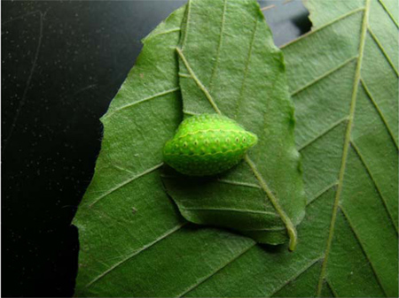
Figure 1. Photograph of a penultimate instar Lithacodes fasciola (Lepidoptera: Limacodidae) caterpillar. Typical body length of these caterpillars ranges from 10-15 mm in penultimate to ultimate instars. Photo credit: John T. Lill.
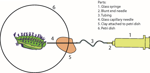
Figure 2. Injection needle setup. A glass syringe is connected to a capillary needle via a blunt end needle and plastic tubing (panels 1-4). A piece of plastic clay is used to rest the capillary needle (5). The caterpillar is placed in a Petri dish for injection with the collection solution (6).
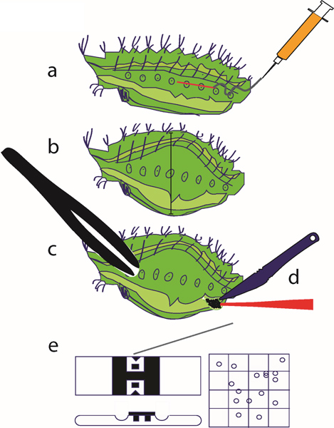
Figure 3. Hemocyte extraction method. (a) The chilled caterpillar is injected with the collection solution; (b) following injection, the caterpillar’s body will swell slightly, and the injected insect is allowed to rest on ice for 30 min; (c, d) a cut is made using a clean scalpel, the caterpillar is squeezed using a pair of plastic forceps to force the hemolymph out of the body through the incision, and hemocytes are collected with a pipette; (e) hemocytes are counted using a hemocytometer.
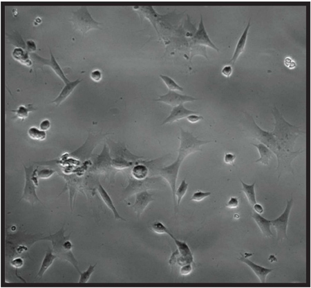
Figure 4. Microscopy picture showing extracted hemocytes from Euclea delphinii (Lepidoptera: Limacodidae) caterpillars (magnification: 20x).
Methods for extracting hemocytes from medically important insects and lepidopteran model insects have previously been reported 9, 14. Hemocyte extraction methods are adapted according to the insect species, the developmental stage of the insect and its morphological features. For example, hemolymph isolation from Manduca larvae can be readily performed by snipping the curved horn near the end of the abdomen 15. Because slug caterpillars (Limacodidae) lack this abdominal structure, we have developed a distinct protocol for hemolymph extraction from small-sized field-collected caterpillars. This method proves efficient for isolating sufficient hemolymph volumes containing large numbers of hemocyte cells that can be stored or processed for further experimentation. In addition, hemocyte collection from adult mosquitoes includes a time-consuming step that involves the careful removal of legs, wings and the tip of the abdomen 16. A similar step is not required in our protocol, thus isolation of hemocytes from slug caterpillars is a simpler and quicker process. Limitations of our technique may include: 1) as with all hemocyte extraction methods, insects must be sacrificed on the day of the assay, and 2) hemocyte numbers may vary significantly between larvae of the same species and developmental stage, which necessitates the use of several individuals in order to calculate mean numbers of hemocytes per volume of extracted hemolymph.
We intend to utilize this method in future studies to morphologically characterize hemocyte cell types from different developmental stages of forest-harvested caterpillars. This assay can also provide the first critical step for isolating healthy hemocytes to examine in detail several physiological aspects of the insect cellular immune response, including the effect of various developmental stages and nutritional conditions on the total number of hemocytes in naïve or infected caterpillars obtained from their natural environments. Given the chronic exposure of lepidopteran larvae to a range of different pathogens in nature, as well as the potentially continuous presence of their microbial symbionts, it is anticipated that these insects possess types of hemocytes with different properties compared to immune cells found in laboratory-reared insects. In addition, this hemocyte extraction protocol can also be applied to studies with other groups of insects, allowing immunological-related assays to be incorporated into ecological field studies, thus increasing our understanding of cellular defense mechanisms in non-model insect species of agricultural or ecological importance. In conclusion, our method will provide a valuable resource to insect biologists, ecologists and those interested in understanding the functional interplay between hemocyte abundance and the ability of wild insect populations to withstand microbial or parasitic infections.
The authors have nothing to disclose.
TMS was supported by the Harlan Trust Fellowship (GWU) during this study. Funding for field collection and breeding L. fasciola and E. delphinii was provided by NSF grant DEB 0642438 to JTL.
Name of the reagent | Company | Catalogue number | Comments |
Micropipette puller | Sutter Instruments | P-1000 | |
Borosilicate glass tubes | Sutter Instruments | B100-50-10 | OD: 1.0, ID: 0.50 mm |
Grace’s Medium | Sigma | G8142 | |
Fetal Bovine Serum | Thermo Fisher Scientific | SH3007102 | Heat inactivated |
Dumont #5 Forceps | Fine science tools | 11252-40 | |
Neubauer hemocytometer | Hausser Scientific | 3200 | |
Plastic tubing | Tri-Tech | TT-3-32OD | OD: 3/32”, ID: 1/32′ |
Glass medical syringe | Fortuna Optima | D-97877 | 50 ml volume |
Blunt end needle | Small Parts | NE-162PL-25 | 16 Gauge x 1″ length |