An adaptable reverse genetic method for zebrafish to assess gene function during later stages of development and physiological homeostasis such as tissue regeneration using intraventricular injections of gene-specific morpholinos.
The zebrafish is an important model to understand the cell and molecular biology of organ and appendage regeneration. However, molecular strategies to employ reverse genetics have not yet been adequately developed to assess gene function in regeneration or tissue homeostasis during larval stages after zebrafish embryogenesis, and several tissues within the zebrafish larva are difficult to target. Intraventricular injections of gene-specific morpholinos offer an alternative method for the current inability to genomically target zebrafish genes in a temporally controlled manner at these stages. This method allows for complete dispersion and subsequent incorporation of the morpholino into various tissues throughout the body, including structures that were formerly impossible to reach such as those in the larval caudal fin, a structure often used to noninvasively research tissue regeneration. Several genes activated during larval finfold regeneration are also present in regenerating adult vertebrate tissues, so the larva is a useful model to understand regeneration in adults. This morpholino dispersion method allows for the quick and easy identification of genes required for the regeneration of larval tissues as well as other physiological phenomena regulating tissue homeostasis after embryogenesis. Therefore, this delivery method provides a currently needed strategy for temporal control to the evaluation of gene function after embryogenesis.
Regeneration of organs and appendages is fundamentally important for survival and fitness; however, several vertebrates including man have limited regenerative abilities. While several animal models exist that have extensive regenerative capacity, reverse genetic techniques to assess gene function during organ and appendage regeneration remain very limited or non-existent. Therefore, new approaches are required to dissect the molecular biology of regeneration in these model organisms.
The zebrafish is a well-established model for understanding the cell and molecular biology of organ and appendage regeneration1, not only because of its significant ability to regenerate multiple organs, tissues and appendages, but also because several transgenic fish lines exist to track cells and to overexpress gene constructs2,3. However, gene inhibition in larval zebrafish is mainly limited to the overexpression of dominant-negative constructs, which are not available to all genes of interest or whose transgene product can acquire gain-of-function effects that do not reflect the endogenous activity of the gene. Thus, an alternative method to specifically remove gene expression by knockout or knockdown is needed to overcome these issues.
Gene-specific targeting using TALENS exists as a reverse genetic means to knockout gene function; however, this knockout strategy is very frequently limited to functional assessments during early embryogenesis, because initial requirements of the gene prevent further progression of embryonic development. Thus, studying later phenomena such as regeneration or organ homeostasis after development using TALENS is precluded4,5. Therefore, an alternate gene removal strategy is needed that targets gene function after early development to assess gene requirements in fully formed organs and structures.
Morpholino injection has been shown to be effective in targeting genes in a few adult organs and the adult regenerating fin6-8, but these methods require electroporation and many internal organs are difficult to electroporate either due to their location or due to their sensitivity to electrical disruption. Furthermore, some tissues in the larva are difficult to inject directly, because direct injection may disrupt their structural integrity or because their size is limiting. The caudal fin of the larva is one such structure, because direct injection into the finfold is not possible. Thus, an alternative to electroporation and direct injection was needed to target genes in tissues that are either too small to inject or can't be electroporated.
In order to target and inhibit the function of specific genes during the regeneration of the larval caudal fin, we have modified existing morpholino technologies allowing the assessment of gene function during caudal fin regeneration in late-staged larva. This method employs intraventricular delivery9 of fluorescein-tagged morpholinos together with Endo-Porter transfection reagent10. Once in the ventricle, the morpholino-Endo-Porter mixture quickly spreads throughout the larva via the vasculature and enters tissues that have been previously impossible to target. This injection method may be modified to target genes in specific tissues and quite possibly can be applied in other animal models that currently lack reverse genetic methods to inhibit gene function. Thus, it offers a quick and easy method with the potential for broad range use to immediately study gene function during general organ homeostasis and regeneration at larval stages.
1. Preparation of the Glass Needles (Figure 1)
2. Preparation of the Morpholino Solution
3. Preparation of the Morpholino Injection (Figure 2)
4. Injections (Figure 3)
5. Analyses of Injections (Figure 4)
6. Assessment of Regenerative Outgrowth
The sharp, beveled injection needle is easily placed in the cardiac ventricle of the zebrafish larva when approached dorsally (Figure 3A). The heart continues pumping and blood flow is maintained despite the presence of the needle (Figures 3B and 3C). Careful injections do not disrupt the morphology of the ventricle or cardiac contractions (Figure 3D) despite injection of the morpholino into the heart (Figure 3E).
Within minutes, the morpholino-Endo-Porter solution distributes throughout the body via the vasculature (Figures 4A and 4B). The morpholino is then distributed from the vasculature into different tissues such as the finfold and brain for at least 12 hr or more as observed from the fluorescence (Figure 4C).
In this approach, we remove the distal tip of the finfold, the distal tip of the spinal cord, distal trunk muscle, the distal notochord and pigment cells (Figures 4D and 4H); all of which are difficult to specifically target by direct injection due to compactness (muscle), small size (spinal cord and notochord) and thinness (finfold), but appear to incorporate the morpholino after serial ventricular injections, which is seen by fluorescence within these tissues (Figure 4E) compared to uninjected animals (Figures 4F and 4G). Thus, this method relatively easily promotes the delivery of morpholino to tissues that are difficult to individually target, and it permits the assessment of gene function in several tissues in the regenerating fin at once. To assess the importance of specific genes involved in regeneration, one surgically partially amputates the larval caudal fin (Figure 4H), resecting all the tissues that have incorporated the morpholino (Figure 4I). The larval finfold regenerates its structure within a few days post amputation (dpa) (Figure 4J). Morpholino targeting a gene required for regeneration (unpublished data) results in a perturbed regeneration response (Figure 4K) compared to uninjected (Figure 4J) and mismatch morpholino controls (Figure 4L). The total effect of these morpholino experiments on regenerative outgrowth can be measured using standard morphometric tools in the publically available Fiji programs12 by tracing the regenerated tissues and comparing the areas (Figures 4J-4L). Thus, this gene-targeting method provides a quick and relatively easy assay to test the importance of genes in the regeneration process.
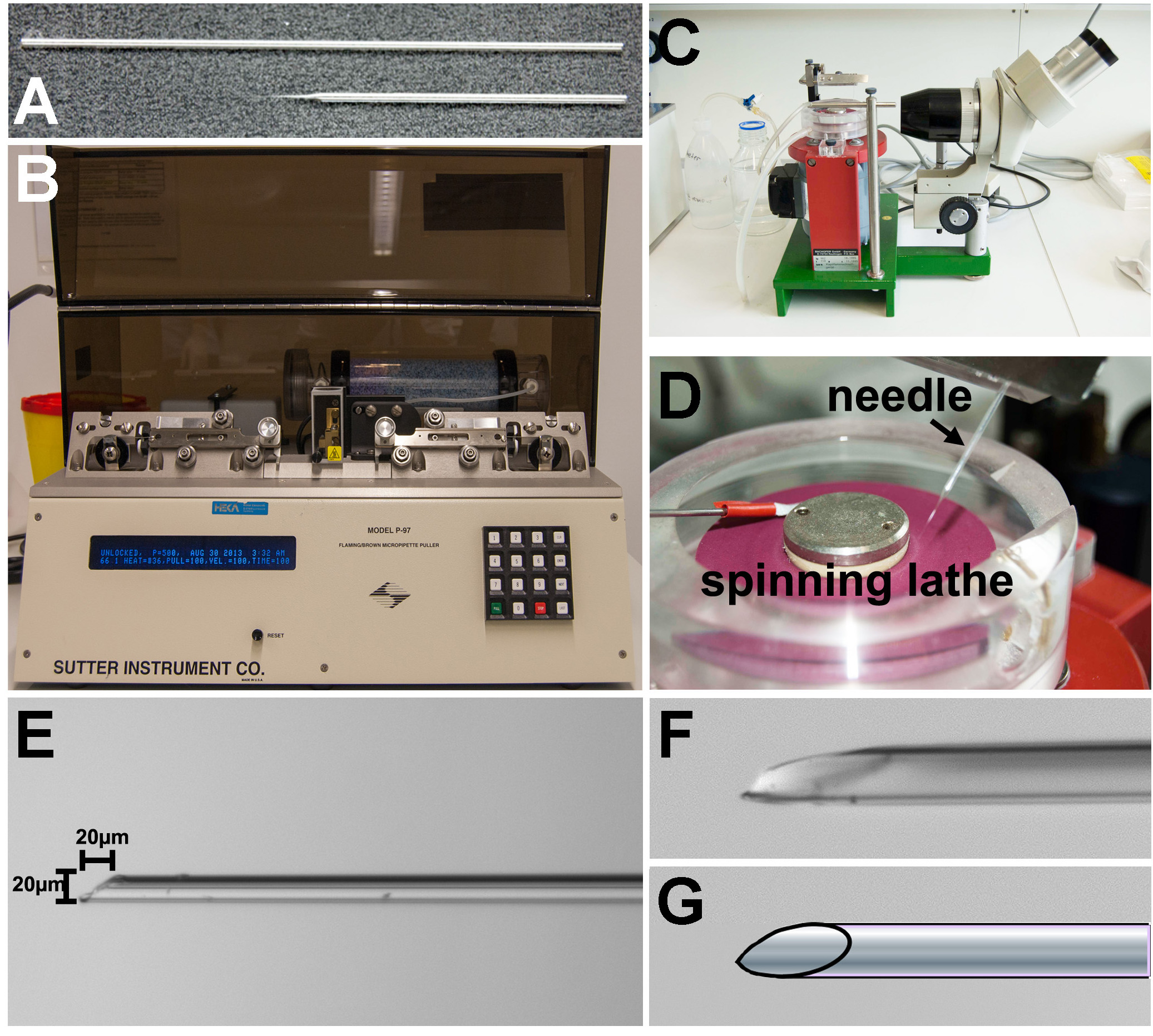
Figure 1. Preparation of glass capillary needles for injection. A) Glass capillary tube before pulling (above) and pulled needle (below). B) Needle pulling apparatus. C) Lathe for beveling and sharpening the glass needle. The needle is viewed through the binoculars. D) The lathe is a wetted rubber spinning disc. The needle is slowly lowered onto the disc. E) Sharpened needle must have a short bevel that is no longer (20 µm) than is wide (20 µm) to minimize the space needed to insert the entire end of the needle into the larval cardiac ventricle. F) Higher magnification image showing the tip of the needle. G) Schematic view of the needle tip showing the shape of the bevel after sharpening.
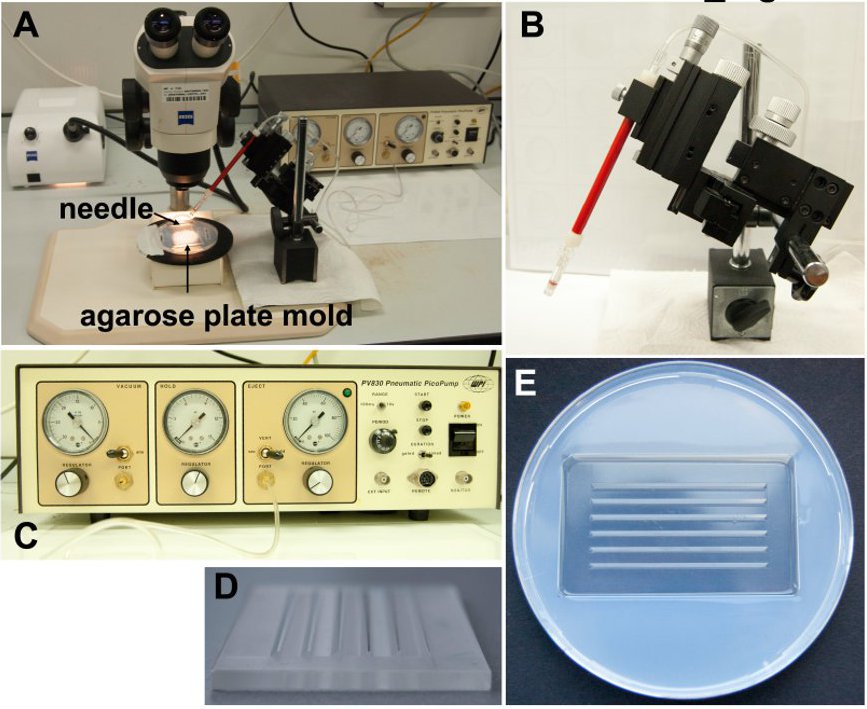
Figure 2. Injection apparatus set up. A) Complete apparatus for injection of morpholino into zebrafish larva. B) Stand to control the placement of the needle during injections. C) The pico pump that regulates pneumatic pressure for injections. D) Form press used to create agarose mold. E) Agarose mold used to stabilize larva for injection.
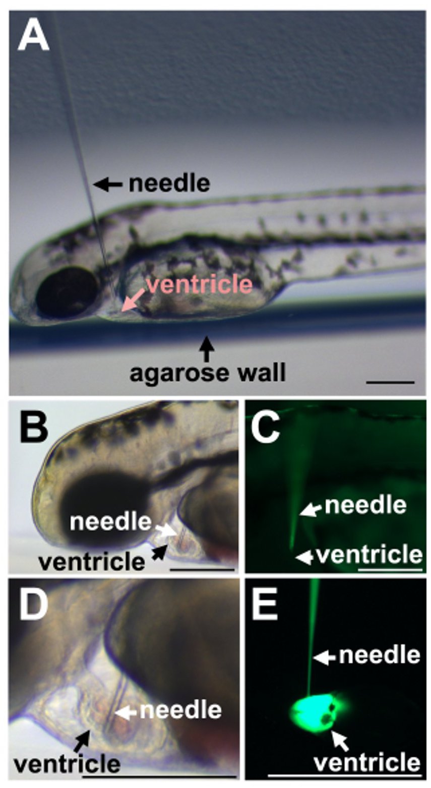
Figure 3. Injection of fish larva. A) Placement of the needle in relation to the zebrafish larva. B) Insertion into cardiac ventricle. C) Fluorescence of the morpholino in the needle but absent in the ventricle before injection. D) Brightfield image shows the size relationship between the glass needle and the larval ventricle. E) Injection of the fluorescent morpholino into the heart ventricle. Scale bars equal 100 µm.
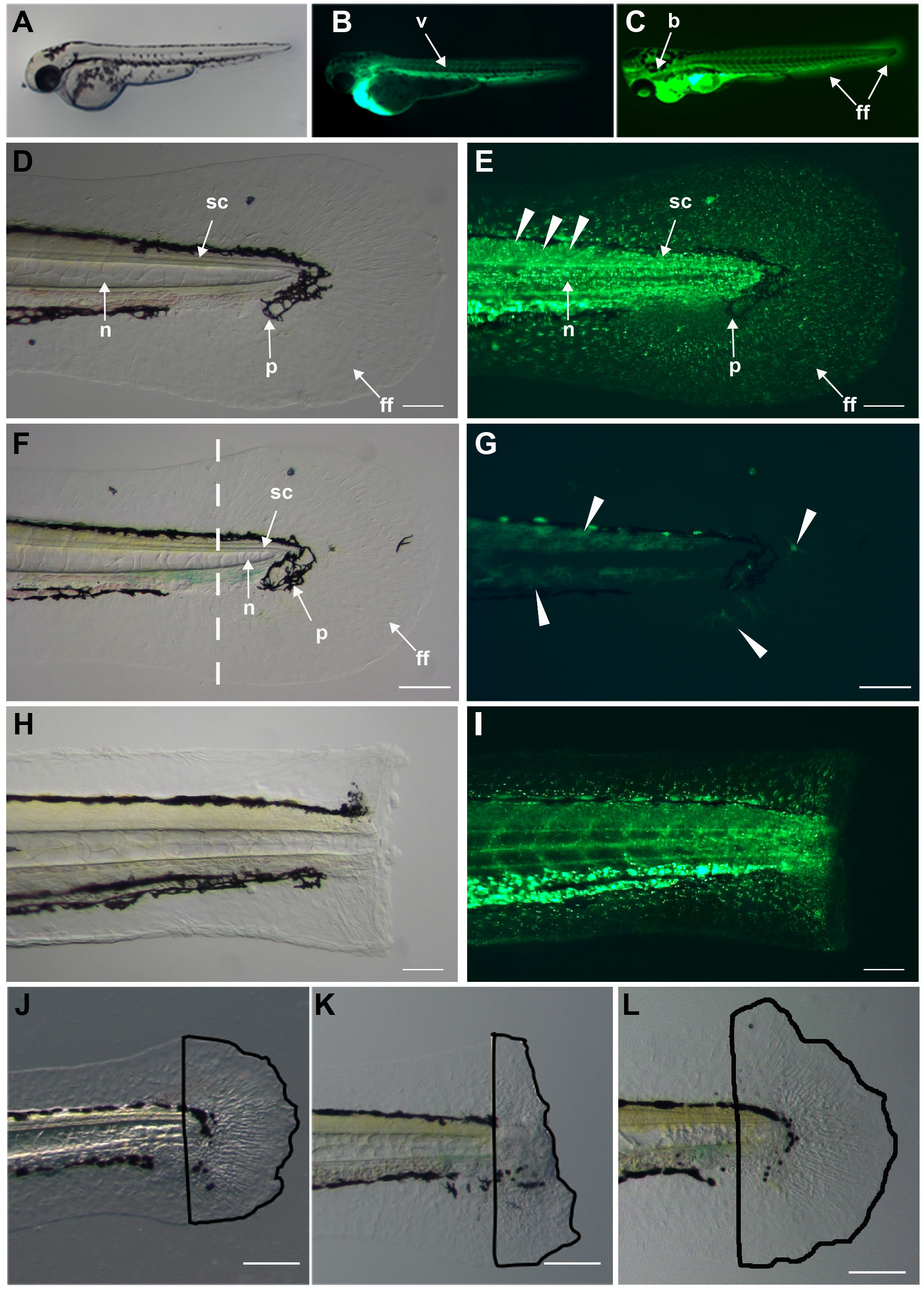
Figure 4. Dispersion of morpholino throughout fish. A) Zebrafish 3 day old larva. B) Fluorescence of fluorescein-tagged morpholino throughout the body vasculature (v) 5 min post ventricular injection. C) Dispersion of the morpholino throughout the animal including finfold (ff), brain (b) in older larva after injections of morpholino every 12 hr. D) Tissues in the larval caudal fin and trunk: notochord (n), spinal cord (sc), pigment (p), and finfold (ff). E) Dispersed morpholino several minutes after injection into larval trunk tissues including the finfold. F) Brightfield image of uninjected larval trunk and caudal fin. Dashed line indicates the prospective amputation plane. G) Uninjected fin imaged with the GFP filter showing autofluorescence in green (arrowheads). The dendritic shape of the fluorescing structures suggests that these are pigment cells. H) Brightfield image of a surgically amputated larval caudal fin through the notochord, muscle and spinal cord. I) Fluorescent image showing morpholino distribution in the stump tissues. Morpholino incorporation into muscle, spinal cord, notochord and finfold. J) Regenerated caudal finfold structures of wild-type larva. Black line traces the regenerated tissues for quantification analysis. K) Morpholino-mediated inhibition of regeneration. Black line tracing clearly highlights the reduced regeneration response after morpholino knockdown. L) Regenerated caudal finfold of larva injected with a mismatch control morpholino showing a similar regeneration response as in uninjected larva (J). Scale bars equal 100 µm.
The intraventricular injection provides a quick and reliable assessment method for testing gene function at later stages of development or of body homeostasis without affecting the gene function during embryogenesis. To ensure the success of this technique, one should be aware of four critical points: 1) needle size, 2) drying out of the needle, 3) minimizing volume, and 4) minimal exposure time in the sedation solution. Needles that are too small will clog frequently, while needles that are too large will damage the ventricle and cause excessive bleeding. While 20 µm is recommended, the needle should never be more than one-fifth the size of the ventricular chamber. A second critical point is to keep the needle from drying out and clogging. The small size of the needle and the inevitable frequent pauses to sedate, transfer and position the larva can provide enough time to allow the morpholino at the needle tip to dry out and clog the needle. To prevent this, one can immerse the needle in a Petri dish of 1x PBS. A third critical point is to minimize bloating of the ventricle by too high of a volume. If the dosage is limited by the size of the needle, then increasing the number of injections is always preferable to increasing the volume: larger volumes can cause stretch injury to the heart. A fourth critical point is the extent of sedation. The sedation time is based on the length of time needed for the procedure. Larvae can remain in the sedation solution and remain sedated for several minutes, because they are rather robust and resuscitate rather easily. However, maintaining the fish too long in the anesthetic will impair its recovery or too short will cause them to revive during the injection. Therefore, one should sedate only the number of larva that one can inject within a 4 min period.
One limitation of this current method is that the morpholino does not target gene function in a tissue-specific manner. Two different modifications can provide tissue specificity: One modification is to use caged morpholinos. After activation by laser light from a confocal microscope, these morpholinos inhibit their gene target, so timed confocal exposure can provide temporal and spatial control to morpholino activity. Caged morpholinos have been used to target genes during early development after injection into the one-cell embryo. While delivery at single-cell stage can work to target genes during embryogenesis, the concentration of the morpholino will likely be too low at later larval stages due to the number of successive cell divisions as development progresses to the larval stage13. The amount of morpholino one can inject at the one-cell-stage is also limited by toxicity13-15. A second possible modification is to directly inject a morpholino into the organ or tissue to be studied when size and imaging allow. The zebrafish larva is translucent, so viewing most internal organs after they have developed should not be a problem. The fluorescein-tagged morpholinos work with this protocol; therefore, fluorescence can be used to track the distribution and tissue-specific localization of the morpholino. The incorporation of the fluorescein-tagged morpholino into tissue cells requires the presence of the Endo-Porter reagent, so another modification would be to limit the spatial distribution of the Endo-Porter reagent by injecting it at lower amounts directly into the target tissue. Thus, spatial resolution should be possible with this technique.
There are transgenic overexpression methods that provide for temporal and spatial control of exogenous gene expression by combining tissue-specific promoters with heat-shock or tamoxifen-inducible promoters which enables the analysis of gene function at later stages of development or in certain tissues by using tissue specific promoters16-19. This includes the transgenic expression of dominant-negative constructs designed to create a product that can outcompete the endogenous gene. This knockdown strategy is susceptible to off-target effects when a transgene produces disproportionate expression levels of a transgenic product that acquires gain-of-function effects20 or when it integrates into a gene and disrupts the gene's endogenous function21,22. While morpholinos can also generate off-target effects, one can more easily control and limit these effects by using mismatch control morpholinos with similar but slightly altered target sequences that do not bind the target mRNA, by using multiple antisense morpholinos that target different sequences within the same mRNA and by testing different morpholino concentrations13-15. Moreover, morpholinos can be produced and tested much faster and cheaper than designing, raising and testing different dominant-negative transgenic lines. Therefore, the intraventricular injection of morpholinos provides a useful tool to knockdown genes at later stages of development, when dominant-negative transgenesis is not possible. When a dominant-negative transgenic strategy is possible, intraventricular morpholino injection is an alternative approach to confirm the specificity of the overexpressed dominant-negative transgene.
It is likely that effective concentrations between different morpholinos will vary, as they do when using them for gene knockdown during embryogenesis13,15. For the gene targeted in this study, we did not observe an effect on regeneration at doses lower than 3.5 mM, and non-specific effects were not observed in mismatch controls with this morpholino concentration.
Intraventricular injection allows for the distribution of small molecules to assess activity and efficacy in a whole animal system in real time when efficacy is limited by penetration through the skin or digestive system or by the amounts of compound available. It also allows the targeting of tissues when they can´t be targeted by direct injections, as is the case for the finfold. This method does not only provide a tool to study gene function during fin regeneration at larval stages. Since the morpholino integrates into the larval finfold comprising mesenchymal cells and epidermis, it might also provide a suitable technique to investigate the role of genes that play part in the process of wound healing or the occurrence of skin cancer. The injection of morpholino together with Endo-Porter may also be applied to target organs or tissues in adult zebrafish, which are amenable to surgery and translucent adult zebrafish lines are available. Furthermore, this method is not limited to zebrafish; dispersion by intraventricular injection can be used on other vertebrate models as well, especially those models that still lack effective recessive gene methods such as Xenopus, Axolotl and newt, which are also important models for regeneration studies.
Thus, intraventricular injection in the zebrafish offers the ability to temporally control gene expression with a relatively quick protocol to target multiple tissues, and the versatility of this method allows for its use with zebrafish transgenic lines as well as for other organisms that lack functional transgenesis or gene knockout strategies.
The authors have nothing to disclose.
This work was supported by the Deutsche Forschungsgemeinschaft (DFG), we wish to thank Ayele Tsedeke Taddese for technical support.
Company | Catalog Number | Comments/Description | |
Reagent/Material | |||
Morpholino with 3´fluorescein tag | Gene Tools Inc. | Customized | More information on www.genetools.com |
Endo-Porter | Gene Tools Inc. | More information on www.genetools.com | |
Agarose | Serva | 120623 | For pouring plates to place the fish during injections and imaging |
Tricane (L-Ethyl-m-amino-benzate-methane sulfonate) | Applichem | For anesthesia | |
E3 medium (5 mM NaCl; 0.17 mM KCl; 0.33 mM CaCl2; 0.33 mM MgSo4) | For larval husbandry | ||
Petri Dishes | Sarstedt | 821.472 | For placing the fish during injections and imaging |
Equipment | |||
Thin Wall Glass Capillaries | World Precision Instruments, Inc. | TW100F-3 | For preparing injection needles |
Microloader pipette tips | Eppendorf | 5,242,956,003 | For loading the needle |
Pasteur Pipettes (3 ml/1 ml) | Brand | 747760/ 74775 | For transfering larva |
Flaming / Brown needle puller | Sutter | More information on www.sutter.com | |
Watchmaker (Dumont) Tweezers (size 5) | World Precision Instruments, Inc. | 501985 | To brake the needle before sharpening |
Dissecting microscope | Olympus, Leica, Zeiss | Varies with the manufacturer | Part of the whole injection setup |
Fluorescence camera | Olympus, Leica, Zeiss | Varies with the manufacturer | To image the fluorescence after injeciton |
PicoNozzle kit (microinjector holder) | World Precision Instruments | 5430-12 | For microinjections |
Pneumatic PicoPump (microinjector) | World Precision Instruments | SYS-PV820 | For microinjections |