Here we describe a protocol for producing harvesting needles that can be used to collect full-thickness skin tissue without causing donor site scarring. The needles can be combined with a simple collection system to achieve high-volume harvesting.
This manuscript describes the production process for a laboratory apparatus, made from off-the-shelf components, that can be used to collect microcolumns of full-thickness skin tissue. The small size of the microcolumns allows donor sites to heal quickly without causing donor site scarring, while harvesting full-thickness tissue enables the incorporation of all cellular and extracellular components of skin tissue, including those associated with deeper dermal regions and the adnexal skin structures, which have yet to be successfully reproduced using conventional tissue engineering techniques. The microcolumns can be applied directly into skin wounds to augment healing, or they can be used as the autologous cell/tissue source for other tissue engineering approaches. The harvesting needles are made by modifying standard hypodermic needles, and they can be used alone for harvesting small amounts of tissue or coupled with a simple suction-based collection system (also made from commonly available laboratory supplies) for high-volume harvesting to facilitate studies in large animal models.
Autologous skin grafting is the mainstay of wound repair, but it is limited by donor site scarcity and morbidity, leading to concerted efforts in recent decades to develop new therapeutic options to replace conventional skin grafting1,2. We recently developed an alternative method of harvesting skin to harness the benefits of full-thickness skin grafting while minimizing donor site morbidity. By collecting full-thickness skin in the form of small (~0.5 mm diameter) "microcolumns", donor sites are able to heal rapidly and without scarring under normal circumstances (for potential exceptions, see the discussion section below)3. Microcolumns can be applied directly into wound beds to accelerate wound closure, reduce contraction3, and restore a diverse range of epidermal and dermal cell types and functional adnexal structures4, many of which are lacking in conventional split-thickness skin grafting or current bioengineered skin substitutes5. The ability of microcolumns to augment healing and of their donor sites to heal without scarring have both been independently validated by other research groups6,7.
We have previously developed a laboratory harvesting system to enable the collection of microcolumns at scale8; however, this system is composed of many customized components that are not widely available. Here, we describe in detail the process for producing harvesting needles, as well as simple collection systems, made from mostly off-the-shelf components, that can be used to achieve high-volume harvesting. The apparatus described in this manuscript is suitable for in vitro and animal work, but not for use in humans. A clinical device with FDA clearance for applying this technique in humans is commercially available but will not be discussed in detail here.
All work involving live animals and animal tissue samples have been approved by the Massachusetts General Hospital Institutional Animal Care and Use Committee (IACUC).
1. Production of Harvesting Needles
2. Skin Tissue Harvesting
The harvesting needles should be able to collect microcolumns of full-thickness skin tissue with approximately a 80-90% success rate, and each microcolumn should contain epidermis, dermis, and some subcutaneous fat (Figure 4). If the success rate of harvesting is low, or if it becomes difficult to insert a needle into tissue, then a new needle is likely needed. If the success rate for harvesting is consistently low, even with new needles, then the needles are probably too short.
If used in vivo, donor sites should heal quickly, as re-epithelialization typically occurs within a few days3. Microcolumns can be applied directly to wound beds to augment wound healing3,4, or they may be combined with different matrix materials to produce combination constructs. Microcolumns can also be maintained in culture for in vitro studies9.
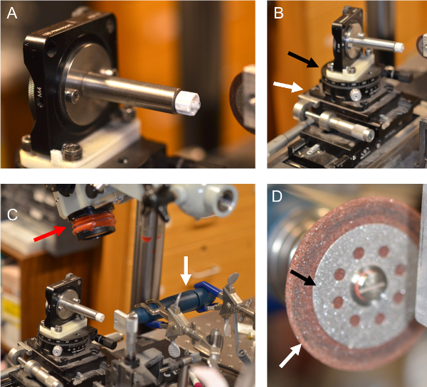
Figure 1: Needle making apparatus. (A) A male luer lock connector secured via a mounting post onto a vertically placed rotation stage, so that the luer lock is at the center of the stage. (B) The vertical rotation stage is mounted perpendicularly onto a second, horizontal rotation stage (black arrow). The horizontal rotation stage is secured to a two-axis translation stage (white arrow). (C) Positioning of the rotary tool parallel to the breadboard (white arrow), and the dissecting microscope over the needle-making apparatus (red arrow). (D) Concentric cut-off wheels mounted onto rotary tool, with a diamond wheel on the inside (black arrow) and stone wheel on the outside (white arrow). Please click here to view a larger version of this figure.
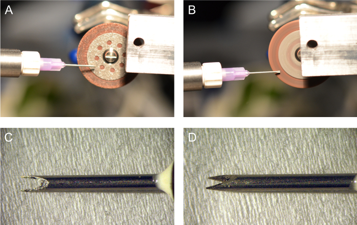
Figure 2: After cutting the needle to the desired length, the rotary tool is used to grind new needle tips. (A) First, the diamond cut-off wheel is used to make "rough" cuts to form the new cutting tips and surfaces. (B) After the new cutting tips are formed with the diamond wheel, the needle is moved to the stone wheel for fine polishing. (C) Finished harvesting needle viewed from the front and (D) from the side. Please click here to view a larger version of this figure.
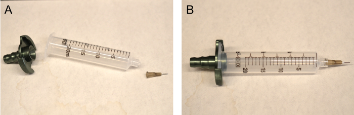
Figure 3: Assembly for high-volume harvesting. (A) Individual components of the assembly, including (left to right) the suction adapter, 20 mL syringe with luer lock nozzle, and harvesting needle. (B) Shown is the completed assembly, ready to connect to negative pressure source. Please click here to view a larger version of this figure.
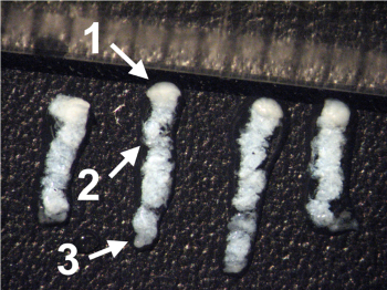
Figure 4: Representative skin microcolumns harvested using the apparatus described in this manuscript. Each microcolumn contains the epidermis (1), full dermis (2), and some subcutaneous fat (3). Checkmarks in the figure represent 1 mm. This figure has been modified from Tam et al.4 in accordance with the terms of the corresponding creative commons license. Please click here to view a larger version of this figure.
The methods described here are intended to enable the collection of tissue microcolumns in sufficient quantities for in vivo large animal studies, using tools made from commercially available laboratory supplies. This apparatus has been used previously in harvesting tissue from excised human skin4,9 as well as live swine skin3. The specific parameters described are those that were found to be most suited for use in swine. It is expected that the same apparatus can be modified and adapted for collecting tissue from rodents and other small animals, but this has not been tested in our laboratory.
Critical steps in this protocol include ensuring the harvesting needles are of sufficient length (inefficient harvesting is usually due to needles being too short), keeping the microcolumns submerged in liquid throughout the harvesting process to prevent desiccation, and flushing the system with saline at least intermittently (otherwise there is a higher likelihood of microcolumns clogging the needle bore). The main limitation of this technique is speed; for example, in our demonstration, the suction-assisted apparatus can generally harvest 120 mg of tissue per minute, which is sufficient for smaller wound sizes typically used in animal experiments. It would likely be logistically challenging to use this approach for very large wounds (e.g., in major burn injury models). Needle gauge is another limitation – the smaller the needle gauge, the more susceptible it is to buckling, which is the main failure mode of this technique (in contrast, the technique is relatively insensitive to needle dulling during the procedure). For swine skin, we normally use 19-gauge needles, which are mechanically robust enough that they rarely buckle. Futhermore, each animal experiment (typically involving about 3,000-5,000 microcolumns) usually requires only 2 to 3 needles. 25-gauge is the smallest needle size we have used with this technique.
The ability of small skin wounds to stimulate tissue regeneration is the underlying principle behind clinical procedures such as fractional laser resurfacing12 and microneedling13. These treatments are known to improve the cosmetic appearance of photoaged skin, and more recently, shown to induce scar remodeling and improve the function and cosmesis of skin scars14,15. The extensive clinical experience with these techniques also provides further validation that skin is able to heal without scarring after these microinjuries in the vast majority of cases, with certain exceptions (fractional laser resurfacing reportedly has a 3.8% incidence of scarring, almost always as a result of infection16, highlighting the importance of post-procedural skin care). In addition, people with a history of keloids or hypertrophic scarring may be susceptible to scarring even with these smaller injuries; thus, treatments involving the production of microinjuries may be counterindicated.
While our previous investigations have focused on directly applying microcolumns into skin wounds to enhance healing, the ability to collect significant amounts of tissue without causing other long-term donor site morbidities (scarring, contracture, etc.) may be useful for a broad range of other applications. Skin microcolumns may serve as the tissue source for approaches involving culture expansion or dissociation and dispersion of autologous skin cells10. Furthermore, microcolumns provide the additional benefits of minimizing donor site morbidity and including dermal cell types, such as those associated with adnexal structures and the various stem/progenitor cell populations that reside in deeper parts of the dermis11 (which are not available with conventional methods that utilize split-thickness skin as the starting material). Autologous microcolumns may also be used in ex vivo assays to study tissue response to various stimuli such as drugs or cosmetic products in which, unlike conventional cell culture-based assays, the cellular and extracellular structures in each microcolumn are maintained in their respective natural organization formats. More generally, the microcolumn harvesting approach may also be broadly applicable to providing autologous cells and tissues for various tissue engineering/regenerative medicine purposes (for skin and other organs), as the underlying principle of small donor wounds undergoing complete and scarless healing is likely to be generalizable to other tissue types.
The authors have nothing to disclose.
This work was supported in part by the Army, Navy, NIH, Air Force, VA and Health Affairs to support the AFIRM II effort, under Award No. W81XWH-13-2-0054. The U.S. Army Medical Research Acquisition Activity, 820 Chandler Street, Fort Detrick, MD 21702-5014 is the awarding and administering acquisition office. Opinions, interpretations, conclusions, and recommendations are those of the author and are not necessarily endorsed by the Department of Defense.
Diamond wheel | Dremel | 545 | |
Hypodermic needle (19G) | Fisher Scientific | 14-840-98 | Other needle sizes could be used, depending on experimental needs |
Stome wheel | Dremel | 540 | |
Syringe (20mL with luer lock) | Fisher Scientific | 22-124-967 | |
Suction adapter | Tulip Medical | PA20BD | Optional, for high volume harvesting |
Suction canister | Fisher Scientific | 19-898-212 | Optional, for high volume harvesting. Sterilize before use. |
Suction tubing | Medline | DYND50216H | Optional, for high volume harvesting |