The goal of the protocol is to compare different extracellular matrix (ECM) coating conditions to assess how differential coating affects the growth rate of induced pluripotent stem cells (iPSCs). In particular, we aim to set up conditions to obtain optimal growth of iPSC cultures.
This study focuses on understanding how growing iPSCs on different ECM coating substrates can affect cell confluence. A protocol to assess iPSC confluence in real time has been established without the need to count cells in single cell suspension to avoid any growth perturbation. A high-content image analysis system was used to assess iPCS confluence on 4 different ECMs over time in an automated manner. Different analysis settings were used to assess cell confluence of adherent iPSCs and only a slight difference (at 24 and 48 hours with laminin) has been observed whether a 60, 80 or 100% mask was applied. We also show that laminin lead to the best confluence compared to Matrigel, vitronectin and fibronectin.
Induced pluripotent stem cells (iPSCs) are obtained from somatic cells and can be differentiated into different cell types. They are often used as a system to model disease pathogenesis or perform drug screening, and also offer the potential to be used in the context of personalized medicine. Since iPSCs have great potential, it is important to fully characterize them for use as a reliable model system. We previously showed the importance of growing iPSCs in a hypoxic environment as these cells rely on glycolysis and an aerobic environment can cause redox imbalance1. iPSCs are also vulnerable to other culture conditions, particularly the extracellular environment. Optimization of culture conditions is a key issue to keep them healthy and proliferating. A healthy iPSC culture will lead to healthy differentiated cells that generally are the endpoint of the model used to understand molecular, cellular and functional features of specific human disorders or cellular processes.
In this study, a simple protocol has been used to test the confluence of iPSCs using different coating conditions in separate wells. iPSCs require a feeder layer of murine embryonic fibroblasts (MEF) in order to properly attach, but the coexistence of iPSCs and MEF makes it difficult to perform analysis like RNA or protein extraction since two populations of cells are present. In order to avoid the feeder layer, different proteins belonging to the extracellular matrix (ECM) have been used to recreate the natural cell niche and to have feeder free iPSC culture. In particular, Matrigel is a solubilized basement membrane preparation extracted from the Engelbreth-Holm-Swarm (EHS) mouse sarcoma, which is enriched in extracellular matrix proteins (i.e., laminin, collagen IV, heparan sulfate proteoglycans, entactin/nidogen, and growth factors)2,3. The other used coating conditions are instead purified proteins with known relevance in building the ECMs: laminin-521 is known to be secreted by human pluripotent stem cells (hPSCs) in the inner cell mass of the embryo and it is one of the most common laminins in the body after birth4,5,6,7,8,9,10,11; vitronectin is a xeno-free cell culture matrix known to support growth and differentiation of hPSC12,13,14,15,16; fibronectin is an ECM protein important for vertebrate development and the attachment and maintenance of embryonic stem cells in a pluripotent state17,18,19,20,21,22,23,24,25. Since different coating conditions are available, we compare them in terms of their effect on iPSCs’ confluence.
1. Coating 96 well plates
NOTE: Different coatings were tested in the same plate but separate wells (see Supplemental File).
2. Maintenance of iPSCs in culture
NOTE: iPSCs were purchased commercially. The iPSCs were derived from healthy human fibroblasts and reprogrammed using episomal technology.
3. Characterization of cell confluence
4. Statistical analyses
5. Characterization of the cytoskeletal microfilaments
In this study, we investigated iPSCs confluence when grown on different coating conditions. Using a cytometer, we were able to obtain readily informative results in triplicates in 5 days. Since iPSCs hardly attach to plastic vessels and a coating is necessary to support their proliferation, we decided to monitor the confluence of human iPSCs as it is indicative of the health of the cell culture and it may reflect on their differentiation potential. After in vitro expansion, we seeded the iPSCs on different ECM substrates and analyzed cells by observation of the sample images acquired in bright-field and using phalloidin staining (used for staining actin filaments, also known as F-actin) in order to understand their adhesion to the vessels (Figure 1). In fact, phalloidin staining allows visualization of the degree of cell adhesion to the surface of the vessel and therefore to the specific coating used for the vessel. Cells that are adherent to the coating showed clearly visible cytoskeletal microfilaments instead of collapsed microfilaments. The observation of the brightfield in combination with phalloidin staining document a good level of adhesion of the iPSCs to the coated surface.
To investigate the confluence, we seeded the iPSCs with Matrigel, LN-521, vitronectin and Hu-fibronectin in triplicates, and performed the experiment three times. In order to avoid the light refraction due to the edge of the well, we applied three types of analysis setting with a mask of 60, 80 and 100%, and observed that they are similar in picking the cells and avoiding the background (Figure 2). The results obtained show that iPSCs seeded on LN-521 show a high rate of cell proliferation in a linear fashion during time, comparing it with the other coatings and that these differences are statistically significant (asterisks in Figure 3A-C). Cells seeded on Matrigel, Vitronectin or Hu-Fibronectin show a linear proliferation rate in the first 96 hours but they also show an increased slope of the confluence curve in the last 24 hours (independently of the mask used, 60%, 80% or 100%, Figure 3A-C). Since the initial difference at 24 h for the different coatings can be due to differences in cell attachment, cell growth has been normalized to the 24 h for the later time points (from 48 to 120 h) (Figure 3D-F). The graphs obtained using the 60, 80 and 100% mask show that no differences exists in terms of confluence among the different coatings and that the differences observed with LN-521 are most probably due to an increased ability of the iPSCs to adhere to this coating when passaged.
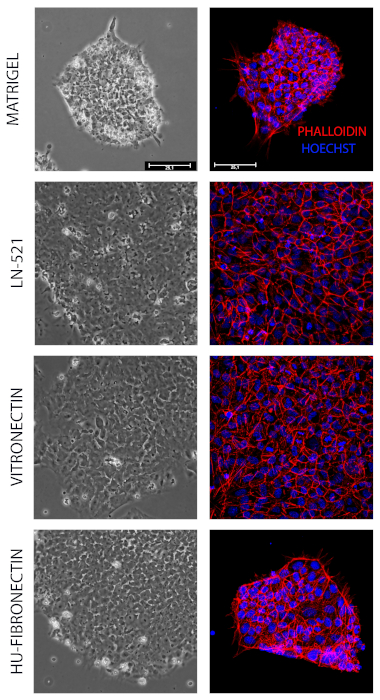
Figure 1. Representative bright-field images and Phalloidin staining of iPSCs seeded on different ECM coatings after 3 days. Bright-field images showing that the cells are healthy on the coating used and that they are well attached to the vessels as documented by the phalloidin staining showing clearly visible cytoskeletal microfilaments. Scale bar: 25 µm. Please click here to view a larger version of this figure.
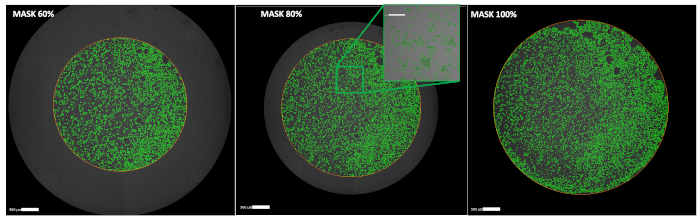
Figure 2. Representative bright-field images showing three different analyses setting for the masks used to perform confluence analyses. Mosaic obtained with a cytometer using bright-field images (16 images/well from a 96 well plate). In green the analysis segmentation displays clearly the different mask applied (60, 80 100%) to avoid or include the round edge of the well. Scale bar: 500 µm. Please click here to view a larger version of this figure.
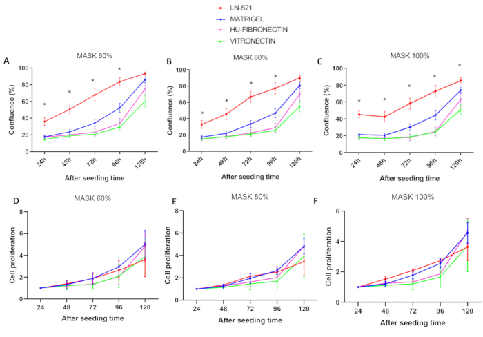
Figure 3. Cell confluence analysis of iPSCs seeded on differently coated vessels. Graph representing the cell confluence of iPSCs seeded on differently coated vessels. Data were analyzed after acquisition with the appropriate software using a (A) 60% mask (B) 80% (C) 100% for 5 days (120 h). Normalization of the confluence of the 48 h to 120 h time points to the first time point (24 h) is shown in (D, E, F). The data were obtained from three independent experiments. Data are represented as mean ± SEM. n= 3 * p<0.05. Please click here to view a larger version of this figure.
Coating compound | Initial Concentration | Final Concentration |
HU-Fibronectin | 1 mg/mL | 10 µg/cm2 |
Laminin 521 | 100 µg/ml | 20 µg/mL |
Matrigel | * | 0.111111111 |
Vitronectin XF | 250 µg/mL | 10 µg/mL |
Table 1. List of coating compounds used to analyze the confluence. The name, initial and final concentration of different coatings used are reported. * The initial concentration of Matrigel is variable, depending on the batch.
Supplemental File. Please click here to download this file.
The use of iPSCs for disease modeling and future drug screening together with their possible application in precision medicine makes it a technology of great relevance and for this reason we believe that it is necessary to clearly understand the in vitro culturing condition that better resemble the physiological situation of embryonic stem cells. In this context, we tested different ECM coatings using wild type iPSCs in order to understand the conditions that allow the cells to remain in a healthy and undifferentiated state. In addition to this, a critical point is the culturing of iPSCs in xenogenic components of MEFs and Matrigel that may account for the experimental variability among triplicates and this hinder the ability to perform mechanistic studies26.
In this study, we tested the iPSCs’ confluence on xenogeneic-free substrates (i.e., LN-521, Vitronectin, Hu-Fibronectin) using a high-content image analyzer cytometer. The reason for using the automated image-analysis system is due to the fact that counting cells, using the Trypan blue exclusion method would necessitate to make single cell suspensions and this is not recommended when manipulating iPSCs as they should be propagated in cell clusters to avoid cell death. The data obtained with the high-content image-analysis allow us to follow cell confluence without perturbing cells as they are simply imaged every day for 5 days. This technology may be considered as the election method to characterize iPSC lines and it can be included to perform quality control panels of human iPSCs. While we used a commercial software package, the methodology here described can be successfully used by means of equivalent high-content/high-throughput image analysis platforms and similar analytic software packages. The data obtained show that the iPSCs seeded on LN-521 present a linear confluence during 5 days in culture without splitting the cells and is therefore the best xenogeneic–free substrate tested in this study. One limitation of this protocol is that the results obtained need to be normalized to the first time point in order to consider differences in iPSC attachment to different substrates. Interestingly, the data obtained are most probably driven by an increased cell attachment rate of iPSCs to LN-521. In fact, when normalizing the results for the first time point, no difference is observed among the different substrates.
Based on the results obtained with the study, it would be interesting to better understand the biology of pluripotent stem cells in terms of knowing the major cell surface receptors that mediate cell-ECM contacts and that may be responsible for the maintenance of their self-renewal ability rather than spontaneous differentiation into specific cell types. Interestingly, there are studies showing that the matrix elasticity of the culture surface influenced the differentiation toward different cell types and this is probably dependent on the cell-ECM interactions that activated some intracellular cell-signaling pathway relevant for cell-type specific differentiation27. In addition to this, Vigilante et al.28 explored the genetic contribution to changes in iPSC behavior, by combining computational approaches with gene expression and cell biology datasets. The work by Vigilante et al.28 is, therefore, a major advance in attempting to map genetic variation to phenotypic variation. These studies may lead to the development of standardized methodologies to be used to perform iPSCs experiments in light of their future possible use in clinics.
The authors have nothing to disclose.
The study was supported by grants from the Fondazione Bambino Gesù and Ricerca Corrente (Italian Ministry of Health) to C.C. We would like to thank Dr Enrico Bertini (Department of Neuroscience, Unit of Neuromuscular and Neurodegenerative Diseases, Laboratory of Molecular Medicine, Bambino Gesù Children's Research Hospital), Dr Stefania Petrini (Confocal Microscopy Core Facility, Research Laboratories, Bambino Gesù Children's Research Hospital), Giulia Pericoli (Department of Onco-hematology, Gene and Cell Therapy, Children’s Research Hospital Bambino Gesù) and Roberta Ferretti (Department of Onco-hematology, Gene and Cell Therapy, Children’s Research Hospital Bambino Gesù) for scientific discussions and technical help. Maria Vinci is recipient of a “Children with Cancer UK fellowship”.
10 mL Stripette Serological Pipets, Polystyrene, Individually Paper/Plastic Wrapped, Sterile | Corning | 4488 | Tool |
15 mL high-clarity polypropylene (PP) conical centrifuge tubes | Falcon | 352097 | Tool |
1x PBS (With Ca2+; Mg2+) | Thermofisher | 14040133 | Medium |
1x PBS (without Ca2+; Mg2+) | Euroclone | ECB4004L | Medium |
5 mL Stripette Serological Pipets, Polystyrene, Individually Paper/Plastic Wrapped, Sterile | Corning | 4487 | Tool |
Cell culture microplate, 96 WELL, PS, F-Bottom | Greiner Bio One | 655090 | Support |
Cell culture plate, 6 well | Costar | 3516 | Support |
DMEM (Dulbecco's Modified Eagle's Medium- high glucose) | Sigma | D5671 | Medium |
EDTA | Sigma | ED4SS-500g | Reagent |
Epi Episomal iPSC Reprogramming Kit | Invitrogen | A15960 | Reagent |
FAST – READ 102 | Biosigma | BVS100 | Tool |
Fetal Bovine Serum (FBS) | Gibco | 10270106 | Medium |
Fibronectin | Merck | FC010 | Coating |
Glycerol | Sigma | G5516 | Reagent |
H2O | MILLIQ | ||
Hoechst | Thermofisher | 33342 | Reagent |
Laminin 521 | Stem Cell Technologies | 77003 | Coating |
L-Glutamine (200 mM) | Gibco | LS25030081 | Reagent |
Matrigel | Corning Matrigel hESC-Qualified Matrix | 354277 | Coating |
Mouse embryonic fibroblasts (MEF) | Life Technologies | A24903 | Coating |
MTESR1 Medium | Stem Cell Technologies | 85851 | Medium |
MTESR1 Supplement | Stem Cell Technologies | 85852 | Medium |
Penicillin-Streptomycin (10,000 U/mL) | Gibco | 15140122 | Reagent |
Phalloidin | Sigma | P1951 | Reagent |
Vitronectin | Stem Cell Technologies | 7180 | Coating |
Y-27632 | Sigma | Y0503 | Reagent |