1. Chip design and micromachining parameters
- Design the microfluidic chip layers with FreeCAD open-source design software; refer to Table 1 for the dimensions of the channels. Include four 2.54 mm diameter holes in the design to use a custom-made aligner for a correct layer superposition.
| Length (μm) | Width (μm) | |
| Lower chamber | 28,400 | 800 |
| Upper chamber | 31,000 | 800 |
Table 1: Dimensions of the upper and lower channels of the device.
- Cut 95 µm-thick, adhesive, transparent vinyl sheets into 30 cm x 30 cm squares to fit in the plotter properly.
- Use Brother CanvasWorkspace software to create a 30 cm x 30 cm workspace, and fill it with the designed patterns for the different layers of the chip (Figure 1A). Store it in a .svg file.
- Cut the 30 cm x 30 cm vinyl sheets with the edge plotter (Figure 1B-D).
- Stick the vinyl sheet to a low tack adhesive mat, and eliminate all the air bubbles if necessary.
- Upload the .svg file to the plotter, and set the cutting parameters: cutting blade: level 3; cutting pressure: level 0; cutting speed: level 1. Place the adhesive mat with the vinyl into the plotter, and start the cutting process.
- Cut the top channel pattern on 12 µm-thick double-sided tape vinyl by following the previous steps.
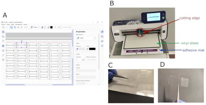
Figure 1: Chip design and micromachining process. (A) Software layout showing the working space filled with both the top and bottom patterns designed for the chip. (B) Edge plotter during cutting process; cutting blade, whole vinyl sheet, and adhesive mat are shown. (C) Patterned vinyl being detached from the cut sheet. (D) Sample of an adhesive vinyl layer patterned with the top channel design. Please click here to view a larger version of this figure.
2. PDMS layer fabrication
- Mix the PDMS and curing agent in a ratio of 10:1 (v/v), and place the mixture under vacuum for 15 min to remove air bubbles. Pour 55 mL of the mixture into a 55 cm2 square culture dish to obtain a 2 mm-thick layer. Remove bubbles with a needle.
- Cure the mixture (step 2.1) in an oven for 1 h at 80 °C. Unmold the PDMS and cut it into rectangles with the chip dimensions. Make holes for the tubing with an 18 G syringe needle.
3. Chip assembly
NOTE: For better understanding, see Figure 2.
- Assemble the whole device using an aligner to adjust channels, inlets, and outlets properly. Pile up four vinyl layers (with the corresponding bottom micropattern) for assembling the lower channel, keeping the cover tape of the bottom layer to avoid sticking to the aligner.
- Cut and place the polycarbonate (PC) porous membrane on top of the lower channel to separate it from the upper one. Be careful not to cover the inlets of the lower channel.
- Add ten vinyl layers with the upper chamber design. Stick a double-sided tape vinyl layer with the top-channel pattern on top. Remove the chip from the aligner and stick it on the glass slide.
- Place a 2-mm-thick PDMS sheet on top of the double-sided tape vinyl layer to provide appropriate anchoring for the tubing and to avoid leakage. Leave a weight on top of the chip overnight to ensure the chip is completely watertight. Sterilize the chip by flushing 70% v/v ethanol for 5 min, and then wash with distilled H2O.
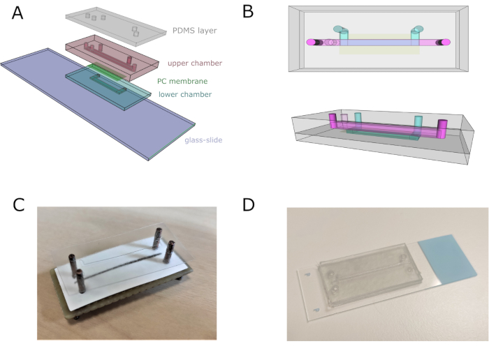
Figure 2: Microfluidic chip assembly. (A) General scheme of the assembly of the device. Lower and upper chambers are composed of four and eleven superimposed vinyl sheets, respectively. (B) Top and lateral views of the microfluidic chip. Top and bottom channels are represented in pink and blue, respectively. (C) Image of the chip assembly using a custom-made aligner. (D) Chip image after complete assembly. Please click here to view a larger version of this figure.
4. Pump connections
NOTE: The graphical representation of pumps connections is shown in Figure 3.
- Connect Pump 1 to the Upper Chamber Inlet 1 (UCi1).
- Connect Pump 2 to the Upper Chamber Inlet 2 (UCi2).
- Connect Pump 3 to the Lower Chamber Inlet (LCi).
- Connect Upper Chamber Outlet (UCo) and Lower Chamber Outlet (LCo) to a waste tube.
- Connect the syringes to each inlet using polytetrafluoroethylene (PTFE) tubes and 18 G stainless steel connectors.
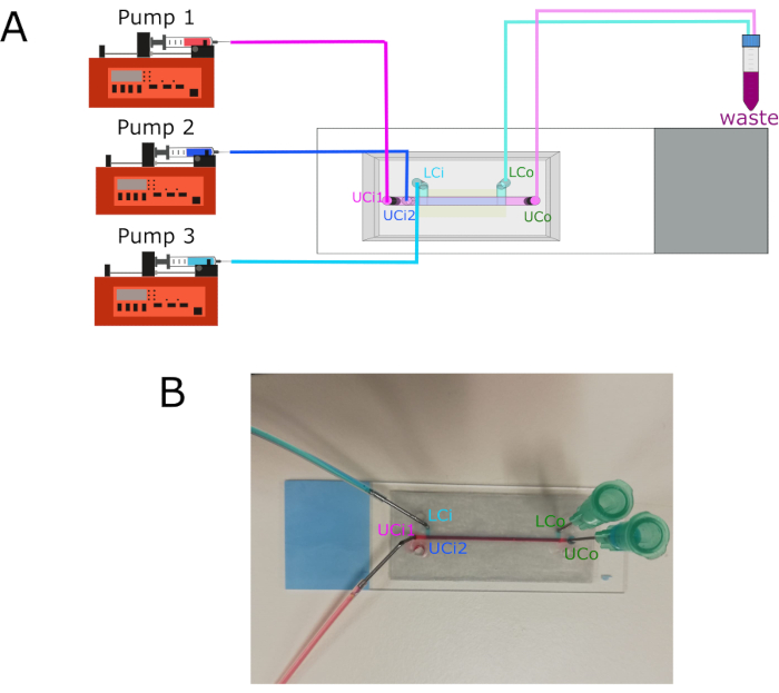
Figure 3: Pump connections and inlets/outlets location. (A) Diagram showing the connection of the three different pumps to their respective inlets. Outlets connect to a waste container. (B) Chip image with labeled inlets and outlets. Abbreviations: LCi = lower chamber inlet; LCo = lower chamber outlet; UCi1 = upper chamber inlet 1; UCi2 = upper chamber inlet 2; UCo = upper chamber outlet. Please click here to view a larger version of this figure.
5. Cell culture
NOTE: The HaCaT cell line has a commercial origin. Human primary fibroblasts come from healthy donors and were obtained from the collection of biological samples of human origin registered in "Registro Nacional de Biobancos para Investigación Biomédica del Instituto de Salud Carlos III".
- Work in a cell culture hood, previously sterilized under ultraviolet light and wiped with ethanol.
- Thaw H2B-GFP-HaCaT cells (human immortalized skin keratinocytes, hKCs) and GFP-human primary fibroblasts (hFBs) at 37 °C, add 2 mL of culture medium, and centrifuge for 7 min at 20 °C at 250 × g.
NOTE: H2B-GFP-HaCaT cells are human immortalized keratinocytes modified to express a hybrid histone H2B-green fluorescent protein (GFP), providing their nuclei with green fluorescence. GFP-hFBs are human primary fibroblasts transformed with the vector pLZRS-IRES-EGFP to express cytoplasmic green fluorescence. These cells were modified following previously published protocols30,31 - Culture both hKCs and hFBs in 1x DMEM supplemented with 10% fetal bovine serum and 1% of antibiotic/antimycotic solution. Pre-warm the culture medium at 37 °C before use.
- Detach the cells by washing them with 1x phosphate-buffered saline (PBS), adding 2 mL of trypsin/ethylenediamine tetraacetic acid (EDTA) and incubating them for 10 min at 37 °C.
- Inactivate trypsin adding 4 mL of culture medium. Resuspend the cells, and transfer them to a 15 mL tube. Remove 10 µL to count cells on a Neubauer chamber and determine the appropriate concentration.
- Centrifuge the 15 mL tube for 7 min at 20 °C at 250 × g. Remove the supernatant, and resuspend the pellets at the desired concentration: hFBs at 50,000 cells/mL and hKCs at 5·× 106 cells/mL.
6. Fibrinogen pre-gel preparation
- Activate thrombin by adding 1mL of CaCl2 (1% w/v in NaCl) to the vial.
- Add the following components to obtain 1 mL of a fibrin hydrogel at a final concentration of fibrin of 3.5 mg/mL: 59 µL of activated thrombin (10 NIH units/mL), 59 µL of tranexamic acid (Table of Materials, 100 mg/mL), 764 µL of culture medium containing 50,000 hFBs/mL, 118 µL of fibrinogen (20 mg/mL in NaCl (0.9% w/v)).
NOTE: Fibrinogen must be added at the last moment.
7. Parallel flow protocol
- Pump 1x PBS with pump 3 through the LCi at 50 µL/min during the whole process.
- Pump sacrificial fluid (1x PBS) with pump 2 through the UCi2 at 100 µL/min.
- Load the syringe with the pre-gel, rapidly place it into pump 1, and run it at 200 µL/min (Figure 4A).
- Stop pumps 1 and 2 once the pre-gel exits the UCo.
- Leave the chip without removing the tubing at 37 °C for at least 10 min to allow gelation.
- Pump culture medium at 50 µL/h with pump 3 through UCi2 overnight.
- Block UCi1 with a cap.
8. hKCs monolayer seeding
- Check under the microscope that hFBs are spread 24 h after the generation of the dermal compartment.
- Introduce the hKCs with pump 2 through UCi2 at 5 ×·106 cells/mL at 40 µL/min for 1 min (Figure 4B).
- Leave the chip overnight at 37 °C in a humidity-saturated incubator for cell attachment.
- Pump fresh culture medium with pump 3 only through LCi at 50 µL/min (Figure 4C).
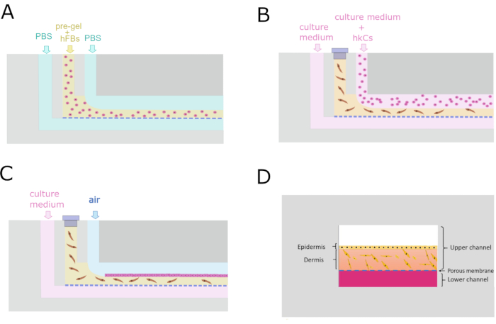
Figure 4: Microfluidic protocol for the generation of the dermo-epidermal construct. (A) Transverse cross-section showing the parallel flow process to generate the dermal compartment. (B) Keratinocyte monolayer seeding 24 h after dermal compartment generation. (C) Cell culture maintenance inside the microfluidic device. (D) Cross-sectional recreation of the skin inside the chip. Please click here to view a larger version of this figure.
9. Cell viability assay
NOTE: Live/Dead kit stains cells with green or red fluorescence depending on their live or dead state. For proper viability differentiation, non-fluorescent hKCs and hFBs must be used in this step. All the steps in the procedure are carried out through UCi2 with pump 2.
- Wash the top channel with 1x PBS for 5 min at 50 µL/min to remove culture medium.
- Pump air to remove the 1x PBS at 50 µL/min.
- Prepare Calcein AM/Ethidium homodimer-1 Kit (Live/Dead) solution by following the manufacturer's instructions.
- Pump Live/Dead solution at 50 µL/min for 2 min.
- Incubate 30 min at 37 °C in the dark.
- Wash top channel by pumping 1x PBS at 50 µL/min for 2 min to remove any remaining reagent.
- Observe the sample under the confocal microscope. Use an excitation wavelength of 495/590 nm and an emission wavelength of 519/617 nm for live and dead cells, respectively.
The designed chip is composed of two fluidic chambers separated by a 5 µm pore size PC membrane that allows the growth of the cell by allowing the passage of growth-promoting molecules from the lower chamber. The upper chamber holds the tissue construct, in this case, a monolayer of hKCs on a fibrin hydrogel containing hFBs.
The height of the channels is determined by the number of adhesive sheets added to each channel. The lower chamber is composed of 4 layers (380 µm) and the upper one of 10 one-sided tape layers and a double-sided one (962 µm). The dimensions of the chip are 4 cm x 2 cm, which enhances its manipulation. The adhesive vinyl sheets provide water-tightness and transparency for visual inspection of the device. The PDMS layer was useful for the proper anchoring of llthe tubing to avoid any leakage from the holes where the tubes were fixed.
According to published literature, the injectability of cell-containing hydrogels into microfluidic chambers using syringe pumps has not been reported to date. For this reason, the injectability of the fibrin pre-gel had to be assessed. We observed that under a flow of 50 µL/min, the syringe was blocked. However, flow rates higher than 200 µL/min could damage the cells. Rheological studies were performed to test the shear thinning behavior of the fibrin pre-gel, obtaining viscosities from 10 to 50 cP within the selected flow rate range (50-200 µL/min). These results helped to establish the working conditions of this system.
In this work, a parallel flow method has been developed based on the generation of two superimposed laminar flows the lower one being the pre-gel, while the upper one was sacrificial fluid (PBS). Numerically solving Navier-Stokes equations and imposing the appropriate boundary conditions, we found that there were multiple possible solutions to obtain the desired height. Considering the shear rate limits established earlier, to achieve a hydrogel of approximately 500 µm height, the flow rates were 104 and 222 µL/min for the sacrificial PBS and the pre-gel, respectively. In practice, microflow rates of 100 and 200 µL/min were used for simplicity. When re-introduced in the model, these values were found to result in a gel height of 576 µm, very similar to the expected values (Figure 5B). To experimentally check the functioning of the proposed method, the height of the hydrogel along the upper chamber was measured. An average height of 550 µm was observed (Figure 5A), quite similar to the prediction of our mathematical model.
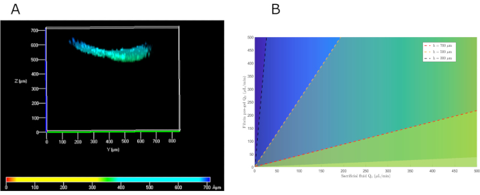
Figure 5: Parallel flow mathematical solutions to choose the appropriate pre-gel and sacrificial fluid flows to obtain the desired dermal height. (A) Front view of the confocal image of hKCs seeded on top of the fibrin gel to assess its height. (B) Mathematical solution for different heights depending on the pre-gel and PBS flow rates. Abbreviations: hKCs = human immortalized skin keratinocytes; PBS = phosphate-buffered saline. Please click here to view a larger version of this figure.
When establishing the protocol at the beginning, PBS was not flushed through the lower channel, leading to discrepancies between the theoretical height of the gel and the one measured. This difference in height was ~40% compared with the estimated one (Figure 6). Once the protocol was optimized and PBS pumped through the lower chamber, this loss in height was resolved (Figure 5).
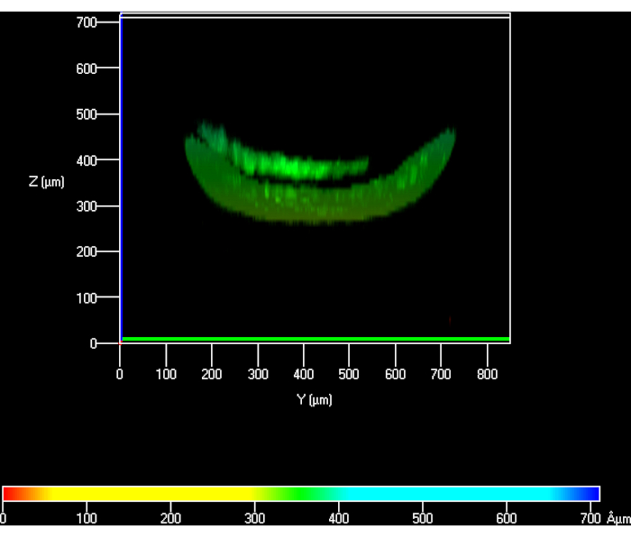
Figure 6: Confocal view of hKCs seeded on top of the hydrogel to measure its height when PBS was not pumped through the lower chamber. Abbreviations: hKCs = human immortalized skin keratinocytes; PBS = phosphate-buffered saline. Please click here to view a larger version of this figure.
Figure 7A shows a fluorescent top view image of the upper chamber containing a fibrin hydrogel with embedded GFP-hFBs, demonstrating that 24 h after loading, cells are uniformly distributed along the chamber and well spread. Figure 7B shows confluent GFP-hKCs seeded on top of the hydrogel.
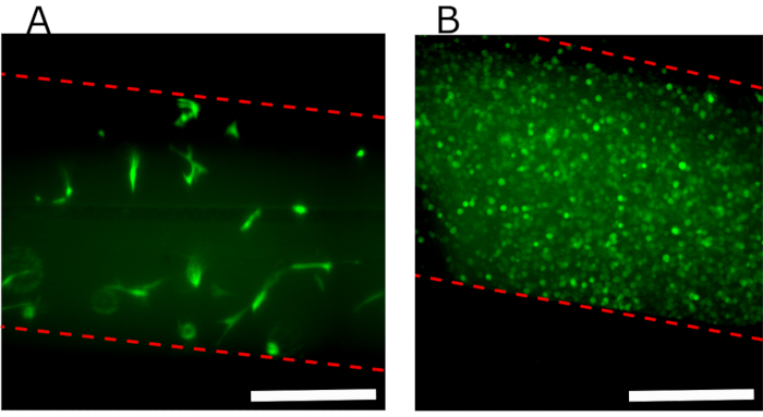
Figure 7: Fluorescence images of the upper channel showing different cells seeded in the device. (A) Top view of the upper chamber 24 h after parallel flow protocol showing the hFBs embedded in the fibrin gel. (B) Confluent GFP-hKCs seeded on top of the hydrogel 24 h after hydrogel generation. Dashed red line indicates channel walls. Scale bars: 400 µm. Abbreviations: hFBs = human primary fibroblasts; GFP-hKCs = green fluorescent protein-expressing human immortalized skin keratinocytes. Please click here to view a larger version of this figure.
It is important to keep the system closed overnight until the hKCs sediment and attach to the hydrogel surface. When the tubing is removed before cell attachment, air bubbles enter the channel and displace cells, leading to a nonuniform confluent monolayer, as shown in Figure 8.
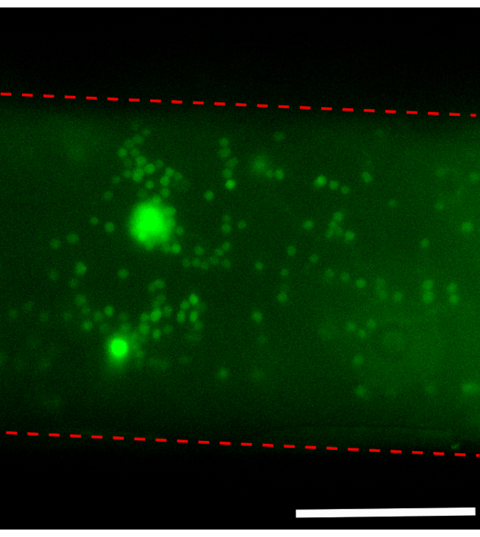
Figure 8: Top view of the hydrogel surface when removing tubing immediately after hKCs seeding. Dashed red line indicates channel walls. Scale bar: 400 µm. Abbreviations: hKCs = human immortalized skin keratinocytes. Please click here to view a larger version of this figure.
The cell viability test performed on hFBs embedded in the hydrogel was carried out 24 h after loading using the parallel flow method, showing a cell viability of ~95%. The same test performed on hKCs cells 24 h after seeding them on the fibrin hydrogel showed similar results. The next step was to generate a dermo-epidermal construct and study its structure using confocal microscopy after 24 h (Figure 9).
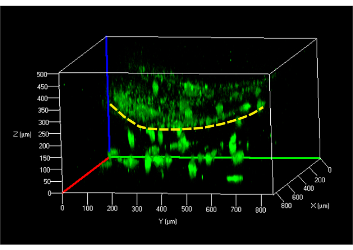
Figure 9: Reconstructed 3D confocal image of the undifferentiated skin model in the microfluidic chip. Yellow dashed line indicates the surface of the hydrogel separating hKCs (top) from the hFBs embedded in the gel (bottom). Abbreviations: hFBs = human primary fibroblasts; hKCs = human immortalized skin keratinocytes. Please click here to view a larger version of this figure.
It is crucial to find an equilibrium between the shear thinning behavior of the fibrinogen gel and its gelation time: if it takes too long to establish the parallel flow, it coagulates and blocks the system; if the gelation process is too slow, hFBs in the hydrogel will sediment as shown in Figure 10. The behavior of this transient state can be regulated by varying the thrombin concentration.
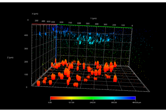
Figure 10: Confocal image of a fibrin hydrogel showing sedimented hFBs (in red) due to a slow fibrin gelling time. A hKCs monolayer (in blue) is shown on top of the gel. Abbreviations: hFBs = human primary fibroblasts; hKCs = human immortalized skin keratinocytes. Please click here to view a larger version of this figure.
During the device planification and posterior experimental practices, some complications arose that had to be solved to obtain an optimally functioning device and well-structured tissue. These problems are shown in Table 2, along with the solutions for troubleshooting.
| Problems | Solutions |
| Introducing a 3D hydrogel in the upper chamber, leaving free space above to seed keratinocytes on top after dermal generation | Apply a parallel flow of two fluids (PBS and pre-gel) with different viscosities to create a dermal compartment with a controlled height. |
| Channel misalignment during vinyl sheet stacking | Use of a custom-made aligner to pile up all the sheets in the correct place |
| Achieve a confluent keratinocyte monolayer on top of the fibrin gel to simulate the epidermis | Allow cell attachment to the gel prior to removing the tubes from the chip, preventing air bubbles from entering the channel and displacing the cells. |
| Hydrogel height loss along the channel due to pre-gel leaking to the lower channel during parallel flow protocol through the porous membrane. | Pump PBS through the lower channel during parallel flow establishment. |
| Fibroblast sedimentation inside the gel | Thrombin concentration was slightly increased to accelerate fibrin gelation |
Table 2: Issues found during the development of the current work and solutions applied.
