Fusion is a common phenomenon in tissue or organ development. Examples include dorsal closure and thorax closure in Drosophila1 and palate morphogenesis, neural tube morphogenesis, and heart morphogenesis in mice and chicken2. CRISPR and RNAi have been applied to investigate the roles of genes in the process of fusion2,3,4.
Spodoptera litura (S. litura, Lepidoptera: Noctuidae) is a detrimental polyphagous pest that is widely distributed in tropical and subtropical areas of Asia, including China4,5,6. The wide distribution of S. litura is partly attributed to its powerful reproductive capability, which is relevant to gonad development. Male infertility is one approach to control this pest. As shown in the schematic figure of testicular structure, the testes are enclosed by the testicular sheath, including the external sheath (peritoneal sheath) and inner basal lamina. The basal lamina extends internally to form the follicular epithelium and separates the inner area of the testis into four chambers named follicles (Figure 1).
In the follicles, spermatogonia develop into spermatozoa after mitosis and meiosis, and then the spermatozoa in the sperm sacs align in the same direction to form sperm bundles7. During spermatogenesis, the primary spermatocytes differentiate into eupyrene sperms or apyrene sperms. Spermatocytes in the larval phase develop into eupyrene sperm with a long tail connected to a head of an elongated nucleus; these can fertilize eggs. Conversely, spermatocytes in the mid-pupal phase develop into apyrene sperm with a discarded nucleus; these sperm assist the survival, motion, and fertilization of eupyrene sperm9,10. The 6th day of the pupa is the period during which the testes have abundant eupyrene and apyrene sperm bundles.
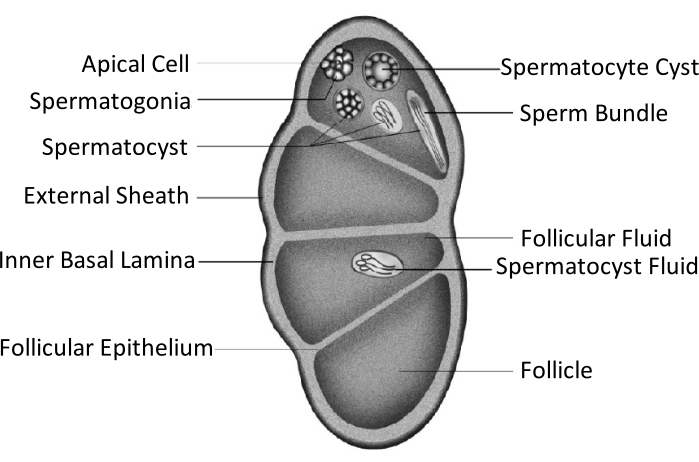
Figure 1: Schematic diagram of the testicular structure of Lepidoptera insects11. Please click here to view a larger version of this figure.
Testicular fusion occurs in most insects of the Lepidoptera order11,12, especially in those species that are agricultural pests. Testicular fusion refers to a pair of testes growing bilaterally in the larval phase, approaching and adhering to each other, eventually integrating into a single gonad11. In Spodoptera litura, it happens during metamorphosis from the larval to the pupal stage. From day 1 of the 5th instar (L5D1) to day 4 of the 6th instar (L6D4), the pair of testes grows gradually in size, and the color turns light yellow from ivory-white. It becomes faint red as it reaches the prepupal phase (L6D5 to L6D6). Two bilateral symmetrical testes approach each other during the prepupal stage, fuse into one, and twist anticlockwise (doral view) to produce a single testis in the pupal and adult phases11. This phenomenon does not occur in silkworms, which have considerable economic importance and have been domesticated for 5000 years13. Thus, it is assumed that the testes' fusion improves reproductive capability.
To determine the significance of Spodoptera litura testicular fusion, it is important to investigate the effects of blocking the process. In this protocol, aluminum foil was microsurgically inserted between the testes to keep them separated, and the consequent changes in the development of the insects and their testes were studied.
1. Insect rearing and maintenance
- Culture the Spodoptera litura larvae in environmental simulation chambers with an artificial diet until they reach day 4 of the 6th instar (L6D4). Select male larvae when the worms enter the first day of the 6th instar (L6D0) based on the inverse triangle-shaped structure on the eighth abdomen14.
NOTE: Larvae rearing and maintenance techniques were published previously4,14.
2. Presurgical preparation
- Trim the aluminum foil into rectangular pieces with rounded corners (1 mm x 2 mm, Figure 2).
- Sterilize the surgery platform and related items (table surface, microscope, icebox, insect box, wax tray, pins, and thread) by spraying 75% alcohol on their surface and wiping them down.
- Sterilize surgical tools (including the aluminum foil) with a high-pressure steam sterilizer for 30 min, and place them in a heating and drying oven at 120 °C.
- Ensure that the operators wear clean laboratory clothes, surgical masks, and sterile gloves.
3. Microsurgical placement of a barrier between the testes
NOTE: The general work-flow is as follows: Freezing → Fixing → Disinfection → Incision → Barrier Placement → Suturing→ Postoperative Feeding and Inspection
- Place male larvae (L6D4) on ice for 10-30 min to keep them anesthetized during the operation.
- Place a larva on the wax tray with the dorsal side up, and then fix the head and the tail of the larva with pins and threads, showing the surgical area that is the dorsal surface on the 9th body segment (Figure 3A).
- Disinfect the surgical area by applying 3% iodine tincture with a cotton swab to the epidermis (9th body segment), followed by 70% alcohol to remove the iodine (Figure 3B).
NOTE: Focus on the larva through coarse and fine adjustment of the surgical microscope (Figure 3C). Place the wax tray on a larger culture dish filled with ice to keep the anesthesia. - Make a 2 mm-long incision on the dorsal epidermis of the 9th body segment. Next, use a sterile cotton swab to remove any leaking hemolymph and fat bodies and obtain a clear view of the surgical area.
NOTE: It is important to avoid the heart during the procedure. This can be done by making the incision slightly next to the mid-line in the 9th body segment or at the joint between the 9th and 10th body segments to prevent the testes from popping out due to the larval internal pressure. While using the scalpel, make a vertical slit with the blade first (Figure 4A), and then turn it 45° towards the epidermis before evenly and continuously cutting through the epidermis (Figure 4B). - Use surgical tweezers to insert a piece of aluminum foil between the testes (Figure 5).
- At the end of the surgery, close the incision to avoid infection, and allow the larvae to recover from the surgery.
- Close the epidermis with a running suture (Figure 6).
- Use a needle holder and surgical tweezers to tie a surgical square knot, requiring two opposing mirror-image simple knots (Figure 6D,E).
- Use scissors to cut the excess suture from the loop tails, leaving a 2 mm thread behind.
- After suturing, gently lay the larva in the rearing box and maintain them in a clean environmental simulation chamber. Continue observing the larvae.
NOTE: The wound stops leaking hemolymph, and the larvae gradually recover after the surgery. The worms continue to complete their metamorphosis.
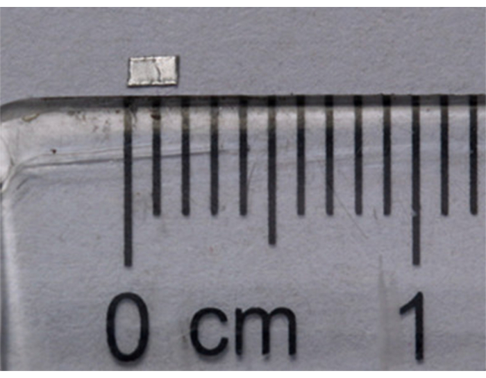
Figure 2: Physical barrier prepared using aluminum foil (1 mm x 2 mm). Please click here to view a larger version of this figure.
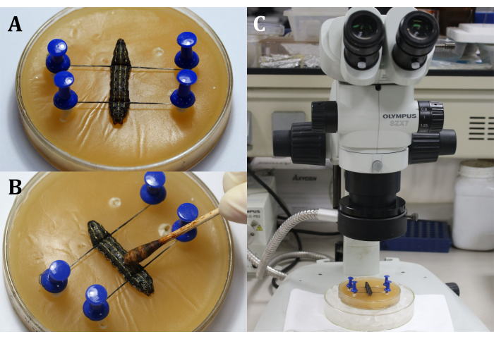
Figure 3: Before incision. (A) Fixing the larva. (B) Disinfection of the epidermis of the surgical area. (C)Performing surgery under the microscope. Please click here to view a larger version of this figure.
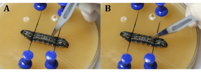
Figure 4: Incision. (A) Slit the larvae vertically with the blade. (B) Turn the blade 45° toward the epidermis before cutting through. Please click here to view a larger version of this figure.
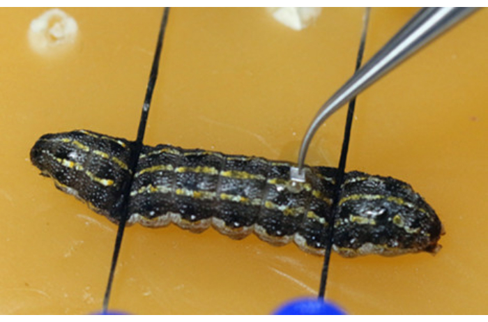
Figure 5: Inserting the physical barrier (aluminum foil) between the testes. Please click here to view a larger version of this figure.
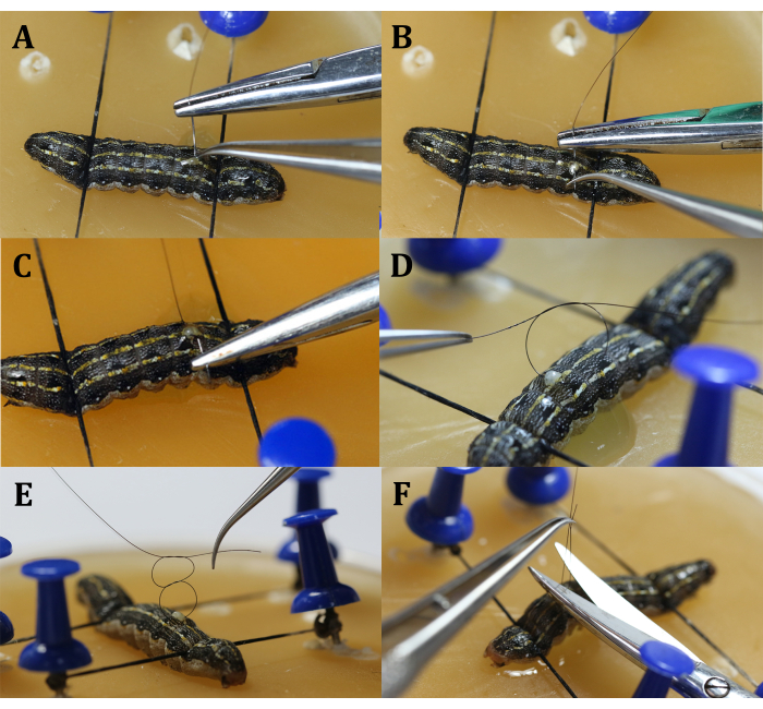
Figure 6: Suturing. (A) Insert the needle. (B) Withdraw the needle. (C) Withdraw and clamp the needle. (D) Tie the first simple knot. (E) Tie the opposing mirror-imaged simple knot. (F) Cut excess suture thread. Please click here to view a larger version of this figure.
The effects of microsurgery on Spodoptera litura growth and development
The microsurgery left a 2 mm-long wound on the dorsal larval epidermis that eventually stopped leaking hemolymph and healed. The larvae went through prepupal and pupal stages and eclosed, indicating that the microsurgery had no major impact on growth and development. When the larvae molted into pupae, the suture threads were discarded along with the epidermis. There were no obvious differences in the appearance of the pupae that did and did not undergo surgery. After eclosion, adult females successfully mated with the adult males previously operated on, resulting in fertilized eggs and hatching larvae (Figure 7).
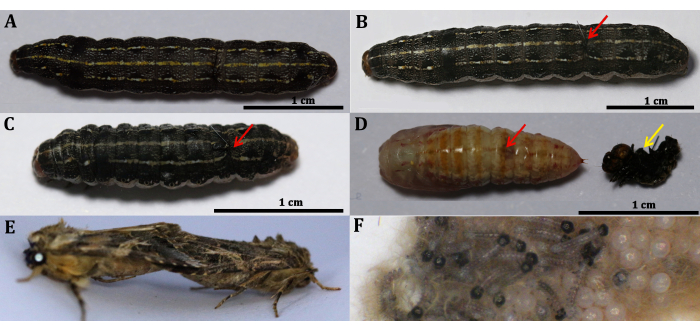
Figure 7: Spodoptera litura Development after microsurgery. (A) Male larva at L6D4. (B) L6D4 larva immediately after surgery. (C) Pre-pupa (L6D6). (D) P0, the red arrow indicates the location of the surgery; the yellow arrow shows the discarded epidermis with suture thread. (E) Mating adults. (F) Eggs and hatched larvae from a female adult mating with a male that underwent surgery. Scale bars = 1 cm. Please click here to view a larger version of this figure.
The larvae went through the pupal stage and eclosed after the microsurgical placement of aluminum foil between the testes. Detailed results following this operation have been published previously11. Although the barrier stopped the testes from fusing in some larvae, most larvae underwent testicular fusion during larval to pupal metamorphosis.
In this research, individuals were grouped by three treatments: Experimental (Exp), Sham-operation (Ctl-sham), and no operation (Ctl). Individuals of the Exp group underwent microsurgery to insert a physical barrier, and their testes remained separated during the pupal and adult stages. Individuals of the Ctl-sham group underwent the same microsurgery; however, their testes were not blocked and fused for unknown reasons. The Ctl group contained the larvae that grew naturally without surgery; their two testes fused normally during the prepupal stage.
The microsurgery group contained two subgroups: larvae that underwent microsurgery to place a barrier between the two testes (Group A) and those that underwent microsurgery to remove one testis (Group B: left testis removed in Group B-1; right testis removed in Group B-2). Table 1 shows the numbers of operation larvae, larval mortality rates, numbers of pupae, percentage of pupation, numbers of adults, percentages of adult emergence, percentages of successful mating, and percentages of successful operations in different groups. Group A includes larvae that underwent microsurgery to insert a barrier between the testes. The success of this procedure could only be determined after dissection, which is when they were further divided into the Exp and Ctl-sham groups.
As shown in Figure 8, the larval mortality rate was slightly higher in the surgical group, whereas the percentages of pupation, adult emergence, and successful mating were slightly lower in the surgical group than the control group. However, none of the differences were significantly different, indicating that the microsurgery did not markedly influence the growth and development of Spodoptera litura larvae.
| Group | Number of Larvae | Larval Mortality Rate/% | Number of Pupae | Percentage of Pupation/% | Number of Adult | Percentage of Adult Emergence/% | Percentage of Successful Mating/% | Percentage of Successful Operation/% | |||||
| Microsurgery group A-1* | 79 | 35.4 | 39 | 76.5 | N | N | N | 10.3 | |||||
| Microsurgery group A-2* | 117 | 12.8 | 102 | 100 | N | N | N | 11.8 | |||||
| Microsurgery group A-3* | 73 | 13.7 | 57 | 90.5 | N | N | N | 10.5 | |||||
| Microsurgery group A-4 | 101 | 4 | 97 | 96 | 29 | 29.9 | N | 26.9 | |||||
| Microsurgery group A-5 | 176 | 20.1 | 140 | 79.5 | 28 | 20 | 44.4 | 25 | |||||
| Microsurgery group A-6 | 434 | 12.4 | 376 | 98.9 | 209 | 55.6 | 26.8 | 14.3 | |||||
| Microsurgery group A-7 | 260 | 10.8 | 135 | 58.2 | 66 | 48.9 | 47 | 48.4 | |||||
| Microsurgery group A-8 | 49 | 24.5 | 37 | 100 | 21 | 56.8 | 81 | 58.8 | |||||
| Microsurgery group B-1 | 117 | 29.1 | 71 | 85.5 | 30 | 42.3 | 23.3 | N | |||||
| Microsurgery group B-2 | 188 | 6.9 | 172 | 98.3 | 115 | 66.9 | 35.7 | N | |||||
| Average of Microsurgery Group (mean± SD) | 159 | 17±10.1 | 123 | 88.3±13.7 | 71 | 45.8±16.3 | 43±20.8 | 25.8±18.5 | |||||
| Control Group 1* | 40 | 17 | 37 | 100 | N | N | N | N | |||||
| Control Group 2 | 300 | 0 | 281 | 93.7 | 184 | 65.5 | N | N | |||||
| Control Group 3 | 354 | 11 | 305 | 96.8 | 127 | 41.6 | N | N | |||||
| Control Group 4 | 679 | 2.7 | 638 | 96.5 | 534 | 83.7 | 41.2 | N | |||||
| Control Group 5 | 448 | 4.2 | 399 | 93 | 232 | 58.1 | 60 | N | |||||
| Control Group 6 | 490 | 7.1 | 448 | 98.5 | 355 | 79.2 | 48 | N | |||||
| Average of Control Group (mean± SD) | 385 | 5.4±6.2 | 351 | 96.4±2.7 | 286 | 65.6±15.1 | 50±9.5 | N | |||||
Table 1: The effects of microsurgery on Spodoptera litura development. Microsurgery groups B-1 and B-2 underwent microsurgery to remove unilateral testis (left in Microsurgery Group B-1 and right in Microsurgery Group B-2). Note: Microsurgery groups A-1 to A-8 underwent microsurgery to insert a barrier between the testes; Microsurgery groups B-1 and B-2 underwent microsurgery to remove unilateral testis (left in Microsurgery Group B-1 and right in Microsurgery Group B-2); the rates and percentages are given as Mean ± SD. Asterisks indicate that the individuals in the group were dissected at the pupal stage, and there were no statistics on the number of adults, percentage of adult emergence, or percentage of successful mating; N indicates no data.
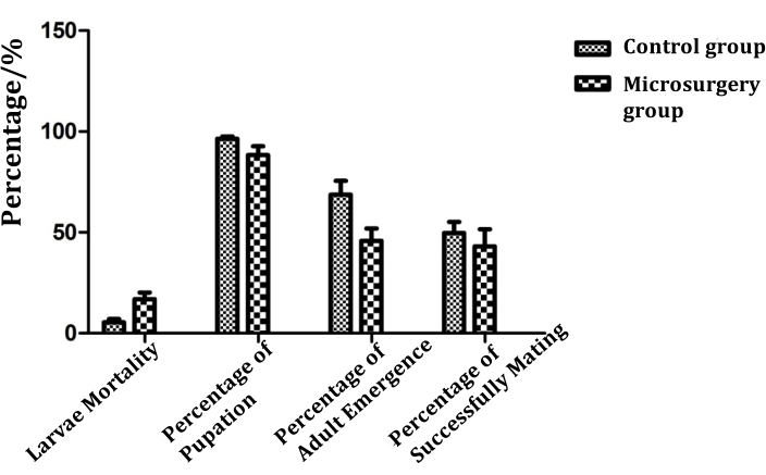
Figure 8: The influence of microsurgery on Spodoptera litura growth and development of (n ≥ 6). Please click here to view a larger version of this figure.
The influence of microsurgery on the number of sperm bundles of Spodoptera litura
Microsurgery was performed to insert a physical barrier to stop the testes fusion or remove unilateral testis in Spodoptera litura. Eupyrene and apyrene sperm bundles were counted to calculate the percentage of eupyrene sperm bundles on the sixth day of the pupal stage. The individuals were grouped by treatment, as described above. The numbers of sperm bundles (eupyrene sperm bundles, apyrene sperm bundles, and total) were significantly lower in the Exp group than in the Ctl-sham and Ctl groups. The mean number of eupyrene sperm bundles from two separated testes in the Exp group was 2082 ± 599. In the Ctl-sham and Ctl groups with fused testes, the number of eupyrene sperm bundles ranged from 4652 to 6200.
The number of apyrene sperm bundles in the Exp group was 1602 ± 703, while it ranged from 3299 to 4632 in the Ctl-Sham and Ctl groups. The total of sperm bundles in the Exp group was 3684 ± 985; it ranged from 9284 to 10832 in the Ctl-Sham and Ctl groups. Thus, the percentages of eupyrene sperm bundles ranged from 50% to 60%, with no significant differences among all three groups. Figure 9 shows that when fusion is prevented, the amount of eupyrene and apyrene sperm bundles decreased, whereas the percentage of eupyrene sperm bundles was unchanged.
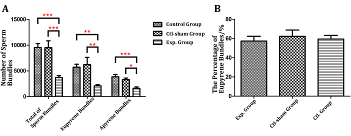
Figure 9: The numbers of sperm bundles and percentages of eupyrene sperm bundles in different groups. (A) The number of sperm bundles in the Exp group was significantly lower than in the Ctl-sham and Ctl groups. (B) The percentages of eupyrene sperm bundles were not significantly different among the three groups. Asterisk indicates a significant difference when compared with Ctl. P < 0.05, Mean ± SD (n ≥5). Please click here to view a larger version of this figure.
After removing a unilateral testis of the larvae, the numbers of eupyrene and apyrene sperm bundles were counted to calculate the percentage of eupyrene sperm bundles on the sixth day of the pupal stage. The number of eupyrene and apyrene sperm bundles ranged from 1286 to 1638 and 720 to 850, respectively, which means the total number ranged from 2006 to 2488, corresponding to a eupyrene sperm bundle percentage of 63% to 65%. Figure 10 shows that the number of sperm bundles decreased significantly after unilateral testis removal (reduced by 60% to 70%), without much influence on the percentage of eupyrene sperm bundles.
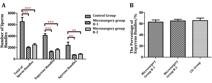
Figure 10: The numbers of sperm bundles and percentages of eupyrene sperm bundles after removing unilateral testis. (A) The numbers of sperm bundles in pupae that underwent unilateral testis removal were significantly different among the three groups (left and right testis removed in Microsurgery Group B-1 and Microsurgery Group B-2, respectively) (B) The percentage of eupyrene sperm bundles in pupae that underwent unilateral testis removal was not significantly different compared with Ctl. The asterisk indicates a significant difference compared with Ctl. P < 0.05, Mean ± SD (n ≥8). Control group = no surgery, the testes fused naturally during the prepupal stage; Ctl-Sham group = operation unsuccessful and testes fused after microsurgery; Exp. Group = microsurgery performed to insert a physical barrier between the two testes. Please click here to view a larger version of this figure.
