This article describes encapsulation of human pluripotent stem cells (hPSCs) using a co-axial flow focusing device. We demonstrate that this microfluidic encapsulation technology enables efficient formation of hPSC spheroids.
Three-dimensional (3D) or spheroid cultures of human pluripotent stem cells (hPSCs) offer the benefits of improved differentiation outcomes and scalability. In this paper, we describe a strategy for the robust and reproducible formation of hPSC spheroids where a co-axial flow focusing device is utilized to entrap hPSCs inside core-shell microcapsules. The core solution contained single cell suspension of hPSCs and was made viscous by the incorporation of high molecular weight poly(ethylene glycol) (PEG) and density gradient media. The shell stream comprised of PEG-4 arm-maleimide or PEG-4-Mal and flowed alongside the core stream toward two consecutive oil junctions. Droplet formation occurred at the first oil junction with shell solution wrapping itself around the core. Chemical crosslinking of the shell occurred at the second oil junction by introducing a di-thiol crosslinker (1,4-dithiothreitol or DTT) to these droplets. The crosslinker reacts with maleimide functional groups via click chemistry, resulting in the formation of a hydrogel shell around the microcapsules. Our encapsulation technology produced 400 µm diameter capsules at a rate of 10 capsules per second. The resultant capsules had a hydrogel shell and an aqueous core that allowed single cells to rapidly assemble into aggregates and form spheroids. The process of encapsulation did not adversely affect the viability of hPSCs, with >95% viability observed 3 days post-encapsulation. For comparison, hPSCs encapsulated in solid gel microparticles (without an aqueous core) did not form spheroids and had <50% viability 3 days after encapsulation. Spheroid formation of hPSCs inside core-shell microcapsules occurred within 48 h after encapsulation, with the spheroid diameter being a function of cell inoculation density. Overall, the microfluidic encapsulation technology described in this protocol was well-suited for hPSCs encapsulation and spheroid formation.
There is considerable interest in 3D cultures of human pluripotent stem cells (hPSCs) due to the improved pluripotency and differentiation potential afforded by this culture format1,2,3. hPSCs are typically formed into spheroids or other 3D culture formats by means of bioreactors, microwells, hydrogels, and polymeric scaffolds4,5,6. Encapsulation offers another means for organizing single hPSCs into spheroids. Once encapsulated hPSC spheroids may be handled with ease and transferred into microtiter plates for differentiation, disease modeling, or drug testing experiments. Encasing hPSCs in a hydrogel layer also protects cells against shear damage and allows to culture spheroids in a bioreactor at high rates of stirring7.
Our methodology for stem cell encapsulation evolved over time. First, we focused on solid hydrogel microparticles and demonstrated successful encapsulation and cultivation of mouse embryonic stem cells (mESCs)8. However, it was noted that human embryonic stem cells (hESCs) had low viability when encapsulated in such hydrogel microparticles, presumably due to the greater need for these cells to re-establish cell-cell contacts after the encapsulation. We reasoned that heterogeneous microcapsule, possessing an aqueous core, may be better suited for encapsulation of cells that rely on rapid re-establishment of cell-cell contacts. The concept of co-axial flow focusing microfluidic device for making aqueous core/hydrogel shell microcapsules was adapted from He et al.9, but instead of alginate employed in the original approach, a PEG-based hydrogel was incorporated into the shell. We first demonstrated successful encapsulation and spheroid formation of primary hepatocyte in core-shell microcapsules10 and most recently described encapsulation of hES and iPS cells7. As outlined in Figure 1A, capsules are fabricated in a flow focusing device where the shell and core flow streams transition from side-to-side to co-axial flow before ejection into the oil phase. The core flow contains cells and additives that increase the viscosity of the solution (non-reactive PEG MW 35kD and iodixanol – commercial name OptiPrep) while the shell stream contains reactive molecules (PEG-4-Mal). Continuous co-axial flow stream is discretized into droplets that retain core-shell architecture. The core-shell structure is made permanent by exposure to di-thiol crosslinker (DTT), which reacts with PEG-4-Mal via click chemistry and results in formation of a thin (~10 µm) hydrogel skin or shell. After the emulsion is broken and capsules are transferred into an aqueous phase, molecules of PEG diffuse from the core and are replaced by water molecules. This results in aqueous core and hydrogel shell microcapsules.
Provided below are step-by-step instructions on how to make microfluidic devices, how to prepare cells, and how to carry out encapsulation of hPSCs.
1. Device fabrication
2. Preparation of solutions
3. Experimental setup
4. hPSC culture and analysis in microcapsules
By following the above-mentioned protocol, the reader will be able to fabricate microfluidic devices and produce cell-carrying microcapsules. Figure 3A shows examples of optimal and suboptimal microcapsules fabricated using microfluidic droplet generation. Different formulations of PEG-4-Mal resulted in capsules of varying morphologies – wrinkled capsules were associated with poor gelation, low mechanical integrity, and did not withstand cultivation in a stirred bioreactor. Smooth capsules observed at PEG content of >6% represented the desired capsule morphology and were robust enough for cell cultivation. The core-shell structure was confirmed by incorporation of microbeads into the core and fluorescent moieties into the shell of microcapsules. Aggregation of beads in the center of the capsules as seen in Figure 3B was used as indication that the core was aqueous, and the beads were free to move about. Fluorescent annulus observed in Figure 3C indicated the presence of a hydrogel shell and was used to determine shell thickness. We recommend that users employ microbead incorporation and fluorescent labeling in the early stages of establishing the encapsulation process.
Once the encapsulation process is established, one can move onto encapsulation of hPSCs. While this protocol describes encapsulation of H9 cells, we have used a similar strategy for encapsulating another hESC line (HUES-8) and an iPSC line (1016).7
While encapsulation may be carried out using only the encapsulation device, we noted that hPSCs tended to clump in the system resulting in non-uniform encapsulation or capsule occupancy. To address this challenge, we designed a dissociation or filtration device and positioned it upstream of the encapsulation device. This dissociation device comprised a flow channel with an array of triangle-shaped posts measured 200 µm per side and with pitch ranging from 400 µm at the inlet to 30 µm at the outlet such that clumps are either retained or broken up before entering the encapsulation device (see Figure1D)7. The use of the dissociation device allows to significantly improve the uniformity of spheroids and increase cell occupancy of capsules from 57% to >90%7.
Figure 4B,C describes representative results from an encapsulation run. As can be seen from these images, H9 cells have high viability with >95% viability achieved routinely during the encapsulation runs. We compared growth rate (see Figure 4B,C) and differentiation potential of encapsulated vs. unencapsulated spheroids7, and determined that the process of encapsulation has no adverse effects on hPSCs. On the other hand, there are several benefits to encapsulating hPSCs. Encapsulated spheroids are easy to handle and may be dispensed/distributed into microtiter plates for developing differentiation protocols or testing therapies. Our lab is interested in using microcapsules as stem cell carriers for suspension cultivation in stirred bioreactors and has demonstrated that hydrogel shell offers protection against shear damage in such cultures7. This is an additional avenue being pursued by us is in creating bioactive microcapsules where hydrogel shell may be loaded with growth factors for local and continuous delivery of inductive cues during differentiation.
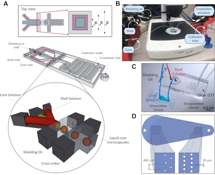
Figure 1: Fabrication of core−shell microcapsules. (A) The microfluidic device for capsule generation consists of four inlet channels (core, shell, oil, and crosslinker oil) and a serpentine channel that leads to a collection tube. The device is fabricated such that core, shell, and oil channels are 120 µm (H1), 200 µm (H2), and 300 µm (H3) in height, respectively. The inset represents a cross-sectional view at the nozzle – the junction between aqueous and oil streams. This cross-sectional view is tilted by 90° to give the reader a better view of coaxial flow channels. A 3D representation of the core-shell microcapsule fabrication process depicts how the coaxial flow is generated upstream of the flow-focusing junction to produce core-shell microcapsule structure. Emulsification is achieved by exposing aqueous droplets to two oil streams, the first of which is designed to stabilize the droplets, and the second to provide the crosslinker DTT, which reacts with PEG-4-Mal. This figure has been modified from reference12. Copyright 2019, American Chemical Society. (B) Experimental setup of the encapsulation system showing locations of syringe pumps, tubing, microfluidic devices and of capsule collection tube. (C) An image of the encapsulation system, which consists of dissociation and encapsulation devices in a sequence. (D) Design of the microfluidic dissociation device used to avoid large cell aggregates entering microencapsulation device. Please click here to view a larger version of this figure.
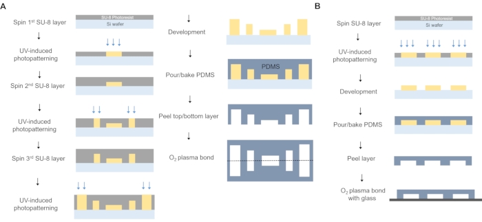
Figure 2: Fabrication of microfluidic devices used for encapsulation. (A) Fabrication of the encapsulation device. Three layers of SU-8 photoresist were spin-coated, and photo patterned to generate core, shell, and oil channels with different heights. Core channels have a width of 220 µm that narrows at the capsule junction to 135 µm. Channels for all phases follow the same principle: shell and oil channels start with a width of 500 µm, narrowing at the junction to 220 µm, and shielding oil starts at 500 µm narrowing to 270 µm. Outlet serpentine has a width of 1.5 mm and a length of ~55 mm. Top and bottom PDMS pieces were then aligned, and plasma bonded. (B) Fabrication of the dissociation device. One layer of SU-8 photoresist was spin-coated, and photo patterned to generate dissociation channels. PDMS piece was then plasma bonded with glass slide. Dissociation device consists of a small chamber with a height of 30 µm, length of 16.5 mm, and width of 5 mm at the inlet and 1.7 mm at the outlet. It has triangle shaped structures (200 µm, equilateral) that are separated from each other by 420 µm of the inlet and become closer on the way to the outlet, with a separation of 50 µm in the last row. Please click here to view a larger version of this figure.
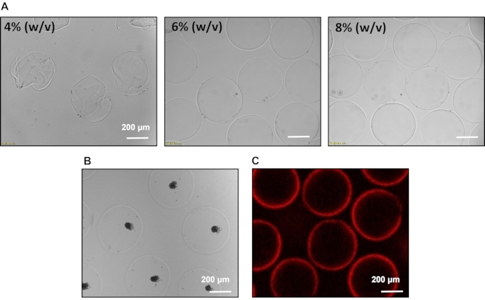
Figure 3: Characterization of microcapsules. (A) Differences in capsule morphology as a function of PEG-4-Mal content in the shell. Smooth capsules with bright edges are associated with desired mechanical integrity. (B) Confirmation of aqueous core of the microcapsules. Entrapped microbeads are free to move in the aqueous core and aggregate in the center of microcapsules. (C) Confirmation of core-shell structure by incorporating Rhodamine B-labeled PEG into the shell of microcapsules. Please click here to view a larger version of this figure.
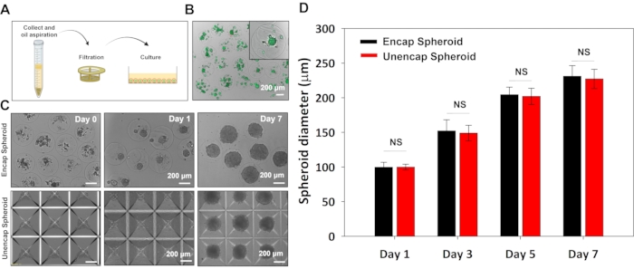
Figure 4: Encapsulation of hPSCs. (A) Microcapsules in the oil phase are collected into a conical tube filled with media. After microcapsules are made to settle to the bottom of the tube, oil and media are aspirated, microcapsules are washed and then transferred to a 6-well plate for cultivation. (B) Live/dead staining 6 h after encapsulating H9 cells. Upper image was taken at 10x, and lower image at 20x. (C) Images comparing spheroid sizes that change over time for H9 cells in capsules and in commercial 3D culture plates (bottom images). (D) Quantification of spheroid diameter that increases for hPSCs in microcapsules and in standard 3D culture plates during 7 days in H9 media. Statistical analysis –t-test, p < 0.05, n = 20. Scale bar: 200 µm. Please click here to view a larger version of this figure.
The encapsulation process described here results in reproducible formation of hPSC spheroids. The microcapsule format makes it easy to dispense spheroids into wells of a microtiter plate for experiments aimed at improving/optimizing differentiation protocols or testing therapies. Encapsulated stem cell spheroids may also be used in suspensions cultures where hydrogel shell protects cells against shear-induced damage7.
There are several critical steps within the protocol. It is important to keep the flow rates of core, shell, and oil streams within the range described in the Protocol section. If the shell flow rate is too low, the flow becomes unstable, resulting in distorted and mechanically unstable capsules. A successful cell encapsulation should look similar to the microcapsules shown in Figure 4C. Microcapsules should be of similar diameter and have a comparable number of cells trapped in the core. Aggregation of single cells into spheroids typically occurs within 48 h and lack of spheroid formation at 72 h is a warning sign. One reason for lack of spheroid formation may be that viscous molecules in the core are not leaching out rapidly enough through the hydrogel shell. We encountered this scenario when adjusting viscosity of the core solution using high molecular weight polymers such as carboxymethyl cellulose (MW 250 kD). One can encapsulate microbeads (see Figure 3B) to test whether these cell-sized objects are free to move about the core and how rapidly components of the core leach out. Lack of spheroid formation was also encountered by us when using PEG-4-Mal concentrations of >8% w/v. In this scenario, porosity and diffusivity of the hydrogel shell are decreased and high viscosity elements in the core do not leach out rapidly enough to allow cells to form into aggregates. It is conceivable that the quality of PEG-4-Mal may vary from batch to batch or manufacturer. Therefore, some adjustment of polymer concentration in the shell may be needed. Once again, bead incorporation method should reveal how rapidly viscous components in the core are displaced by water molecules. In our experience, this should occur within 3 h of transferring microcapsules into the aqueous environment.
The encapsulation protocol described here has worked well for several hPSC lines7, primary hepatocytes10, and multiple cancer cell lines (unpublished results). However, we did encounter difficulties forming spheroids after encapsulation of stem cell-derived (sc)-hepatocytes and sc-β-cells. Troubleshooting with encapsulated microbeads revealed that viscous components were rapidly eluted from the core and that cells were free to move and form cell-cell contacts. Additional troubleshooting involved titrating concentration of di-thiol crosslinker DTT (present in the oil phase) down from 66 mM to 10 mM. Spheroid formation for sc-hepatocytes and β-cells did occur at this lower concentration of DTT. Therefore, the user may want to consider optimizing crosslinker concentration but only if other troubleshooting steps listed above have not been successful. We find that DTT concentration is critical for maintaining mechanical integrity of microcapsules with 66 mM DTT resulting in mechanically robust microcapsules and being non-toxic to vast majority of cell types used to date.
A number of strategies for creating 3D stem cell constructs have been described in the literature. The benefits of forming stem cell constructs within core-shell microcapsules are multiple. Once encapsulated, spheroids may be handled with ease with hydrogel capsules providing protection against shear damage. Encapsulated spheroids may be cultured in suspension with vigorous agitation without damaging stem cells.
There are multiple avenues for expanding the capabilities of microcapsules. Our team has previously described the use of heparin-containing hydrogels for encapsulation of hepatocytes and stem cells8,12, and is presently developing heparin-containing bioactive core-shell microcapsules that serve as depots for storage and local release of inductive cues (GFs) to stem cells. Such microcapsules may help decrease the usage of expensive GFs while improving differentiation outcomes. Microcapsules may be further enhanced by incorporating ECM proteins into the core thus modulating stem cell microenvironment. Core-shell microcapsules may be made degradable to make it easier to retrieve hPSC spheroids13,14. Overall, core-shell microcapsules represent a platform technology that can be modified and enhanced to address the needs of a specific stem cell differentiation protocol.
The authors have nothing to disclose.
This study was supported in part by the grants from the Mayo Clinic Center for Regenerative Medicine, J. W. Kieckhefer Foundation, Al Nahyan Foundation, Regenerative Medicine Minnesota (RMM 101617 TR 004), and NIH (DK107255).
0.22 µm Syringe Filters | Genesee Scientific | 25-244 | |
1 ml syringe luer-lock tip | BD | 309628 | |
1x DPBS | Corning | 23220003 | |
4-arm PEG maleimide, 10kDa | Laysan Inc. | 164-68 | |
5 ml syringe luer-lock tip | BD | 309646 | |
6-WELL NON-TREATED PLATE | USA Scientific | CC7672-7506 | |
Aquapel Applicator Pack | Aquapel Glass Treatment | 47100 | |
CAD software | Autodesk | AutoCAD v2020 | |
CELL STRAINER 100 µm pore size | cardinal | 335583 | |
Chlorotrimethylsilane | Aldrich | 386529-100mL | |
Countess II FL Automated Cell Counter | Life technology | A27974 | |
Digital hot plate | Dataplate | ||
Digital vortex mixer | Fisher Scientific | 215370 | |
Distilled water | Gibco | 15230-162 | |
Dithiotheritol (DTT) | Sigma | D0632-10G | |
DMEM/F12 media | gibco | 11320-033 | |
Falcon 15 mL Conical Centrifuge Tubes | Fisher scientific | 14-959-53A | |
Fisherbrand accuSpin Micro 17 Microcentrifuge | live | 13-100-675 | |
HERACELL VIOS 160i CO2 Incubator | Thermo Scientific | 50144906 | |
Inverted Fluorescence Motorized Microscope | Olympus | Olympus IX83 | |
Laurell Spin Coaters | Laurell Technologies | WS-650MZ-23NPPB | |
Live/Dead mammalian staining kit | Fisher | L3224 | |
Magic tape | Staples | 483535 | |
Micro Medical Tubing (0.015" I.D. x 0.043" O.D.) | Scientific Commodities, Inc | BB31695-PE/2 | |
Micro stir bar | Daigger Scientific | EF3288E | |
MilliporeSigma Filter Forceps | Fisher scientific | XX6200006P | |
Mineral oil | Sigma | M8410-1L | |
mTeSR 1 Basal Medium | STEMCELL TECHNOLOGY | 85850 | |
Needles-Stainless Steel 14 Gauge | CML supply | 901-14-025 | |
Needles-Stainless Steel 15 Gauge | CML supply | 901-15-050 | |
OptiPrep | STEMCELL TECHNOLOGY | 7820 | |
Oven | Thermo Scientific | HERA THERM Oven | |
Penicillin:Streptomycin (10,000 U/mL Penicillin G, 10mg/mL Streptomycin) | Gemini | 400-109 | |
Petri Dish 150X20 Sterile Vent | Sarstedt, Inc. | 82.1184.500 | |
Plasma Cleaning System | Yield Engineering System, Inc. | YES-G500 | |
Pluronic F-127 | Sigma | P2443-250G | |
Poly(ethylene glycol) 35kDa | Sigma | 94646-250G-F | |
PrecisionGlide Needle 27G | BD | 305109 | |
Rock inhibitor Y-27632 dihydrocloride | SELLECK CHEM | S1049-10mg | |
Silicon wafer 100mm | University Wafer | 452 | |
Slide glass (75mm ´ 25mm) | CardinalHealth | M6146 | |
Span 80 | Sigma | S6760-250ML | |
SpeedMixer | Thinky | ARE-310 | |
Spin-X Centrifuge Tube Filter (0.22 µm) | Costar | 8160 | |
SU-8 2025 | Kayaku Advanced Materials | Y111069 0500L1GL | |
SU-8 developer | Kayaku Advanced Materials | Y020100 4000L1PE | |
Surgical Design Royaltek Stainless Steel Surgical Scalpel Blades | fisher scientific | 22-079-684 | |
SYLGARD TM 184 Silicone Elastomer Kit (PDMS) | Dow Corning | 2065622 | |
Syringe pump | New Era Pump System, Inc | NE-4000 | |
Triethanolamine | Sigma-aldrich | T58300-25G | |
TrypLE Express | Gibco | 12604-013 | |
Tygon Tubing (0.02" I.D. x 0.06" O.D.) | Cole-Parmer | 06419-01 | |
Tygon Tubing (0.04" I.D. x 0.07" O.D.) | Cole-Parmer | 06419-04 | |
Ultrasonic cleaner FS20D | Fisher Scientific | CPN-962-152R | |
Vacuum desiccator | Bel-Art | F42025-0000 | |
Zeiss Stemi DV4 Stereo Microscope 8x-32x | ZEISS | 435421-0000-000 | |
μPG 101 laser writer | Heidelberg Instruments | HI 1128 |