Here, we present a simple and effective assay procedure for resorption pit assays using calcium phosphate coated cell culture plates.
Mature osteoclasts are multinucleated cells that can degrade bone through the secretion of acids and enzymes. They play a crucial role in various diseases (e.g., osteoporosis and bone cancer) and are therefore important objects of research. In vitro, their activity can be analyzed by the formation of resorption pits. In this protocol, we describe a simple pit assay method using calcium phosphate (CaP) coated cell culture plates, which can be easily visualized and quantified. Osteoclast precursors derived from human peripheral blood mononuclear cells (PBMCs) were cultured on the coated plates in the presence of osteoclastogenic stimuli. After 9 days of incubation, osteoclasts were fixed and stained for fluorescence imaging while the CaP coating was counterstained by calcein. To quantify the resorbed area, the CaP coating on plates was stained with 5% AgNO3 and visualized by brightfield imaging. The resorption pit area was quantified using ImageJ.
Osteoclasts (OCs) are tissue-specific macrophages derived from hematopoietic stem cells (HSCs), playing a pivotal role in bone remodeling together with osteoblasts1. Sex hormone-induced, immunological, and malignant bone disorders that destroy bone systemically or locally are due to excess osteoclastic activity, including menopause-related osteoporosis2, rheumatoid arthritis3, periodontal disease4, myeloma bone disease5, and osteolytic bone metastasis6. In contrast, defects in OC formation and function can also cause osteopetrosis7. HSCs undergo differentiation into OC progenitors under macrophage colony-stimulating factor (M-CSF, gene symbol ACP5) stimulation. In the presence of both M-CSF and receptor activator of NF-κB ligand (RANKL, gene symbol TNFSF11), OC progenitors differentiate further into mononuclear OCs and subsequently fuse to become multinucleated OCs8,9,10. Both cytokines M-CSF and RANKL are indispensable and sufficient for induction of osteoclastic markers such as calcitonin receptor (CT), receptor activator of nuclear factor κ B (RANK), proton pump V-ATPase, chloride channel 7 alpha subunit (CIC-7), integrin β3, tartrate-resistant acid phosphatase (TRAP, gene symbol ACP5), lysosomal cysteine protease cathepsin K (CTSK), and matrix metallopeptidase 9 (MMP9). Activated OCs form a sealing zone on the bone surface through the formation of an actin ring with a ruffled border11,12. Within the sealing zone, OCs mediate resorption through secreting protons via the proton pump V-ATPase12,13, MMP914, and CTSK15, leading to the formation of lacunae.
For in vitro experiments, OC progenitors can be obtained by expansion of bone marrow macrophages from mice's femur and tibia16,17, as well as by isolation of human peripheral blood mononuclear cells (PBMCs) from blood samples and buffy coats18,19,20, or by differentiation of the immortalized murine monocytic cells RAW 264.721,22.
In the present protocol, we describe an osteoclastic resorption assay in CaP coated cell culture plates using OCs derived from primary PBMCs. The CaP coated cell culture plates method used here are adopted and refined from the method described previously by Patntirapong et al.17 and Maria et al.21. To obtain OC precursors, PBMCs are isolated by density gradient centrifugation and expanded as described previously20.
The protocol was reviewed and approved by the local ethics committee (approval number 287/2020B02).
1. Preparation of calcium phosphate coated cell culture plates
2. Isolation of PBMCs from human peripheral blood
3. Expansion of OC progenitors
4. Induction of osteoclastogenesis in CaP coated plates
5. Fluorescence staining of OCs and CaP coating
6. Quantification of total resorption pit area
7. Quantification of OC number and size, and normalized resorption pit area
The calcium phosphate coating on the bottom of cell culture plates was performed in two coating steps comprising a 3-day pre-calcification and a 1-day calcification step. As shown in Figure 1, uniformly distributed calcium phosphate was obtained on the bottom of the 96-well plates. The coating adhered very well to the bottom after the performed washing steps.
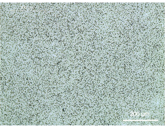
Figure 1: Representative brightfield image of the calcium phosphate coating on 96-well cell culture plates. The coating was carried out in two coating steps comprising a 3-day pre-calcification step and a 1-day calcification step. The scale bar represents 200 µm. Please click here to view a larger version of this figure.
OC precursors derived from human PBMCs were cultured on the CaP coated plates. Resorption pits and large OCs (blank area) were formed in the presence of M-CFS and RANKL after 9 days of culture (Figure 2A). The multinucleated OCs located in the pits of the CaP coating expressed high levels of TRAP (Figure 2B).
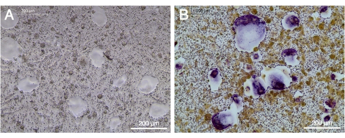
Figure 2: TRAP expression by OCs after maturation on the CaP coated cell culture plates. OC precursors were cultured on the CaP coated plates in the presence of 20 ng/mL M-CSF and 20 ng/mL RANKL for 9 days. (A) Blank area represented resorption pits. (B) Several multinucleated TRAP-positive (purple) OCs were observed within the resorption pits. The scale bar represents 200 µm. Please click here to view a larger version of this figure.
To further characterize the maturation of OCs on the CaP coated plates, cells were stained for actin (red fluorescence). Cell nuclei were stained with Hoechst (blue) and calcein was used for calcium visualization in green. Resorption pits are visible as black areas in the green CaP coating (Figure 3B). Functional mature OCs showed three or more nuclei and the characteristic actin ring which is essential for the osteoclastogenic resorption (Figure 3A,C). The pit area of fluorescence images can be measured using the thresholding tool of ImageJ (Figure 3D). The number and size of OCs can be calculated using the ROI Manager and Polygon selection tool of ImageJ (Figure 3E).
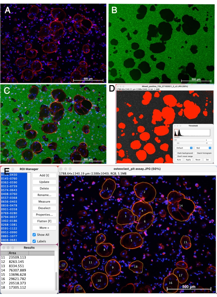
Figure 3: Morphology of mature OCs on the CaP coating. OC precursors were cultured on the CaP coated plates for 9 days in the presence of 20 ng/mL M-CSF and 20 ng/mL RANKL. (A) OCs were stained for actin and nuclei by phalloidin-Alexa Fluor 546 and Hoechst 33342. (B) CaP coating was stained by calcein. Black areas represent resorption pits. (C) Merged image. (D) Quantification of pit area (red area) using the thresholding tool of ImageJ. (E) OC outlining and counting using the ROI Manager and Polygon selection tool of ImageJ. The scale bar represents 500 µm. Please click here to view a larger version of this figure.
For the quantification of the resorption pits on the CaP coated cell culture plates, calcium was stained by incubation with 5% AgNO3 (Von Kossa staining). As shown in Figure 4A, OC precursors did not reach full functionality in the absence of RANKL and were not able to form resorption pits. Osteoclastogenic pits were observed only in the presence of both factors M-CSF and RANKL (Figure 4B). The resorption pit area was quantified with the thresholding tool of ImageJ (Figure 4C).
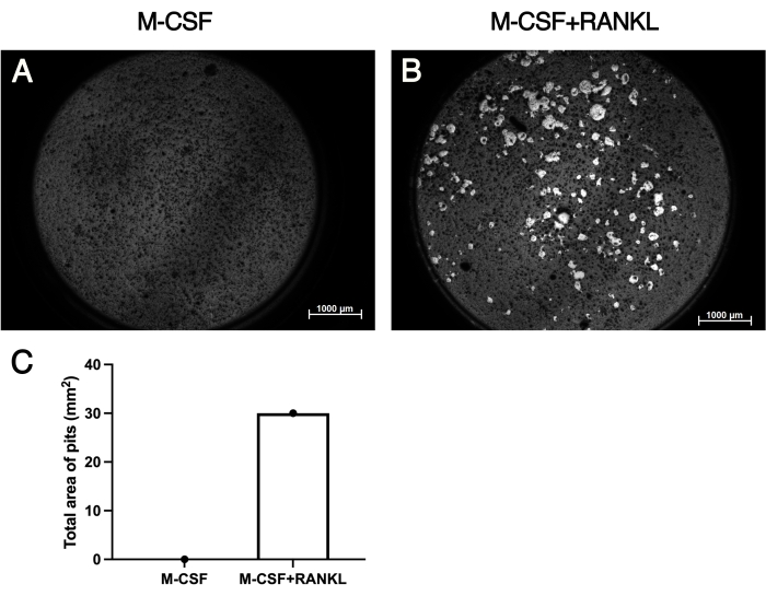
Figure 4: Visualization and quantification of resorption pits. OC precursors were cultured on the CaP coated 96-well cell culture plates in the absence of RANKL (A) and in the presence of both factors M-CSF and RANKL (B). (C) Numbers of formed resorption pits were quantified using the thresholding tool of ImageJ. Scale bar represents 1,000 µm. Please click here to view a larger version of this figure.
Here we describe a simple and reliable method for an osteoclastic resorption assay using OCs derived and expanded in vitro from PBMCs. The used CaP coated cell culture plates can be easily prepared and visualized using lab-available materials. In addition to unsorted PBMCs adopted in this protocol, OCs generated from murine monocytic cells21 and bone marrow macrophage cells17 have also been cultured on similar synthetic substrates for pit assay, thus these cell sources can be moved to this approach referring to the corresponding literature.
For this protocol, we used a simple in-lab fabricated CaP coating instead of expensive commercially available assay plates. Although bone and dentin slices are two common and affordable resorbable materials for resorption pit assay, the availability and complicated preparation are major drawbacks. As an easily prepared synthetic substrate, CaP coating is more convenient and readily available than the two original materials. Another advantage of this pit assay approach is that cells can be visualized under a bright field microscope during culture, whereas cells growing on opaque bone and dentin slices cannot be observed.
This CaP coating method can be flexibly adapted according to the required experimental needs to different cell culture plate sizes.
Unlike previously reported17,21, we incubate the plates with coating solution at 37 °C instead of at room temperature, resulting in the spontaneous growth of apatite nuclei on cell culture plates at body temperature. CaP coated plates are available for experiments immediately after being dried and sterilized by UV radiation, which saves time compared to previously reported methods17,21.
Filtration of the coating solution (Step 1.3.1 and 1.4.2) is a crucial step, which helps to obtain a uniform particle size and reduce uncoated spots within the coating caused by air bubbles and impurities in the solution. Drying the plate immediately (step 1.5.2) is another critical step, which helps to make a uniform CaP coating. Otherwise, it is prone to form a thicker coating in the middle of the well.
It is worth pointing out that the thickness of the coating and the density of calcium phosphate deposits depend on the volume of coating solution within a certain range. Therefore, it is possible to control coating thickness by changing the volume of coating solution in the well. However, it is suggested to add as much volume of coating solution as possible when the 96-well cell culture plate is used because of the small total volume for this culture plate format. The inner wall of the well is also coated by CaP during both coating steps, therefore caution is required to not touch the bottom nor the wall with the pipette in order to protect the coating from damage.
According to our observation, most of OC precursors expanded from PBMCs are able to attach to the CaP coating without any problems. However, soaking the coated plate with FBS before cell seeding improves the cell adhesion efficiency, as previously reported21.
Detaching the OC precursors gently using a cell scraper (step 4.2) is also important because most of the OC precursors were still adherent, and minimizing the mechanical injury is favorable for cell viability.
A limitation of this method is that the bulk PBMCs that we used as a source of OC precursors is a highly mixed cellular source, of which only a small fraction of cells (CD14+ cells) are actual OC precursors. Since other cells capable of affecting osteoclastogenesis (such as stromal cells and lymphocytes) are present amongst isolated PBMCs, this may impact assay results in ways that may be hard to predict, and might confound assay interpretation. To better investigate direct effects of growth factors, cytokines or glucocorticoids on resorptive activity of OCs, methods of OC precursor purification (e.g., fluorescence-activated cell sorting (FACS) and magnetic beads purification20 based on specific cell-surface markers23,24,25,26) are recommended.
The limitations of the CaP substrate in obtaining brightfield images of both cells and underlying pits for the same area is caused by the incompatibility of TRAP and Von Kossa staining, so that normalized resorption area of the entire well is not available. But instead, fluorescence imaging can be used to visualize resorption area and cell number to obtain normalized resorption data. These data may provide important insight into whether the increase in total resorption area under experimental conditions is due to increased osteoclast number alone or increased resorption capacity of individual cells. In addition, it allows the study of the relative effects of osteoclast number, size, nucleus number, and resorption capacity.
In summary, we describe a useful and simple protocol for a resorption pit assay using a two-step calcium phosphate coating and OCs derived from primary human PBMCs. This protocol provides an easy method to establish an in vitro bone resorption model for studies related to osteoclastic resorption, and could be applied to study treatments for bone disorder diseases.
The authors have nothing to disclose.
This work was partially funded by the China Scholarship Council [CSC No. 201808440394]. W.C. was financed by CSC.
AgNO3 | SERVA Electrophoresis GmbH | 35110 | Silver nitrate |
a-MEM | Gibco | 32561-029 | MEM alpha, GlutaMAX, no nucleosides |
amphotericin B | Biochrom | 03-028-1B | Amphotericin B Solution |
CaCl2 | Sigma-Aldrich | 21097-50G | Calcium chloride Dihydrate |
Calcein | Sigma-Aldrich | C0875 | Calcein |
FBS | Sigma-Aldrich | F7524 | fetal bovine serum |
Ficoll | Cytiva | 17144002 | Ficoll Paque Plus |
Fixation buffer | Biolegend | 420801 | Paraformaldehyde |
HCl | Merk | 1.09057.1000 | Hydrochloric acid |
Hoechst 33342 | Promokine | PK-CA707-40046 | Hoechst 33342 |
M-CSF | PeproTech | 300-25 | Recombinant Human M-CSF |
MgCl2 | Sigma-Aldrich | 7791-18-6 | Magnesium chloride |
Na2HPO4 | AppliChem GmbH | A2943,0250 | di- Sodium hydrogen phosphate anhydrous |
NaCl | Merk | S7653-250G | Sodium chloride |
NaHCO3 | Merk | K15322429 | Bicarbonate of Soda |
PBS | Lonza | 17-512F | Dulbecco's Phosphate Buffered Saline (1X), DBPS without Calcium and Magnesium |
Pen-Strep | Lonza | DE17-602E | Penicillin-Streptomycin Mixture |
Phalloidin-Alexa Fluor 546 | Invitrogen | A22283 | Alexa Fluor 546 Phalloidin |
RANKL | PeproTech | 310-01 | Recombinant Human sRANK Ligand (E.coli derived) |
Tris | Sigma-Aldrich | 93362 | Tris(hydroxymethyl)aminomethan |
Triton X-100 | Sigma-Aldrich | T8787 | Alkyl Phenyl Polyethylene Glycol |
TrypLE Express | Gibco | 12605010 | Recombinant cell-dissociation enzymes |