This protocol provides technical information for vessel reconstruction using the cuff technique in mouse orthotopic liver transplantation.
Mouse orthotopic liver transplantation is an effective methodology for investigating the underlying mechanisms of liver ischemia and reperfusion injury. However, the technical challenges pose a barrier to utilizing this valuable experimental model and passing on these skills to the next generation. The most challenging aspect of this procedure is vascular reconstruction, including the portal vein (PV), infrahepatic inferior vena cava (IHIVC), and suprahepatic inferior vena cava. The use of plastic cuffs, rather than sutures, allows for smoother PV and IHIVC reconstruction. Vessels are reconstructed by attaching a cuff made from an intravenous catheter to the tip of the graft vessel and interposing the cuff into the recipient vessel. The two most crucial aspects are properly visualizing the inner lumen of the vessel and avoiding the use of excessive force. Our aim is to provide a technical overview of vascular reconstructions using the cuff technique in recipient surgery. These technical tips for the cuff technique are expected to help microsurgeons facilitate vascular reconstruction and advance their research.
Mouse orthotopic liver transplantation (MOLT) is an effective experimental method first reported in 19911. This experimental model, which utilizes genetically modified mice and various research reagents, has played a pivotal role in investigating warm and cold ischemia and reperfusion injuries. However, the model's high technical complexity has hindered the development of basic medicine for liver transplantation2. MOLT involves three major steps: (1) retrieval of the liver from the donor mouse, (2) back table surgery, and (3) implantation of the liver into the recipient. Among these recipient procedures, vascular anastomosis poses the greatest challenge. While the suprahepatic inferior vena cava anastomosis is typically completed by hand stitching2, the infrahepatic inferior vena cava (IHIVC) and portal vein (PV) can be reconstructed more efficiently using plastic cuffs in place of hand-sewn sutures.
The anhepatic period signifies the interval between the removal of the recipient's native liver and the graft implantation. To ensure consistent results, it is imperative to limit the anhepatic time to less than 20 min. Consequently, studies employing this model have been confined to specific institutions3,4,5,6,7,8,9. Among the various stages of MOLT, achieving smooth PV and IHIVC reconstruction is crucial for minimizing anhepatic time and ensuring successful transplantation.
PV and IVC reconstruction is generally carried out using vascular cuffs since the cuff technique simplifies vascular anastomosis compared to hand-sewn sutures2,5,8. The technique involved in vascular cuff preparation and secure attachment of the cuff significantly impacts the complexity of recipient vascular reconstruction. Our objective is to provide detailed visual guidance for numerous technical tips related to the cuff method, thereby reducing the learning curve. These video clips will provide a clear understanding of how to attach the cuff to the vessels and reconstruct the PV and IHIVC during recipient surgery.
The experimental protocol was approved by the Animal Experimentation Committee at Kyoto University. The study utilized C57BL/6 mice, aged over 10 weeks and weighing between 25 g and 30 g, obtained from a commercial source (see Table of Materials). All animals were anesthetized with 2.5% isoflurane (following institutionally approved protocols), maintained under specific pathogen-free conditions, and all experimental procedures were conducted in compliance with Kyoto University's Regulations on Animal Experimentation. The appropriate instruments used for the study are listed in the Table of Materials and depicted in Figure 1 and Figure 2.
1. Animal selection
2. Cuff making
3. Cuff attachment
4. Portal vein reconstruction
5. Infrahepatic inferior vena cava reconstruction
6. Postoperative care
PV reconstruction is successful when, upon unclamping the portal vein, there is no tortuosity, and the liver is uniformly perfused. Anhepatic time should be under 20 min, as anhepatic times exceeding 25 min increase the risk of mouse mortality. IHIVC reconstruction is deemed successful if there is no blood regurgitation from the graft.
Storing the graft at cold temperatures for 1 h using organ preservation solution results in a serum alanine aminotransferase level of approximately 2,000 U/L at 6 h after reperfusion (Figure 7A). The survival rate is 100% at 7 days after transplantation (Figure 7B). However, in our model without hepatic artery anastomosis, recipient mice may succumb to intrahepatic biloma-related issues approximately 30 days post-transplantation
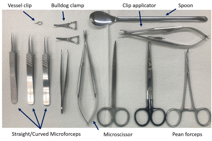
Figure 1: Instruments for vascular reconstruction. Straight or curved forceps are used as needed for the situation and the operative field. Please click here to view a larger version of this figure.
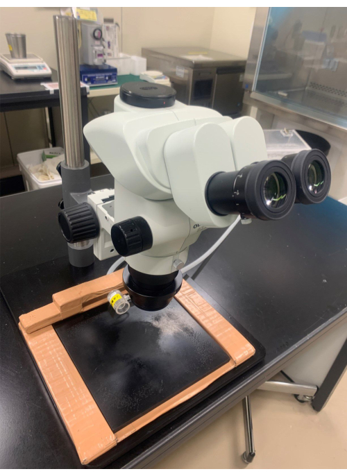
Figure 2: The microscope. The microscope is equipped with an objective lens with 10x magnification and an eyepiece lens with at least 0.8x magnification. Please click here to view a larger version of this figure.
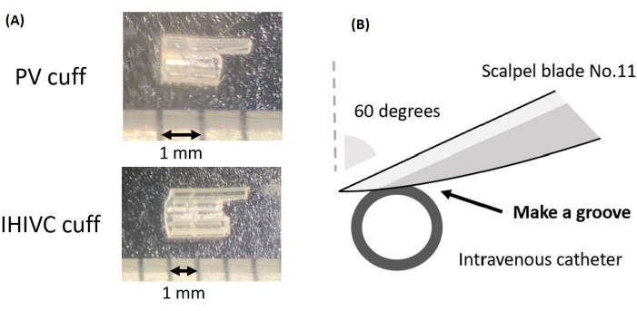
Figure 3: Preparation of the cuff. (A) The cuff for the PV and IHIVC. (B) A schematic representation of the process of creating a groove on the cuff. Please click here to view a larger version of this figure.
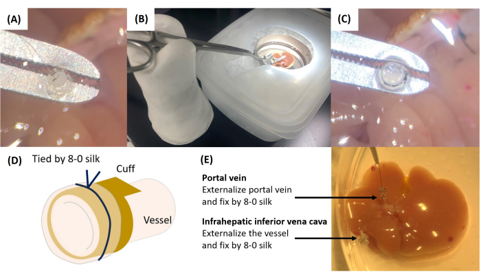
Figure 4: Cuff attachment. (A) The thread attached to the PV is passed into the cuff. (B) Instrument setup for cuff attachment. (C) Securing the cuff with 8-0 silk ligations. (D) Schematic illustration of vascular externalization and fixation to the cuff. (E) Completed cuff attachment. Please click here to view a larger version of this figure.
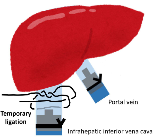
Figure 5: Temporary ligation of the IHIVC root. Please click here to view a larger version of this figure.
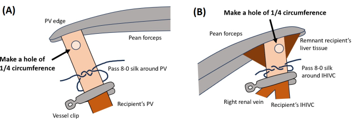
Figure 6: Vascular reconstruction. This illustration shows the locations for creating holes for (A) PV and (B) IHIVC. Please click here to view a larger version of this figure.
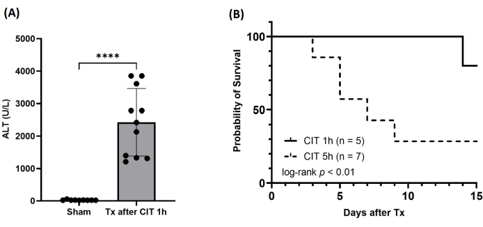
Figure 7: Postoperative results. (A) Serum levels of alanine aminotransferase (ALT) at 6 h after reperfusion. CIT, cold ischemic time; Tx, transplantation. ****p < 0.0001. (B) Survival of the recipient after MOLT. Please click here to view a larger version of this figure.
Learning vascular reconstruction is the most challenging aspect of achieving successful MOLT. The cuff’s quality significantly influences the reconstruction’s difficulty, given the small size of mice5. This article provides a detailed protocol for cuff preparation, attachment, and reconstruction.
While there are no major differences from previous reports regarding cuff preparation and connection2,5, some minor points should be considered. First, the cuff handle’s thickness should be limited to approximately 1/6 of the total circumference. If it’s too thin, the handle can easily bend and become hard to grasp. If it’s too thick, it may be challenging to handle in the recipient’s abdomen. Identifying your personal preference early on is essential.
Second, visibility of the lumen at the back table is crucial. A 10-0 nylon thread is used, attached to the donor’s splenic vein as a landmark to confirm the PV lumen. The nylon thread is placed at 8 o’clock to prevent excessive twisting. The vessel wall is externalized by grasping it at 3 o’clock and 9 o’clock with two forceps and pressing it against the cuff wall at 6 o’clock. Fixation with 8-0 silk thread requires careful movements. Adequate fixation is achieved by securing it in the groove. After attachment, lift the cuff to confirm it doesn’t come off.
Third, during reconstruction, it’s important not to create too large a hole in the recipient vessel because the hole naturally enlarges as the cuff is inserted. Making a large hole can lead to vessel splitting, the most common cause of failure. Particularly in IHIVC reconstruction, smaller holes should be made since the shorter length of IHIVC makes it easier to pull out than a PV. When passing saline solution through the lumen, support cuff insertion by applying upward force rather than downward.
Common causes of failure and precautions have been discussed in detail. The most crucial points include ensuring clear visualization of the inner vessel lumen and avoiding unnecessary force during cuff insertion to prevent vessel splitting. These technical tips for the cuff method are believed to elevate skills and advance research.
The authors have nothing to disclose.
This work was supported by 2022 JST basic research (The Japanese Society for Transplantation).
16 G intravenous catheter | TERUMO | SR-FF2032 | IHIVC cuff |
20 G intravenous catheter | TERUMO | SR-FF1651 | PV cuff |
8-0 braid silk | Natsume Seisakusho | CR9-80B2 | 8-0 silk |
Belzer UW Cold Storage Solution | Astellas | Organ preservation fluid | |
Bulldog clamp | B BRAUN | FB329R | Bulldog clamp |
C57BL/6 mice | Oriental Bio Service | ||
Isoflurane inhalation solution | Viatris | Anesthesic | |
Micro Blunted Tips 0.1 mm x 0.06 mm | F.S.T | 11253-20 | Straight microforceps |
Micro Serrefine Clamp Applicator with Lock | F.S.T | 18056-14 | Vessel clip applicator |
Micro Serrefines | F.S.T | 18055-4 | Vessel clip |
No.11 Spare Blades | FEATHER Safety Razor | 11 | Blades |
Ophthalmic scissor, round handle | B BRAUN | FD103R | Microscissor |
Plastic rectangular-shaped container | Daiso | 10 cm long, 15 cm wide and 6 cm high | |
SuperGrip Tips | F.S.T | 00649-11 | Curved microforceps |
SZX7 | Olympus | SZX7 | Microscope |