Summary

레이저 캡처 미세 절 RNA-시퀀싱 용 공간 및 측두 조직 -특정 유전자 발현 분석

Published: August 05, 2020
doi:

Summary

여기에 제시된 것은 식물 조직의 레이저 포획 미세 절(LCM)을 위한 프로토콜입니다. LCM은 오염없는 방식으로 조직의 영역을 격리하기위한 현미경 기술입니다. 절차는 조직 고정, 파라핀 포함, 단면, LCM 및 RNA 추출을 포함한다. RNA는 다운스트림 조직 특이적, 전사의 현세적으로 해결된 분석에서 사용된다.

Abstract

복잡한 다세포 유기체의 발달은 상이한 전사 단면도가 있는 명백한 세포 모형에 의해 지배됩니다. 발달 과정을 제어하는 전사 적 규제 네트워크를 식별하려면 이러한 개별 세포 유형의 공간 및 측두성 유전자 발현 프로파일을 측정할 필요가 있습니다. 따라서 유전자 발현의 현세-측량 제어에 대한 통찰력은 생물학적 및 발달 과정이 어떻게 규제되는지 이해하는 데 필수적입니다. 여기서는 발아 중에 시간 과정을 통해 3개의 보리 배아 기관에서 소수의 세포를 분리하는 레이저 포획 미세 절(LCM) 방법을 설명하고 전사체 프로파일링을 실시한다. 이 방법은 조직 고정, 조직 처리, 파라핀 포함, 단면, LCM 및 RNA 추출 다음으로 실시간 PCR 또는 RNA-seq로 구성됩니다. 이 방법을 통해 종자 장기 전사의 공간적 및 측두프로파일을 다양한 수의 세포(수십~수백 개)에서 얻을 수 있게 되어 일반적인 벌크 조직 분석보다 훨씬 더 큰 조직 특이성을 제공합니다. 이러한 데이터에서 우리는 전사 적 규제 네트워크를 정의하고 비교할 뿐만 아니라 개별 조직에 대한 후보 규제 전사 요인을 예측할 수 있었습니다. 이 방법은 최소한의 최적화를 가진 다른 플랜트 조직에 적용되어야 합니다.

Introduction

식물 개발 및 성장은 복잡한 세포 환경에 존재하는 다른 세포 내의 전사 규제 네트워크의 조정 된 작용을 포함한다. 이러한 규제 네트워크의 활동을 이해하기 위해, 우리는 발달 단계에 걸쳐 다른 세포 모형 내의 공간 및 측두성 유전자 발현의 지식을 요구합니다. 그러나, 유전자 발현의 분석은 세포의 작은 숫자를 격리하고 분석의 기술적 인 도전 때문에 전체 기관 또는 대량 조직 샘플에서 더 일반적으로 수행됩니다. 여기서 설명하는 방법은 LCM과 RNA-seq를 결합하여 공간 및 측두조직 특이적 전사 분석을 얻을 수 있게 하였다.

LCM은 에머트 벅과 동료에머트벅에 의해 이십 년 전에 개발되었다 1 . 이 기술을 통해 연구자들은 좁은 빔 레이저1을사용하여 직접 현미경 시각화 및 조작을 사용하여 단일 세포 또는 세포 클러스터를 환경으로부터 정확하게 분리할 수 있었습니다. 그 이후로 이 방법은 암 생물학 및 병리학2,,3에서널리 사용되고 있다. ,많은 식물 연구 그룹은 또한 다른 식물 종 및 다른 조직 유형,4,,5,,6,7,8,9,10,11과함께 사용하기 위해,LCM을적응시켰다. 최근에는, 몇몇 논문은 또한 종자 발달 및 발아 도중 배아, 내배식 및 그밖 종자 구조물을 공부하기 위하여 eudicot 및 단장화장 씨앗에LCM을 이용했습니다10,,12,13. 마이크로 파이펫팅, 세포 선별, 자기 분리 및 미세 유체 플랫폼과 같은 다른 일반적으로 사용되는 단일 세포 격리 방법의 대부분은 세포를 해리하기 위해 효소 소화 또는 기계적 균질화에 의존한다. 이는 데이터해석(14,15)을혼동하는 기술 적 유물을 도입하여 유전자발현을방해할 수 있다. 이러한 방법은 또한 해리된 세포를 공간 위치 및 진정한 세포 유형에 연관시키기 위해 각 세포 유형에 대한 마커 유전자에 대한 이전 지식을 요구합니다. 기술의 추가 그룹은 전체 세포 대신 세포 체형 구조의 친화성 기반 절연에 따라 달라집니다, 예를 들어 INTACT (세포 유형에서 태그 핵의 격리) 및 TRAP (리보솜 친화 정화 번역)16,,17. 그러나 핵 이나 리보좀의 선호도 라벨링 및 정제는 잘 확립 된 변형 프로토콜이없는 식물 종에서 기술적으로 도전적입니다. LCM은 짧은기간동안 이산 세포를 단기간에 격리할 수 있도록 하는 정상 조직/장기 문맥 내에서 세포의 직접 시각화에 의한 성적증명서 수준 및 종래의 조직학적 식별을 보존하기 위해 빠른 조직 고정을활용한다.19

여기에 제시된 프로토콜은 조직학적으로 식별될 수 있는 대부분의 세포에 적용될 수 있는 시리얼 씨앗의 조직 섹션에서 특정 세포 또는 세포 유형의 분리를 위한 최적화된 방법입니다. LCM은 접촉없는 세포 격리 방법을 제공하여 오염을 크게 줄이고 회수 된 RNA의 무결성을 증가시킵니다. 더욱이, 이 방법은 소량의 생물학적 물질로 시작하는 대규모 게놈 넓은 연구에 대한 LCM의 힘을 보여줍니다. 우리는 또한 다운스트림 전사/전사 분석을 위한 충분한 입력 물질을 생성하기 위한 RNA의 선형 증폭을 기술합니다.

이 LCM RNA-seq 프로토콜에는 조직 샘플, 탈수, 파라핀 침투, 임베딩, 단면, LCM, RNA 추출, RNA 증폭, RNA 정량화 및 qRT-PCR 및/또는 RNA-seq(그림1)의고정을 포함하는 공간 및 측두조직 특이적 전사에 대한 10가지 주요 단계가 있습니다.

Figure 1
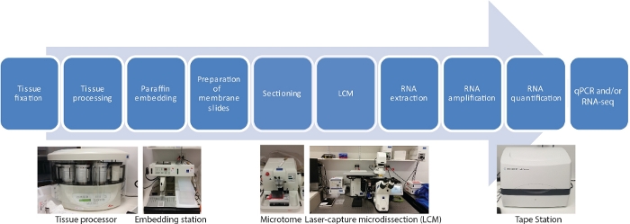
그림 1: LCM의 순서도는 RNA-seq 또는 qRT-PCR이 그 뒤를 따릅니다. LCM은 현미경 시각화 하에서 레이저 빔을 사용하여 고정 조직 섹션에서 세포를 수집하는 공간적으로 정확하고 접촉이 없는 기술입니다. 이 과정은 조직 샘플의 고정으로 시작하여 에탄올과 자일렌의 그라데이션 시리즈를 사용하여 탈수로 시작하여 파라핀 침투로 완성됩니다. 이 과정은 티슈 프로세서를 사용하여 완전히 자동화될 수 있습니다. 조직이 파라핀으로 침투하면 포함 스테이션을 사용하여 용융 파라핀이있는 금형에 내장됩니다. 절제는 원하는 두께로 설정된 마이크로토메를 사용하여 수행됩니다. 슬라이드는 RNA가 포획된 세포에서 추출되기 직전에 제조되고 LCM이 수행됩니다. RNA 추출은 qRT-PCR 및/또는 RNA-seq 이전에 RNA 증폭의 2라운드에 의해 직접 뒤따릅니다. 이 그림의 더 큰 버전을 보려면 여기를 클릭하십시오.

Protocol

최종 제품은 RNA이기 때문에 RNases와 함께 작업을 오염하지 않도록주의하십시오. 장갑을 착용하는 것은 필수입니다. 다이틸 파이로카보네이트(DEPC) -처리된 물, 완충제 등을 사용하십시오. 자동 클래브 버퍼와 사용하기 전에 유리 제품을 굽습니다. 1. 조직 고정 종 과 조직 유형에 따라 선택의 고정을 준비; 보리 씨앗의 경우 농부의 고정 (75 % 에탄올, 25 % 빙하 아세트산 (v …

Representative Results

우리는 우리의 LCM RNA-seq 프로토콜10을사용하여 발아 하는 동안 보리 씨앗에서 공간 및 측두 조직 특이적인 전사를 생성 했다. 이 연구는 발아 중 48시간 코스(0-48h, 7시간 포인트)를 통해 3개의 배아 기관(plumule, radicle tip, scutellum)에서 소수의 세포에 LCM RNA-seq를 적용하여 수행하였다(도2A,B). <img alt="Figure…

Discussion

많은 조직 별 유전자 발현 연구는 시간이 많이 소요되는, 노동 집약적인 시료의 손 해부에 의해 제한되었으며, 오염위험이 높으며 인간 수술이 수확하기에 충분히 손재주적이라는 시료만 활용할 수 있습니다. LCM은 현미경 시각화 하에서 기계적으로 작동하는 레이저 빔을 사용하여 고정 조직 섹션에서 세포를 수집하는 정확하고 접촉없는 기술입니다.

좋은 샘플 준비는 LCM에 ?…

Declarações

The authors have nothing to disclose.

Acknowledgements

이 작품은 식물 에너지 생물학 (CE140100008)에서 JW에 우수의 호주 연구 위원회 센터에 의해 지원되었다. M.G.L은 라 트로브 대학 시작 보조금에 의해 지원되었다. 우리는 높은 처리량 시퀀싱 및 데이터 분석에 대한 지원에 대한 La Trobe 유전체학 플랫폼에 감사드립니다. 우리는 우리의 실험실에서 LCM을 설립에 대한 전문가의 조언을 준 교수 매튜 터커 에게 감사드립니다.

Materials

Acetic acid 100 % ACS/R. AnalaR NORMAPUR (BioStrategies) VWRC20104.323
AdhesiveCap 200 opaque Zeiss 415190-9181-000
Clear base moulds 8 X 10 Leica 3803015
Diethyl pyrocarbonate Sigma-Aldrich 40718-25ML
High Sensitivity RNA ScreenTape Agilent 5067-5579
Low­profile disp.blades DB80LS Leica 14035843489
MembraneSlide 1.0 PEN Zeiss 415190-9041-000
MessageAmp II aRNA Amplification Kit Ambion (ThermoFisher) AMB17515
On-Column DNase I Digestion Set Sigma-Aldrich DNASE70
Ovation RNA-Seq System V2 NuGen (Integrated Science) 7102-08
Paraffin (Surgipath Paraplast) Leica 39601006
PicoPure RNA Isolation Kit ABI (ThermoFisher) KIT0214
RNaseZap RNase Decontamination Solution Ambion (ThermoFisher) AM9780
Xylene AnalaR NORMAPUR (BioStrategies) VWRC28975.360
Leica Benchtop Tissue Processor Leica Biosystems TP1020
Leica Heated Paraffin Embedding Module Leica Biosystems EG1150H
Leica Cold Plate Leica Biosystems EG1150C
Safemate Class 2 Biological Safety Cabinets LAF Technologies Pty Ltd Safemate 1.5
Leica Fully Automated Rotary Microtome Leica Biosystems RM2265 with PALMRobo v 4.6 software
Zeiss PALM MicroBeam LCM system Zeiss miscroscopy
TapeStation Agilent TapeStation 2200

Referências

  1. Emmert-Buck, M. R., et al. Laser capture microdissection. Science. 274 (5289), 998-1001 (1996).
  2. Alevizos, I., et al. Oral cancer in vivo gene expression profiling assisted by laser capture microdissection and microarray analysis. Oncogene. 20 (43), 6196-6204 (2001).
  3. Cong, P., et al. In situ localization of follicular lymphoma: description and analysis by laser capture microdissection. Blood, The Journal of the American Society of Hematology. 99 (9), 3376-3382 (2002).
  4. Blokhina, O., et al. Laser capture microdissection protocol for xylem tissues of woody plants. Frontiers in Plant Science. 7, 1965 (2017).
  5. Casson, S., Spencer, M., Walker, K., Lindsey, K. Laser capture microdissection for the analysis of gene expression during embryogenesis of Arabidopsis. The Plant Journal. 42 (1), 111-123 (2005).
  6. Chen, Z., et al. LCM-seq reveals the crucial role of LsSOC1 in heat-promoted bolting of lettuce (Lactuca sativa L.). The Plant Journal. 95 (3), 516-528 (2018).
  7. Jiao, Y., et al. A transcriptome atlas of rice cell types uncovers cellular, functional and developmental hierarchies. Nature Genetics. 41 (2), 258-263 (2009).
  8. Kivivirta, K., et al. A protocol for laser microdissection (LMD) followed by transcriptome analysis of plant reproductive tissue in phylogenetically distant angiosperms. Plant Methods. 15 (1), 1-11 (2019).
  9. Li, P., et al. The developmental dynamics of the maize leaf transcriptome. Nature Genetics. 42 (12), 1060-1067 (2010).
  10. Liew, L. C., et al. Temporal tissue-specific regulation of transcriptomes during barley (Hordeum vulgare) seed germination. The Plant Journal. 101 (3), 700-715 (2020).
  11. Matas, A. J., et al. Tissue-and cell-type specific transcriptome profiling of expanding tomato fruit provides insights into metabolic and regulatory specialization and cuticle formation. The Plant Cell. 23 (11), 3893-3910 (2011).
  12. Sakai, K., et al. Combining laser-assisted microdissection (LAM) and RNA-seq allows to perform a comprehensive transcriptomic analysis of epidermal cells of Arabidopsis embryo. Plant Methods. 14 (1), 10 (2018).
  13. Zhan, J., et al. RNA Sequencing of Laser-Capture Microdissected compartments of the maize kernel identifies regulatory modules associated with endosperm cell differentiation. The Plant Cell. 27 (3), 513-531 (2015).
  14. Hwang, B., Lee, J. H., Bang, D. Single-cell RNA sequencing technologies and bioinformatics pipelines. Experimental & Molecular Medicine. 50 (8), 1-14 (2018).
  15. Zeb, Q., Wang, C., Shafiq, S., Liu, L. . Single-Cell Omics. , 101-135 (2019).
  16. Deal, R. B., Henikoff, S. The INTACT method for cell type-specific gene expression and chromatin profiling in Arabidopsis thaliana. Nature Protocols. 6 (1), 56 (2011).
  17. Heiman, M., Kulicke, R., Fenster, R. J., Greengard, P., Heintz, N. Cell type-specific mRNA purification by translating ribosome affinity purification (TRAP). Nature Protocols. 9 (6), 1282 (2014).
  18. Bevilacqua, C., Ducos, B. Laser microdissection: A powerful tool for genomics at cell level. Molecular Aspects of Medicine. 59, 5-27 (2018).
  19. Nelson, T., Tausta, S. L., Gandotra, N., Liu, T. Laser microdissection of plant tissue: what you see is what you get. Annual Reviews in Plant Biology. 57, 181-201 (2006).
  20. Day, R. C., Grossniklaus, U., Macknight, R. C. Be more specific! Laser-assisted microdissection of plant cells. Trends in Plant Science. 10 (8), 397-406 (2005).
  21. Takahashi, H., et al. A method for obtaining high quality RNA from paraffin sections of plant tissues by laser microdissection. Journal of Plant Research. 123 (6), 807-813 (2010).
  22. Schroeder, A., et al. The RIN: an RNA integrity number for assigning integrity values to RNA measurements. BMC Molecular Biology. 7 (1), 3 (2006).
  23. Ferreira, E. N., et al. Linear mRNA amplification approach for RNAseq from limited amount of RNA. Gene. 564 (2), 220-227 (2015).
  24. Schneider, J., et al. Systematic analysis of T7 RNA polymerase based in vitro linear RNA amplification for use in microarray experiments. BMC Genomics. 5 (1), 29 (2004).
  25. Shanker, S., et al. Evaluation of commercially available RNA amplification kits for RNA sequencing using very low input amounts of total RNA. Journal of Biomolecular Techniques. 26 (1), 4 (2015).
  26. Bhattacherjee, V., et al. Laser capture microdissection of fluorescently labeled embryonic cranial neural crest cells. Genesis. 39 (1), 58-64 (2004).
  27. Clément-Ziza, M., Munnich, A., Lyonnet, S., Jaubert, F., Besmond, C. Stabilization of RNA during laser capture microdissection by performing experiments under argon atmosphere or using ethanol as a solvent in staining solutions. RNA. 14 (12), 2698-2704 (2008).
  28. Blokhina, O., et al. Parenchymal Cells Contribute to Lignification of Tracheids in Developing Xylem of Norway Spruce. Plant Physiology. 181 (4), 1552-1572 (2019).
  29. Schad, M., Lipton, M. S., Giavalisco, P., Smith, R. D., Kehr, J. Evaluation of two-dimensional electrophoresis and liquid chromatography-tandem mass spectrometry for tissue-specific protein profiling of laser-microdissected plant samples. Electrophoresis. 26 (14), 2729-2738 (2005).
  30. Schad, M., Mungur, R., Fiehn, O., Kehr, J. Metabolic profiling of laser microdissected vascular bundles of Arabidopsis thaliana. Plant Methods. 1 (1), 2 (2005).
  31. Latrasse, D., et al. The quest for epigenetic regulation underlying unisexual flower development in Cucumis melo. Epigenetics & Chromatin. 10 (1), 22 (2017).
  32. Turco, G. M., et al. DNA methylation and gene expression regulation associated with vascularization in Sorghum bicolor. The New Phytologist. 214 (3), 1213-1229 (2017).
  33. Gomez, S. K., Harrison, M. J. Laser microdissection and its application to analyze gene expression in arbuscular mycorrhizal symbiosis. Pest Management Science: Formerly Pesticide Science. 65 (5), 504-511 (2009).
  34. Roux, B., et al. An integrated analysis of plant and bacterial gene expression in symbiotic root nodules using laser-capture microdissection coupled to RNA sequencing. The Plant Journal. 77 (6), 817-837 (2014).
  35. Tang, W., Coughlan, S., Crane, E., Beatty, M., Duvick, J. The application of laser microdissection to in planta gene expression profiling of the maize anthracnose stalk rot fungus Colletotrichum graminicola. Molecular Plant-Microbe Interactions. 19 (11), 1240-1250 (2006).
check_url/pt/61517?article_type=t

Play Video

Citar este artigo
Liew, L. C., Wang, Y., Peirats-Llobet, M., Berkowitz, O., Whelan, J., Lewsey, M. G. Laser-Capture Microdissection RNA-Sequencing for Spatial and Temporal Tissue-Specific Gene Expression Analysis in Plants. J. Vis. Exp. (162), e61517, doi:10.3791/61517 (2020).

View Video