Summary

성장과 유전자 발현의 이질성을 측정하는 마이크로콜로니의 높은 처리량 라이브 이미징

Published: April 18, 2021
doi:

Summary

효모 성장 표현형은 마이크로콜로 성장하는 고정 된 세포의 매우 병렬 시간 경과 이미징을 통해 정확하게 측정됩니다. 동시에, 스트레스 내성, 단백질 발현 및 단백질 국소화를 모니터링할 수 있고, 통합된 데이터 세트를 생성하여 환경 및 유전적 차이뿐만 아니라 동종 세포 간의 유전자 발현 이질성, 변조 성장을 연구할 수 있습니다.

Abstract

미생물 성장 속도에서 스트레인 내이질성의 정확한 측정은 유전적 및 환경 입력을 스트레스 내성, 병원성 및 기타 피트니스 구성 요소로 이해하는 데 필수적입니다. 이 원고는 실험 당 약 105 Saccharomyces cerevisiae 미세 식민지를 추적하는 현미경 기반 분석서를 설명합니다. 멀티웰 플레이트에 고정된 효모의 자동 타임랩스 이미징 후 맞춤형 이미지 분석 소프트웨어로 미세 콜로니 성장률을 쉽게 분석할 수 있습니다. 각 미세 식민지에 대 한, 발현 및 형광 단백질의 국소화 및 급성 스트레스의 생존또한 모니터링 될 수 있습니다. 이 분석법은 균주의 평균 성장률의 정확한 추정뿐만 아니라 클로나 집단 내에서 성장, 유전자 발현 및 스트레스 내성의 이질성의 포괄적 인 측정을 허용합니다.

Introduction

성장 표현형은 효모 피트니스에 매우 기여합니다. 자연 선택은 108명을 초과할 수 있는 유효 인구 규모의 역에 따라 성장률이 다른 혈통을 효율적으로 구별할 수있다. 더욱이, 인구 내개인의 성장률의 변동성은 2,3,4,5,6과같은 생존 전략의 기초역할을 할 수 있기 때문에 진화적으로 관련된 매개 변수이다. 따라서, 성장 표현형및 그 분포의 고도로 정확한 측정을 허용하는 아스약은 미생물의 연구에 중추적인 이다. 여기에 설명된 미세 식민지 성장 분석은 실험 당 ~105 마이크로 콜로니에 대한 개별 성장 속도 측정을 생성 할 수 있습니다. 이 분석법은 따라서 효모 진화 유전학 및 유전체학을 연구하기 위하여 강력한 프로토콜을 제공합니다. 그것은 유전적으로 동일한 단일 세포의 인구 내의 가변성이 생성되고, 유지되고, 인구 적합성7,8,9,10에기여하는 방법을 테스트하는 데 특히 잘 빌려준다.

여기서 설명된 방법은(도 1)을주기적으로 캡처한, 96-384웰 유리 바닥 플레이트에서 액체 미디어에서 성장하는 세포의 저배율 밝은 필드 이미지를 사용하여 마이크로콜로니로의 성장을 추적한다. 세포는 현미경 판의 바닥을 코팅하고 2 차원 식민지를 형성하는 렉틴 concanavalin A를 부착합니다. 마이크로콜로니가 단층에서 자라기 때문에 마이크로콜로니 영역은 세포 번호7과매우 상관관계가 있다. 따라서, 마이크로콜로니 의 성장 속도와 지연 시간의 정확한 추정은 각 마이크로 콜로니의 영역의 변화의 속도를 추적하는 사용자 정의 이미지 분석 소프트웨어로 생성 될 수있다. 더욱이, 실험용 설정은 이러한 마이크로콜로니에서 발현된 형광 표지 단백질의 풍부하고 세포전 소세포 국소화를 모니터링할 수 있다. 이러한 마이크로콜로니 성장 분석에서 데이터의 다운스트림 처리는 맞춤 분석 또는 기존 이미지 분석 소프트웨어(예: 쉽게(PIE)11,GitHub12를통해 사용할 수 있는 저배율, 브라이트필드 이미지로부터의 견고한 콜로니 영역 인식 및 고처리량 성장 분석을 위한 알고리즘으로 달성될 수 있다.

마이크로콜로니-성장 분석에서 파생된 성장률 추정치는 많은 수의 단일 콜로니 측정에서 생성되기 때문에, 표준 오류로 합리적으로 크기의 실험을 위한 추정치 자체보다 몇 배 더 작은 크기로 매우 정확합니다. 따라서 상이한 유전자형, 치료 또는 환경 조건 간의 성장률 차이를 검출하는 분석의 힘이 높다. 멀티웰 플레이트 포맷은 단일 실험에서 수많은 다른 환경과 유전자형 조합을 비교할 수 있게 해줍니다. 균주가 다른 형광 마커를 구성적으로 표현하는 경우, 동일한 우물로 혼합되고 후속 이미지 분석에 의해 구별될 수 있으며, 이는 잘 함으로써 잘 데이터 정규화를 허용함으로써 전력을 더욱 증가시킬 수 있습니다.

Figure 1
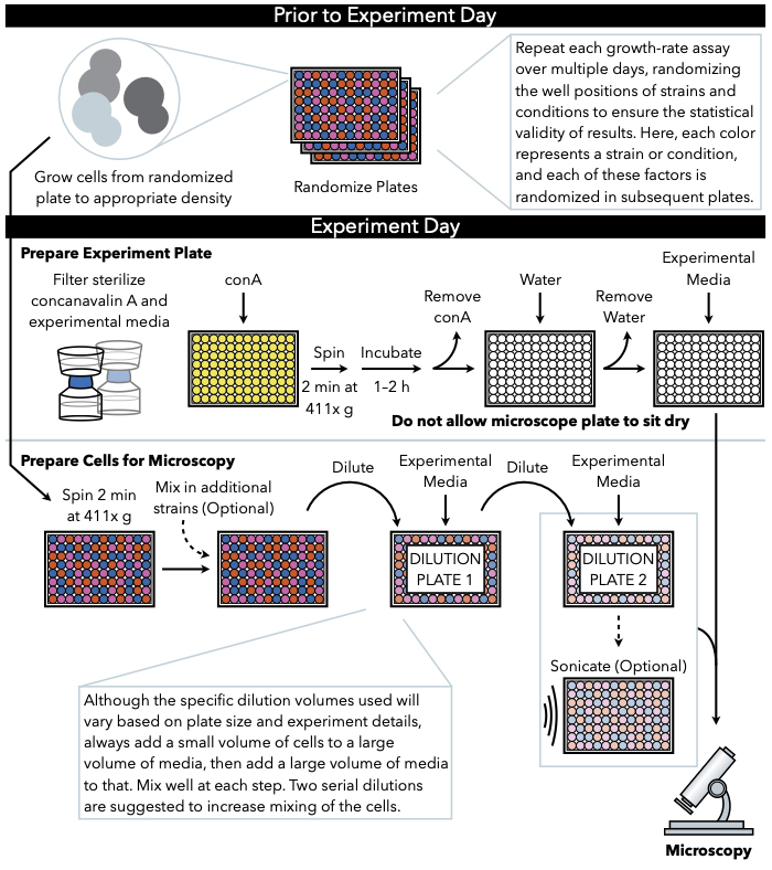
그림 1: 프로토콜의 회로도 표현. 이 프로토콜은 실험 판의 준비및 화상에 세포의 준비인 두 가지 주요 단계를 따릅니다. 플레이트의 무작위화와 세포의 성장은 실험의 날까지 전에 실시되어야한다. 희석 하는 동안 각 단계에서 세포의 반복 혼합 도금 까지 단계에서 필수적 이다, 따라서 먼저 실험 판을 준비 하는 것이 좋습니다 세포 희석의 완료 시 즉시 도금에 대 한 준비가. 이 그림의 더 큰 버전을 보려면 여기를 클릭하십시오.

Protocol

1. 무작위 판 준비 (실험일 이전) 성장 분석으로 테스트할 균주와 조건을 계획합니다. 이 시점에서, 임의로 어떤 우물에 균주와 조건을 할당합니다.참고: 플레이트 설정을 고려할 때, 측정에서 잘 관련된 소음을 설명하기 위해 단일 플레이트에 변형 및 성장 조건당 둘 이상의 복제본을 포함하는 것이 좋습니다. 자세한 내용은 토론을 참조하십시오. 다른 날에 실행되는 플레이트 복…

Representative Results

이 프로토콜의 참신은 시간 경과 이미징(그림 2A)을통해 마이크로 콜로니로의 성장을 추적하여 인구 내의 개별 세포에 대해 성장 속도를 계산할 수 있다는 것입니다. 마이크로콜로니가 파랑자 방식으로 수시간 동안 자라기 때문에, 그 영역은 실험 전반에 걸쳐 추적될 수 있으며, 시간이 지남에 따라 지역의 자연 로그의 변화에 선형적 맞춤을 관찰한 각 개별 식민지에 대한 성…

Discussion

여기서 설명된 프로토콜은 세포 성장 및 유전자 발현이 개별 마이크로콜로니의 수준에서 동시에 모니터링될 수 있도록 하는 다목적 분석이다. 이 두 가지 양식이 결합하면 고유한 생물학적 통찰력을 얻을 수 있습니다. 예를 들어, 이전 연구는 이 분석을 사용하여 TSL1 유전자의 발현과 동종 야생형 세포에서 마이크로콜로니 성장률 사이의 부정적인 상관관계를 동시에7,

Declarações

The authors have nothing to disclose.

Acknowledgements

나오미 지브, 사샤 레비, 슈앙 리(Shuang Li)가 이 프로토콜 개발에 기여한 것에 대해 감사를 표하고, 데이비드 그레샴(David Gresham)은 공유 장비를 제공했으며, 마리사 크놀이 비디오 제작에 도움을 준 것에 대해 감사드립니다. 이 작품은 건강 보조금 R35GM118170의 국가 학회에 의해 지원되었다.

Materials

General Materials
500 mL Bottletop Filter .22 µm PES Sterilizing, Low Protein Binding, w/45mm Neck Fisher CLS431154 used to filter the media
BD Falcon*Tissue Culture Plates, microtest u-bottom Fisher 08-772-54 96-well culture tubes used to freeze cells, pre-grow cells, and dilutions
BD Syringes without Needle, 50 mL Fisher 13-689-8 Used to filter the Concanavalin A
Costar Sterile Disposable Reagent Reservoirs Fisher 07-200-127 reagent reservoirs used to pipette solutions with multichannel pipette
Costar Thermowell Aluminum Sealing Tape Fisher 07-200-684 96-well plate seal for pre-growth and freezing
lint and static free Kimwipes Fisher 06-666A lint and static free wipes to keep microscope plate bottom free of debris and scratches
Nalgene Syringe Filters ThermoFisher Scientific 199-2020 0.2 μm pore size, 25 mm diameter; used to filter concanavalin A solution
Media Components
Minimal chemically defined media (MD; 2% glucose) alternative microscopy media used for yeast pre-growth and growth during microscopy
Synthetic Complete Media (SC; 2% glucose) microscopy media used for yeast pre-growth and growth during microscopy
Yeast extract-peptone-dextrose (YEPD; 2% glucose) medium cell growth prior to freezing down randomized plates
Microscopy Materials
Breathe-Easy sealing membrane Millipore Sigma Z380059-1PAK breathable membranes used to seal plate during microscopy experiment. At this stage breathable membranes are reccomended because they prevent condensation in the wells and allow for better microscopy images
Brooks 96-well flat clear glass bottom microscope plate Dot Scientific MGB096-1-2-LG-L microscope plate
Concanavalin A from canavalia ensiformis (Jack Bean), lyophilized powder Millipore Sigma 45-C2010-1G Make 5x concanavalin A solution and freeze 5ml of 5x concanavalin A in 50 mL conical tubes at -80 °C
Strains Used
MAH.5, MAH.96, MAH.52, MAH.66, MAH.11, MAH.58, MAH.135, MAH.15, MAH.44, MAH.132 Haploid mutation accumulation strains in a laboratory background, described in Hall and Joseph 2010
EP026.2A-2C Progeny of the ancestral Hall and Joseph 2010 mutation accumulation strain, transformed with YFR054cΔ::Scw11P::GFP
Equipment
Misonix Sonicator S-4000 with 96-pin attachment Sonicator https://www.labx.com/item/misonix-inc-s-4000-sonicator/4771281
Nikon Eclipse Ti-E with Perfect Focus System Inverted microscope with automated stage and autofocus system

Referências

  1. Geiler-Samerotte, K. A., Hashimoto, T., Dion, M. F., Budnik, B. A., Airoldi, E. M., Drummond, D. A. Quantifying condition-dependent intracellular protein levels enables high-precision fitness estimates. PloS one. 8 (9), 75320 (2013).
  2. Kussell, E., Leibler, S. Phenotypic diversity, population growth, and information in fluctuating environments. Science. 309 (5743), 2075-2078 (2005).
  3. Thattai, M., van Oudenaarden, A. Stochastic gene expression in fluctuating environments. Genética. 167 (1), 523-530 (2004).
  4. King, O. D., Masel, J. The evolution of bet-hedging adaptations to rare scenarios. Theoretical population biology. 72 (4), 560-575 (2007).
  5. Acar, M., Mettetal, J. T., van Oudenaarden, A. Stochastic switching as a survival strategy in fluctuating environments. Nature genetics. 40 (4), 471-475 (2008).
  6. Avery, S. V. Microbial cell individuality and the underlying sources of heterogeneity. Nature reviews. Microbiology. 4 (8), 577-587 (2006).
  7. Levy, S. F., Ziv, N., Siegal, M. L. Bet hedging in yeast by heterogeneous, age-correlated expression of a stress protectant. PLoS biology. 10 (5), 1001325 (2012).
  8. van Dijk, D., et al. Slow-growing cells within isogenic populations have increased RNA polymerase error rates and DNA damage. Nature communications. 6, 7972 (2015).
  9. Ziv, N., Shuster, B. M., Siegal, M. L., Gresham, D. Resolving the Complex Genetic Basis of Phenotypic Variation and Variability of Cellular Growth. Genética. 206 (3), 1645-1657 (2017).
  10. Li, S., Giardina, D. M., Siegal, M. L. Control of nongenetic heterogeneity in growth rate and stress tolerance of Saccharomyces cerevisiae by cyclic AMP-regulated transcription factors. PLoS genetics. 14 (11), 1007744 (2018).
  11. Plavskin, Y., Li, S., Ziv, N., Levy, S. F., Siegal, M. L. Robust colony recognition for high-throughput growth analysis from suboptimal low-magnification brightfield micrographs. bioRxiv. , (2018).
  12. Ziv, N., Siegal, M. L., Gresham, D. Genetic and nongenetic determinants of cell growth variation assessed by high-throughput microscopy. Molecular biology and evolution. 30 (12), 2568-2578 (2013).
  13. Hall, D. W., Joseph, S. B. A high frequency of beneficial mutations across multiple fitness components in Saccharomyces cerevisiae. Genética. 185 (4), 1397-1409 (2010).
  14. Saleemuddin, M., Husain, Q. Concanavalin A: a useful ligand for glycoenzyme immobilization–a review. Enzyme and microbial technology. 13 (4), 290-295 (1991).
  15. Geiler-Samerotte, K. A., Bauer, C. R., Li, S., Ziv, N., Gresham, D., Siegal, M. L. The details in the distributions: why and how to study phenotypic variability. Current opinion in biotechnology. 24 (4), 752-759 (2013).
  16. Nakagawa, S., Schielzeth, H. Repeatability for Gaussian and non-Gaussian data: a practical guide for biologists. Biological reviews of the Cambridge Philosophical Society. 85 (4), 935-956 (2010).
  17. Bolker, J. A. Exemplary and surrogate models: two modes of representation in biology. Perspectives in biology and medicine. 52 (4), 485-499 (2009).
check_url/pt/62038?article_type=t

Play Video

Citar este artigo
Sartori, F. M. O., Buzby, C., Plavskin, Y., Siegal, M. L. High-Throughput Live Imaging of Microcolonies to Measure Heterogeneity in Growth and Gene Expression. J. Vis. Exp. (170), e62038, doi:10.3791/62038 (2021).

View Video