Summary

Microscopietechnieken voor het interpreteren van schimmelkolonisatie in mycoheterotrofe plantenweefsels en symbiotische kieming van zaden

Published: May 17, 2022
doi:

Summary

Dit protocol is bedoeld om gedetailleerde procedures te bieden voor het verzamelen, fixeren en onderhouden van mycoheterotrofe plantenmonsters, waarbij verschillende microscopietechnieken worden toegepast, zoals scanning- en transmissie-elektronenmicroscopie, licht-, confocale en fluorescentiemicroscopie om schimmelkolonisatie te bestuderen in plantenweefsels en zaden ontkiemd met mycorrhizaschimmels.

Abstract

Structurele plantkunde is een onmisbaar perspectief om de ecologie, fysiologie, ontwikkeling en evolutie van planten volledig te begrijpen. Bij het onderzoeken van mycoheterotrofe planten (d.w.z. planten die koolstof uit schimmels verkrijgen), kunnen opmerkelijke aspecten van hun structurele aanpassingen, de patronen van weefselkolonisatie door schimmels en de morfoanatomie van ondergrondse organen hun ontwikkelingsstrategieën en hun relaties met schimmeldraden, de bron van voedingsstoffen, verlichten. Een andere belangrijke rol van symbiotische schimmels is gerelateerd aan de kieming van orchideeënzaden; alle Orchidaceae-soorten zijn mycoheterotroof tijdens ontkieming en zaailingstadium (initiële mycoheterotrofie), zelfs degenen die fotosynthetiseren in volwassen stadia. Vanwege het gebrek aan voedingsreserves in orchideeënzaden zijn schimmelsymbionten essentieel om substraten te bieden en ontkieming mogelijk te maken. Het analyseren van kiemfasen door structurele perspectieven kan ook belangrijke vragen beantwoorden met betrekking tot de interactie van schimmels met de zaden. Verschillende beeldvormingstechnieken kunnen worden toegepast om fungi-endofyten in plantenweefsels te onthullen, zoals in dit artikel wordt voorgesteld. Vrije hand en dunne delen van plantenorganen kunnen worden gekleurd en vervolgens worden waargenomen met behulp van lichtmicroscopie. Een fluorochroom geconjugeerd aan tarwekiem agglutinine kan worden toegepast op de schimmels en co-geïncubeerd met Calcofluor White om de celwanden van planten te benadrukken in confocale microscopie. Bovendien zijn de methodologieën van scanning- en transmissie-elektronenmicroscopie gedetailleerd voor mycoheterotrofe orchideeën en worden de mogelijkheden onderzocht om dergelijke protocollen toe te passen in verwante planten. Symbiotische kieming van orchideeënzaden (d.w.z. in aanwezigheid van mycorrhizaschimmels) wordt in het protocol in detail beschreven, samen met mogelijkheden om de structuren verkregen uit verschillende stadia van ontkieming voor te bereiden voor analyses met licht-, confocale en elektronenmicroscopie.

Introduction

Structureel onderzoek in de plantkunde, dat betrekking heeft op plantenmorfologie en anatomie, is fundamenteel voor het begrijpen van het hele organisme 1,2, en biedt onmisbare perspectieven om te integreren en bij te dragen aan kennis over de ecologie, fysiologie, ontwikkeling en evolutie van planten3. Methoden in plantenmorfologie en anatomie omvatten momenteel protocollen, apparatuur en kennis die onlangs en meer dan een eeuw geleden zijn ontwikkeld2. De continue uitvoering en aanpassing van klassieke methoden (bijv. Lichtmicroscopie) samen met meer recente technieken (bijv. Confocale microscopie, röntgenmicrotomografie) hebben dezelfde essentiële basis: theoretische kennis die de ontwikkeling van een methodologie mogelijk maakt.

Het belangrijkste hulpmiddel in de anatomie en morfologie van planten is het beeld. Ondanks de misvatting dat dergelijke analyses eenvoudige observaties zijn, ruimte geven aan subjectieve interpretaties2, vereist het analyseren en begrijpen van beelden op dit gebied kennis van de toegepaste methoden (de apparatuur, type analyse, methodologische procedures), celcomponenten, histochemie en het plantenlichaam (weefselorganisatie en -functie, ontogenie, morfologische aanpassingen). Het interpreteren van de beelden verkregen via een verscheidenheid aan methoden kan leiden tot het correleren van vorm en functie, het ontcijferen van de chemische samenstelling van een structuur, het bevestigen van taxa, het begrijpen van infecties door fytopathogenen en andere dergelijke beoordelingen.

Bij het onderzoeken van mycoheterotrofe (MH) planten (d.w.z. niet-fotosynthetische planten die koolstof verkrijgen uit mycorrhizaschimmels 4,5), kunnen opmerkelijke aspecten van hun structurele aanpassingen, de patronen van weefselkolonisatie door schimmels en de morfoanatomie van ondergrondse organen hun ontwikkelingsstrategieën en relaties met schimmeldraden, die de bron van voedingsstoffen zijn, verlichten. De ondergrondse organen van MH-planten vertonen meestal belangrijke aanpassingen in verband met hun associatie met bodemschimmels, daarom is het essentieel om deze anatomische en morfologische onderzoeken uit te voeren6. De luchtorganen van MH-soorten mogen niet worden genegeerd, omdat endofyten ook in deze weefsels aanwezig kunnen zijn, zelfs als het geen mycorrhizaschimmels zijn (persoonlijke waarnemingen, nog niet gepubliceerd).

Naast de gevestigde essentie van mycorrhizaschimmels associëring met MH-soorten gedurende hun hele levenscyclus7, heeft elke orchideeënsoort, zelfs de autotrofe, een initieel verplicht mycoheterotroof stadium in natuurlijke omgevingen. Het komt voor omdat het embryo van de orchideeën ongedifferentieerd is en geen endosperm of zaadlobben heeft, waardoor het niet in staat is zich te ontwikkelen en zich te vestigen in natuurlijke omgevingen zonder de voedingsondersteuning van schimmelpartners 4,8. Gezien het feit dat symbiotische kiemprotocollen niet alleen kunnen worden toegepast op MH-soorten, maar ook op fotosynthetiserende orchideeën, gericht op het onderzoeken van orchidee-schimmelspecificiteit in kieming en protocormontwikkeling, een enorm toegepaste methodologie in initiatieven voor het behoud van bedreigde soorten 9,10,11.

In deze methodeassemblage beschrijven we belangrijke stappen die betrokken zijn bij het verzamelen, fixeren en opslaan van MH-fabrieksmonsters voor anatomisch onderzoek (sectie 1), oppervlakteanalyse en monsterselectie (sectie 2), sectiemethoden (uit de vrije hand: sectie 3, microtomie: sectie 4, cryomicrotomie: sectie 5), kleuring en montage (sectie 6), fluorescentie en confocale microscopie van schimmel-endofyten (sectie 7), scanning-elektronenmicroscopie (sectie 8), en transmissie-elektronenmicroscopie (rubriek 9). Daarnaast beschrijven we een symbiotische kiemingsmethode voor orchideeënzaden (MH en autotroof, sectie 10), omdat de eerder genoemde beeldvormingsmethoden met succes kunnen worden toegepast om schimmelkolonisatie van zaden, protocormen en zaailingen in het kiemproces te analyseren.

Figure 1
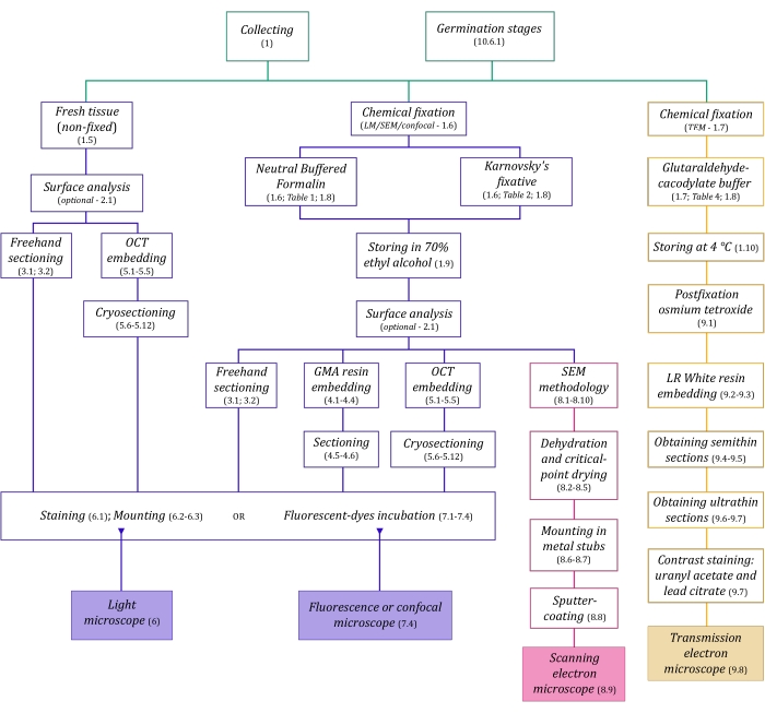
Figuur 1: Schematische samenvatting van beeldvormingsmethoden. De schema’s geven indicaties van protocolstappen waarin ze worden beschreven. Afkortingen: GMA = glycolmethacrylaat, OCT = optimale snijtemperatuurverbinding, SEM = scanningelektronenmicroscopie. Klik hier om een grotere versie van deze figuur te bekijken.

De hier in detail beschreven microscopietechnieken (figuur 1) worden voorafgegaan door de volgende essentiële stappen: verzamelen, fixeren, dehydrateren, insluiten en doorsnijden van monsters. Aangezien de stappen variabel zijn (figuur 1), afhankelijk van de gekozen techniek(en), is het belangrijk om vooruit te denken, rekening houdend met de fixeermiddelen die moeten worden voorbereid en naar de verzamelplaats moeten worden vervoerd, hoe de monsters moeten worden voorbereid voordat ze worden gefixeerd, de te gebruiken dehydratatieprocessen (sectie 1) en verschillende inbeddingsmogelijkheden en sectiemethoden (secties 4, 5, en 9). Figuur 1 geeft een overzicht van alle stappen die nodig zijn voor elke microscopietechniek die hieronder grondig wordt beschreven.

Protocol

1. Verzamelen, bevestigen en onderhouden van monsters OPMERKING: Volledig MH-planten zijn meestal te vinden in het donkere bos onder12,13, voornamelijk in vochtige en strooiselrijke gebieden, terwijl gedeeltelijk MH-planten te vinden zijn in meer open bossen12,13. MH-planten hebben meestal goed ontwikkelde ondergrondse organen in verschillende vormen en maten.</p…

Representative Results

Het volgen van de essentiële stadia van het fixeren van plantenweefsel levert cellulaire structuren op die zo veel mogelijk lijken op de levende toestand, rekening houdend met de morfologie, het volume en de ruimtelijke organisatie van cellulaire componenten en weefsels16. Observeer dergelijke eigenschappen in de monsters na chemische fixatie (figuur 4). Figuur 4C-F vertegenwoordigt voldoende vaste monst…

Discussion

Beeldanalyses in de anatomie en morfologie van planten hebben een belangrijk potentieel om doelstellingen te bereiken en helpen de relaties tussen mycoheterotrofe planten en hun onmisbare schimmel-endofyten te begrijpen, zoals aangetoond door studies van ondergrondse organen 6,40, structurele analyses van symbiotische kieming van zaden39 en lucht- en voortplantingsstructuren41 . Structurele plantkunde, ondanks het f…

Declarações

The authors have nothing to disclose.

Acknowledgements

De auteurs bedanken de financiering van FAEPEX en FAPESP (2015/26479-6). MPP bedankt Capes voor zijn masterbeurs (proces 88887.600591/2021-00) en CNPq. JLSM bedankt CNPq voor productiviteitsbeurzen (303664/2020-7). De auteurs bedanken ook de toegang tot apparatuur en hulp van LME (Laboratory of Electron Microscopy – IB / Unicamp), INFABiC (National Institute of Science and Technology on Photonics Applied to Cell Biology – Unicamp) en LaBiVasc (Laboratory of Vascular Biology – DBEF / IB / Unicamp); LAMEB (UFSC) en Eliana de Medeiros Oliveira (UFSC) voor bijdragen aan cryoprotectieprotocol; LME voor bijdragen aan het TEM-protocol.

Materials

Acetone Sigma-Aldrich 179124 (for SEM stubs mounting)
Agar-agar (AA) Sigma-Aldrich A1296 (for seeds germination tests)
Calcofluor White Stain Sigma-Aldrich 18909 fluorescent dye (detects cellulose)
Citrate Buffer Solution, 0.09M pH 4.8 Sigma-Aldrich C2488 (for toluidine blue O staining)
Conductive Double-Sided Carbon Tape Fisher Scientific 50-285-81 (for SEM)
Confocal Microscope Zeiss (any model)
Copper Grids Sigma-Aldrich G4776 (for TEM)
Critical-point dryer Balzers (any model)
Cryostat Leica Biosystems (any model)
Dissecting microscope Leica Biosystems (= stereomicroscope, any model)
Entellan Sigma-Aldrich 107960 rapid mounting medium for microscopy
Ethyl alcohol, pure (≥99.5%) Sigma-Aldrich 459836 (= ethanol, for dehydration processes)
Formaldehyde solution, 37% Sigma-Aldrich 252549 (for NBF solution preparation)
Formalin solution, neutral buffered, 10% Sigma-Aldrich HT501128 histological tissue fixative
Gelatin capsules for TEM Fisher Scientific 50-248-71 (for resin polymerisation in TEM)
Gelatin solution, 2% in H2O Sigma-Aldrich G1393 (dilute for slides preparation – OCT adherence)
Glutaraldehyde solution, 25% Sigma-Aldrich G6257 (for Karnovsky’s solution preparation)
HistoResin Leica Biosystems 14702231731 glycol methacrylate (GMA) embedding kit
Iodine Sigma-Aldrich 207772 (for Lugol solution preparation)
Lead(II) nitrate Sigma-Aldrich 228621 Pb(NO3)2 (for TEM contrast staining)
Light Microscope Olympus (any model)
LR White acrylic resin Sigma-Aldrich L9774 hydrophilic acrylic resin for TEM
Lugol solution Sigma-Aldrich 62650 (for staining)
Metal stubs for specimen mounts Rave Scientific (for SEM, different models)
Microtome Leica Biosystems manual rotary microtome or other model
Oatmeal agar (OMA) Millipore O3506 (for seeds germination tests)
OCT Compound, Tissue-Tek Sakura Finetek USA 4583 embedding medium for frozen tissues
Osmium tetroxide Sigma-Aldrich 201030 OsO4 (for TEM postfixation)
Parafilm M Sigma-Aldrich P7793 sealing thermoplastic film
Paraformaldehyde Sigma-Aldrich 158127 (for Karnovsky’s solution preparation)
Poly-L-lysine solution, 0.1% in H2O Sigma-Aldrich P8920 (for slides preparation – OCT adherence)
Poly-Prep Slides Sigma-Aldrich P0425 poly-L-lysine coated glass slides
Polyethylene Molding Cup Trays Polysciences 17177A-3 (6x8x5 mm, for embbeding samples in GMA resin)
Polyethylene Molding Cup Trays Polysciences 17177C-3 (13x19x5 mm, for embbeding samples in GMA resin)
Potassium iodide Sigma-Aldrich 221945 (for Lugol solution preparation)
Potato Dextrose Agar (PDA) Millipore 70139 (for seeds germination tests)
Scanning Electron Microscope Jeol (any model)
Silane [(3-Aminopropyl)triethoxysilane] Sigma-Aldrich A3648 (for slides preparation – OCT adherence)
Silane-Prep Slides Sigma-Aldrich S4651 glass slides coated with silane
Silica gel orange, granular Supelco 10087 (for dessicating processes)
Sodium cacodylate trihydrate Sigma-Aldrich C0250 (for glutaraldehyde-sodium cacodylate buffer)
Sodium hydroxide Sigma-Aldrich S5881 NaOH (for Karnovsky’s solution preparation and TEM contrast staining)
Sodium hypochlorite solution Sigma-Aldrich 425044 NaClO (for seeds surface disinfection)
Sodium phosphate dibasic, anhydrous Sigma-Aldrich 71640 Na2HPO4 (for NBF solution and PB preparation)
Sodium phosphate monobasic monohydrate Sigma-Aldrich S9638 NaH2PO4·H2O (for NBF and PB)
Sputter coater Balzers (any model)
Sucrose Sigma-Aldrich S0389 C12H22O11 (for cryoprotection and germination test)
Sudan III Sigma-Aldrich S4131 (for staining)
Sudan IV Sigma-Aldrich 198102 (for staining)
Sudan Black B Sigma-Aldrich 199664 (for staining)
Syringe (3 mL, any brand, for TEM contrast staining)
Syringe Filter Unit, Millex-GV 0.22 µm Millipore SLGV033R PVDF, 33 mm, gamma sterilized (for TEM contrast staining)
Tek Bond Super Glue 793 Tek Bond Saint-Gobain 78072720018 liquid cyanoacrylate adhesive, medium viscosity
Toluidine Blue O Sigma-Aldrich T3260 (for staining)
Transmission Electron Microscope Jeol (any model)
Triphenyltetrazolium chloride Sigma-Aldrich T8877 (for the tetrazolium test in seeds germination)
Trisodium citrate dihydrate Sigma-Aldrich S1804 Na3(C6H5O7)·2H2O (for TEM contrast staining)
Ultramicrotome Leica Biosystems (any model)
Uranyl acetate Fisher Scientific 18-607-645 UO2(CH3COO)2 (for TEM contrast staining)
Vacuum pump (any model)
Wheat Germ Agglutinin, Alexa Fluor 488 Conjugate TermoFisher Scientific W11261 fluorescent dye-conjugated lectin (detects sialic acid and N-acetylglucosaminyl residues)

Referências

  1. Evert, R. F. . Esau’s Plant Anatomy: Meristems, Cells, and Tissues of the Plant Body: Their Structure, Function, and Development. , (2006).
  2. Yeung, E. C. T., Stasolla, C., Sumner, M. J., Huang, B. Q. . Plant Microtechniques and Protocols. , (2015).
  3. Sokoloff, D. D., Jura-Morawiec, J., Zoric, L., Fay, M. F. Plant anatomy: at the heart of modern botany. Botanical Journal of the Linnean Society. 195 (3), 249-253 (2021).
  4. Leake, J. R. The biology of myco-heterotrophic (‘saprophytic’) plants. New Phytologist. 127 (2), 171-216 (1994).
  5. Bidartondo, M. I. The evolutionary ecology of myco-heterotrophy. New Phytologist. 167 (2), 335-352 (2005).
  6. Imhof, S., Massicotte, H. B., Melville, L. H., Peterson, R. L. Subterranean morphology and mycorrhizal structures. Mycoheterotrophy. , 157-214 (2013).
  7. Rasmussen, H. N., Rasmussen, F. N. Orchid mycorrhiza: implications of a mycophagous life style. Oikos. 118 (3), 334-345 (2009).
  8. Rasmussen, H. N., Dixon, K. W., Jersáková, J., Těšitelová, T. Germination and seedling establishment in orchids: a complex of requirements. Annals of Botany. 116 (3), 391-402 (2015).
  9. Zettler, L. W. Terrestrial orchid conservation by symbiotic seed germination: techniques and perspectives. Selbyana. 18 (2), 188-194 (1997).
  10. Stewart, S. L., Kane, M. E. Symbiotic seed germination and evidence for in vitro mycobiont specificity in Spiranthes brevilabris (Orchidaceae) and its implications for species-level conservation. In Vitro Cellular & Developmental Biology – Plant. 43 (3), 178-186 (2007).
  11. Zhao, D. -. K., et al. Orchid reintroduction based on seed germination-promoting mycorrhizal fungi derived from protocorms or seedlings. Frontiers in Plant Science. 12, 701152 (2021).
  12. Selosse, M. A., Roy, M. Green plants that feed on fungi: facts and questions about mixotrophy. Trends in Plant Science. 14 (2), 64-70 (2009).
  13. Merckx, V. S. F. T., Mennes, C. B., Peay, K. G., Geml, J. Evolution and diversification. Mycoheterotrophy: The Biology of Plants Living on Fungi. , 215-244 (2013).
  14. Boon, M. E., Drijver, J. Routine Cytological Staining Techniques: Theoretical Background and Practice. Macmillan International Higher Education. , (1986).
  15. Karnovsky, M. A formaldehyde-glutaraldehyde fixative of high osmolality for use in electron microscopy. Journal of Cell Biology. 27 (2), 137-138 (1964).
  16. Hayat, M. . Fixation for Electron Microscopy. , (1981).
  17. Roland, J. C., Vian, B. General preparation and staining of thin sections. Electron Microscopy of Plant Cells. 1, 675 (1991).
  18. Gerrits, P. O., Horobin, R. W. Glycol methacrylate embedding for light microscopy: basic principles and trouble-shooting. Journal of Histotechnology. 19 (4), 297-311 (1996).
  19. Zhang, Z., Niu, L., Chen, X., Xu, X., Ru, Z. Improvement of plant cryosection. Frontiers in Biology. 7 (4), 374-377 (2012).
  20. BeneŠ, K. On the media improving freeze-sectioning of plant material. Biologia Plantarum. 15 (1), 50-56 (1973).
  21. Fischer, A. H., Jacobson, K. A., Rose, J., Zeller, R. Preparation of slides and coverslips for microscopy. Cold Spring Harbor Protocols. 2008 (5), (2008).
  22. Sakai, W. S. Simple method for differential staining of paraffin embedded plant material using toluidine blue O. Stain Technology. 48 (5), 247-249 (1973).
  23. O’Brien, T., Feder, N., McCully, M. E. Polychromatic staining of plant cell walls by toluidine blue O. Protoplasma. 59 (2), 368-373 (1964).
  24. Ventrella, M. C., Almeida, A. L., Nery, L. A., Coelho, V. P. d. e. M. Métodos Histoquímicos Aplicados às Sementes. Universidade Federal de Viçosa. , (2013).
  25. Pearse, A. G. E. . Histochemistry, Theoretical and Applied. , (1960).
  26. Andrade-Linares, D. R., Franken, P. Fungal endophytes in plant roots: taxonomy, colonization patterns, and functions. Symbiotic Endophytes. , 311-334 (2013).
  27. Wymer, C. L., Beven, A. F., Boudonck, K., Lloyd, C. W. Confocal microscopy of plant cells. Confocal Microscopy Methods and Protocols. , 103-130 (1999).
  28. Marques, J. P. R., Soares, M. K. M. Manual de Técnicas Aplicadas à Histopatologia Vegetal. FEALQ. , (2021).
  29. Navarro, B. L., Marques, J. P. R., Appezzato-da-Glória, B., Spósito, M. B. Histopathology of Phakopsora euvitis on Vitis vinifera. European Journal of Plant Pathology. 154 (4), 1185-1193 (2019).
  30. Marques, J. P. R., et al. Sugarcane cell wall-associated defense responses to infection by Sporisorium scitamineum. Frontiers in Plant Science. 9, 698 (2018).
  31. Jeffree, C. E., Read, N. D. Ambient-and low-temperature scanning electron microscopy. Electron Microscopy of Plant Cells. , 313-413 (1991).
  32. Bozzola, J. J., Russell, L. D. . Electron Microscopy: Principles and Techniques for Biologists. , (1999).
  33. Murray, S. Basic transmission and scanning electron microscopy. Introduction to electron Microscopy for Biologists. , 3-18 (2008).
  34. . Glossary of TEM terms Available from: https://www.jeol.co.jp/en/words/emterms/ (2021)
  35. Seaton, P. T., et al. Orchid seed and pollen: a toolkit for long-term storage, viability assessment and conservation. Orchid Propagation: From Laboratories to Greenhouses—Methods and Protocols. , 71-98 (2018).
  36. Otero, J. T., Ackerman, J. D., Bayman, P. Differences in mycorrhizal preferences between two tropical orchids. Molecular Ecology. 13 (8), 2393-2404 (2004).
  37. Koch, R. A., et al. Marasmioid rhizomorphs in bird nests: Species diversity, functional specificity, and new species from the tropics. Mycologia. 112 (6), 1086-1103 (2020).
  38. Webster, J., Weber, R. . Introduction to Fungi. , (2007).
  39. Sisti, L. S., et al. The role of non-mycorrhizal fungi in germination of the mycoheterotrophic orchid Pogoniopsis schenckii Cogn. Frontiers in Plant Science. 10, 1589 (2019).
  40. Martos, F., et al. Independent recruitment of saprotrophic fungi as mycorrhizal partners by tropical achlorophyllous orchids. New Phytologist. 184 (3), 668-681 (2009).
  41. Alves, M. F., et al. Reproductive development and genetic structure of the mycoheterotrophic orchid Pogoniopsis schenckii Cogn. BMC Plant Biology. 21 (1), 332 (2021).
  42. Merckx, V. S. F. T. Mycoheterotrophy: an introduction. Mycoheterotrophy: The Biology of Plants Living on Fungi. , 1-17 (2013).
  43. Hall, J. L., Hawes, C. . Electron Microscopy of Plant Cells. , (1991).
check_url/pt/63777?article_type=t

Play Video

Citar este artigo
Pena-Passos, M., Sisti, L. S., Mayer, J. L. S. Microscopy Techniques for Interpreting Fungal Colonization in Mycoheterotrophic Plants Tissues and Symbiotic Germination of Seeds. J. Vis. Exp. (183), e63777, doi:10.3791/63777 (2022).

View Video