Augmented reality technology was applied to core decompression for osteonecrosis of the femoral head to realize real-time visualization of this surgical procedure. This method can effectively improve the safety and precision of core decompression.
Osteonecrosis of the femoral head (ONFH) is a common joint disease in young and middle-aged patients, which seriously burdens their lives and work. For early-stage ONFH, core decompression surgery is a classical and effective hip preservation therapy. In traditional procedures of core decompression with Kirschner wire, there are still many problems such as X-ray exposure, repeated puncture verification, and damage to normal bone tissue. The blindness of the puncture process and the inability to provide real-time visualization are crucial reasons for these problems.
To optimize this procedure, our team developed an intraoperative navigation system on the basis of augmented reality (AR) technology. This surgical system can intuitively display the anatomy of the surgical areas and render preoperative images and virtual needles to intraoperative video in real-time. With the guide of the navigation system, surgeons can accurately insert Kirschner wires into the targeted lesion area and minimize the collateral damage. We conducted 10 cases of core decompression surgery with this system. The efficiency of positioning and fluoroscopy is greatly improved compared to the traditional procedures, and the accuracy of puncture is also guaranteed.
Osteonecrosis of the femoral head (ONFH) is a common disabling disease occurring in young adults1. Clinically, it is necessary to determine the staging of ONFH based on X-ray, CT, and MRI to decide the treatment strategy (Figure 1). For early-stage ONFH, hip preservation therapy is usually adopted2. Core decompression (CD) surgery is one of the most frequently used hip preservation methods for ONFH. Certain curative effects of core decompression with or without bone grafting in treating early-stage ONFH have been reported, which can avoid or delay subsequent total hip arthroplasty (THA) for a long time3,4,5. However, the success rate of CD with or without bone grafting was reported differently among previous studies, from 64% to 95%6,7,8,9. The surgical technique, especially the accuracy of drilling position, is important for the success of hip preservation10. Due to the blindness of the puncture and positioning procedure, the traditional techniques of CD have several problems, such as more fluoroscopy time, repeated puncture using Kirschner wire, and injury of normal bone tissue11,12.
In recent years, the augmented reality (AR)-assisted method has been introduced in orthopedic surgery13. The AR technique can visually show the anatomy of the surgical field, guide the surgeons in planning the operating procedure, and consequently reduce the difficulty of the operation. The applications of the AR technique in pedicle screw implantation and joint arthroplasty surgery have been reported earlier14,15,16,17. In this study, we aim to apply the AR technique to the CD procedure and verify its safety, accuracy, and feasibility in clinical practice.
System hardware components
The main components of the AR-based navigation surgical system include the following: (1) A depth camera (Figure 2A) installed directly above the surgical area; the video is shot from this and sent back to the workstation for registration and cooperation with the imaging data. (2) A puncture device (Figure 2B) and a non-invasive body surface marking frame (Figure 2C), both with passive infrared reflectors. A special reflective coating of marking balls (Figure 3) can be captured by infrared equipment to achieve accurate tracking of surgical equipment in the surgical area. (3) An infrared positioning device (Figure 2D) is responsible for tracking markers in the surgical area, matching the body surface marking frame and puncture device with high accuracy (Figure 4). (4) The host system (Figure 2E) is a 64-bit workstation, installed with the independently developed AR-assisted orthopedic surgery system. Augmented reality display of hip joint and femoral head puncture operation can be completed with its assistance.
This study was approved by the ethics committee of the China-Japan Friendship Hospital (approval number: 2021-12-K04). All of the following steps were performed according to standardized procedures to avoid injury to the patients and the surgeons. Informed patient consent was obtained for this study. The surgeon must be skilled in conventional core decompression procedures to ensure that the surgery can be performed in a traditional way in case of inaccurate navigation or other unexpected situations.
1. Preoperative diagnosis and grading of ONFH
2. System registration and accuracy testing
3. Patient and system preparation before puncture
4. Puncture assisted by surgical system
5. Operation evaluation
Operation characteristics
The surgical navigation system was applied in continuative 10 hips of nine patients. The average total positioning time of the surgery was 10.1 min (median 9.5 min, range 8.0-14.0 min). The mean C-ARM fluoroscopies was 5.5 times (median 5.5 times, range 4-8 times). The mean error of puncture accuracy was 1.61 mm (median 1.2 mm, range -5.76-19.73 mm; Table 1). The results show that the positioning time and fluoroscopy times are obviously shortened compared to traditional procedures.
Clinical outcome evaluation
The nine enrolled patients consisted of seven males and two females, with an average age of 41.6 ± 10.0 years.The mean BMI was 23.93 ± 3.08 kg/m2. For the hips evaluated, two hips were in ARCO I stage, four hips were in ARCO IIA stage, and four in ARCO IIB stage. Preoperative and postoperative visual analogue scale and Harris hip score were used to evaluate the outcome (Table 1). The mean preoperative VAS score was 6 and mean postoperative score was 3.75. The average preoperative Harris score was 77.5 and mean postoperative score was 85.5. Hip X-ray was examined 3 months after the surgery. All the patients returned to the ward safely. No postoperative complications such as infection, hematoma, or nerve damage were found. So far, no femoral head collapse occurred in any cases, and the long-term function and success rate of hip preservation are still being followed up. Surgical indicators and scores are shown in Table 2.
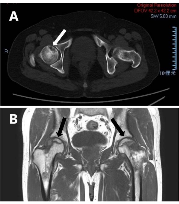
Figure 1: Imaging of early stage of femoral head necrosis. (A) The CT image. (B) The MRI image. Arrows indicate areas of necrosis. Please click here to view a larger version of this figure.
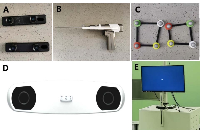
Figure 2: The main components of the AR-based navigation surgical system. (A) Depth camera. (B) The puncture device with a positioning frame. (C) Non-invasive body surface marking frame independently designed and developed. (D) Infrared positioning device. (E) The surgical system workstation. Please click here to view a larger version of this figure.
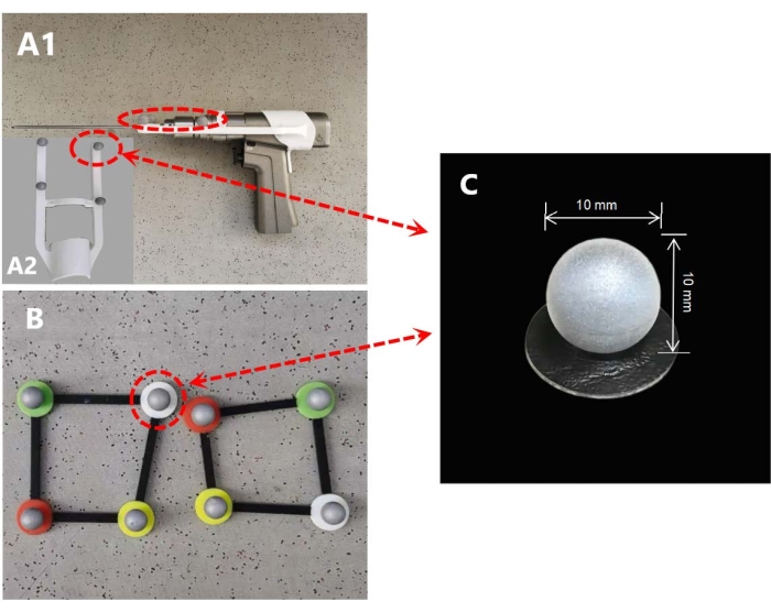
Figure 3: Installation of a passive infrared reflector. (A) Self-designed positioning frame mounted on puncture device. (B) The reflector is mounted at the four corners of the non-invasive body surface marking frame. (C) The specification of the passive infrared reflector is a spherical device with a diameter of 10 mm. Please click here to view a larger version of this figure.
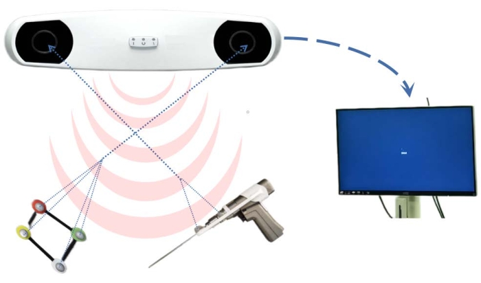
Figure 4: Working principles of infrared positioning device. Infrared radiation emitted by the infrared positioning device is reflected by the passive infrared reflectors; the receivers in that device receive the reflected signal and transmit the movement data to the workstation. Please click here to view a larger version of this figure.
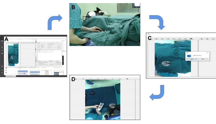
Figure 5: An overview of the preoperative registration process. (A) Operating interface of AR-assisted orthopedic surgery system. (B) The surgical area was planned using a non-invasive body surface marking frame. (C) Tips for successful registration of one of the matching points in the surgical video. (D) After all the matching points were successfully matched, the tracking of surgical instruments was tested. Please click here to view a larger version of this figure.
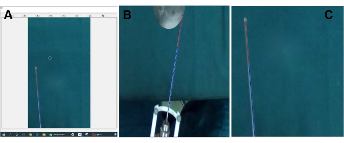
Figure 6: Superposition of virtual Kirschner wire on real Kirschner wire. (A-C) The images show that the virtual Kirschner needle is precisely superimposed on the physical one and moves with it in the screen. Please click here to view a larger version of this figure.
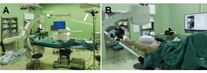
Figure 7: An overview of surgical scenarios. (A) The main components of the AR-based surgical system in the operating room. (B) A patient with necrosis of the femoral head is being treated with the aid of the surgical system. Please click here to view a larger version of this figure.
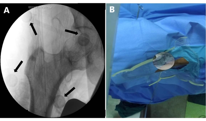
Figure 8: Hip joint imaging and augmented reality display. (A) Radiograph of the hip joint containing a non-invasive body surface marking frame. The black arrow indicates the passive infrared reflectors. (B) Radiograph is processed at the workstation and then superimposed by the surgical system on the surface of the affected hip on the screen. Please click here to view a larger version of this figure.
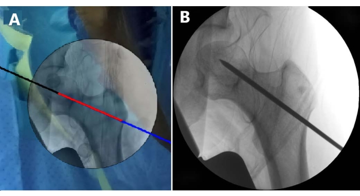
Figure 9: Puncture effect demonstration. (A) Image presents the screenshot after the puncture, the black-red-blue line is a virtual Kirschner wire in the system (step 2.6). (B) Image shows the hip radiograph after completion of puncture, the black line is an image of a real Kirschner wire in X-ray. Please click here to view a larger version of this figure.
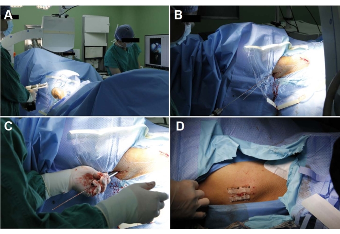
Figure 10: Femoral head puncture guided by the AR-based surgical system. (A) The surgeon is adjusting the position of the puncture device according to the screen display. (B) Kirschner wire punctures the skin and points to the necrosis. (C) Drill into the necrotic area along the Kirschner wire with a 5 mm trephine to fill artificial bone or autologous bone implantation. (D) Close the wound. Please click here to view a larger version of this figure.
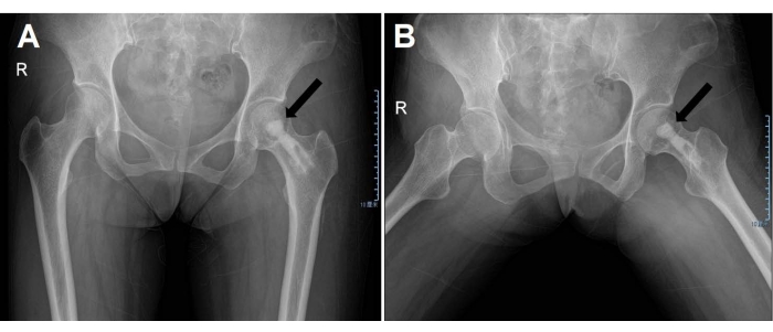
Figure 11: Postoperative radiograph of the hip joint. (A) From the front view. (B) The patient is in frog position. The black arrows indicate artificial bone implants in the femoral head. Please click here to view a larger version of this figure.
Case | Sex | Age | BMI | Disease | ARCO |
1 | M | 22 | 28.40 | ONFH(left) | IIA |
2 | F | 21 | 22.40 | ONFH(right) | IIB |
3 | M | 42 | 19.56 | ONFH(left) | IIB |
4 | M | 51 | 22.10 | ONFH(left) | I |
5 | M | 31 | 24.34 | ONFH(bilateral) | L:IIB |
6 | R:IIA | ||||
7 | M | 46 | 27.24 | ONFH(right) | IIA |
8 | F | 41 | 21.20 | ONFH(left) | IIB |
9 | M | 56 | 22.83 | ONFH(right) | I |
10 | M | 38 | 27.30 | ONFH(left) | IIA |
Table 1: Basic patient information. The table provides the information for the 10 patients enrolled in this study.
Case | Positioning time(min) | Fluoroscopy shots | Positioning error(mm) | Harris Hip Score | Visual Analogue Scale | ||
Before | After | Before | After | ||||
1 | 13 | 6 | 2.83 | 82 | 89 | 6 | 4 |
2 | 9 | 6 | 0.35 | 86 | 85 | 4 | 3 |
3 | 9 | 4 | 2.05 | 88 | 89 | 5 | 3 |
4 | 10 | 5 | -5.01 | 73 | 85 | 7 | 4 |
5 | 8 | 6 | -1.52 | L:84 | L:88 | L:4 | L:3 |
6 | 14 | 4 | -4.13 | R:68 | R:82 | R:6 | R:4 |
7 | 11 | 7 | 3.97 | 74 | 84 | 7 | 4 |
8 | 10 | 5 | 3.55 | 81 | 89 | 5 | 3 |
9 | 9 | 8 | 19.73 | 74 | 82 | 6 | 4 |
10 | 8 | 4 | -5.76 | 62 | 81 | 8 | 5 |
Table 2: Surgical indicators and scores. The positioning time, fluoroscopy time, and puncture accuracy were calculated and are shown. The pre- and post-operative VAS score and Harris score are also shown in this table.
Although THA has developed rapidly in recent years and become an effective ultimate method for ONFH, hip preservation therapy still plays an important role in treating early-stage ONFH18,19. CD is a basic and effective hip preservation surgery, which can release hip pain and delay the development of femoral head collapse20. The puncture positioning of the focal necrosis is the crucial procedure of CD, as it determines the success or failure of the surgery. However, the traditional puncture positioning method still contains some blind spots that may lead to repeated puncture, increase in exposure to fluoroscopy, and increased operation time10,11. Many scholars have also made efforts to improve this aspect, such as using 3D printing, a combination of hip arthroscopy, and the use of a robot-assisted navigation system12,21,22,23. These methods certainly improve the efficiency and accuracy of puncture positioning, however they also have some deficiencies in other aspects, such as adding operative complexity, causing subsidiary injury, and increasing medical cost.
The system shown here can divide the virtual surgical area in the preoperative registering process. In the virtual surgical area, a high-precision trace of the electro-optical target tracking equipment and the virtual display of the Kirschner wire can be achieved. As required, the second film and superposition can also be conducted when adjusting the angle of the hip. The mean registering time is only 10.1 min. When performing other operations in the same areas, repeated registration is not required. The whole process of registering and positioning is non-invasive, thus ensuring a high level of safety and fitting with a less invasive surgical principle.
AR technique superimposes the imperceptible information into the real-time video frame, which achieves the combination of virtuality and reality24. The AR technique has been combined into many orthopedic surgeries, such as fracture reduction, bone tumor resection, etc.25,26,27. To our knowledge, this is the first study applying AR in CD surgery. The greatest advantage of our system is real-time visualization, which can reduce the difficulty of surgery and shorten the learning curve of the surgeons.
There are also some limitations in this study. Firstly, the sample size of this study is very small and therefore, the result is not convincing enough. Secondly, we only report the early clinical outcomes; further follow-up is also required to evaluate the real benefit for the patients. Certainly, there is still some room for development in this system. With the improvement of performance, it will better serve the clinical practice.
The authors have nothing to disclose.
This work was supported by Beijing Natural Science Foundation(7202183), National Natural Science Foundation of China(81972107), and Beijing Municipal Science and Technology Commission(D171100003217001).
AR-assisted Orthopedic Surgery System | Self development | None | An operating software that implements AR for orthopedic surgery |
Depth camera | Stereolabs | ZED depth camera(ZED mini) | shoot video and sent back to the workstation. |
Image processing software | Adobe Systems Incorporated | Adobe Photoshop CS6 | Image processing software |
Infrared positioning device | Northern Digital Inc. | NDI Polaris Spectra optical tracking device | Tracking markers in the surgical area. |
Puncture device | Stryker | Stryker System 7 Cordless driver and Sabo | Insert kirschner wire into the necrotic area. |