Vascularized Composite Allotransplantations (VCA) have become a clinical reality. However, broad clinical application of VCA is limited by chronic multi-drug immunosuppression. The authors present a reliable and reproducible large animal model to translate novel immunomodulatory strategies that can minimize or potentially eliminate the need of immunosuppression in VCA.
Vascularized Composite Allotransplantation (VCA) such as hand and face transplants represent a viable treatment option for complex musculoskeletal trauma and devastating tissue loss. Despite favorable and highly encouraging early and intermediate functional outcomes, rejection of the highly immunogenic skin component of a VCA and potential adverse effects of chronic multi-drug immunosuppression continue to hamper widespread clinical application of VCA. Therefore, research in this novel field needs to focus on translational studies related to unique immunologic features of VCA and to develop novel immunomodulatory strategies for immunomodulation and tolerance induction following VCA without the need for long term immunosuppression.
This article describes a reliable and reproducible translational large animal model of VCA that is comprised of an osteomyocutaneous flap in a MHC-defined swine heterotopic hind limb allotransplantation. Briefly, a well-vascularized skin paddle is identified in the anteromedial thigh region using near infrared laser angiography. The underlying muscles, knee joint, distal femur, and proximal tibia are harvested on a femoral vascular pedicle. This allograft can be considered both a VCA and a vascularized bone marrow transplant with its unique immune privileged features. The graft is transplanted to a subcutaneous abdominal pocket in the recipient animal with a skin component exteriorized to the dorsolateral region for immune monitoring.
Three surgical teams work simultaneously in a well-coordinated manner to reduce anesthesia and ischemia times, thereby improving efficiency of this model and reducing potential confounders in experimental protocols. This model serves as the groundwork for future therapeutic strategies aimed at reducing and potentially eliminating the need for chronic multi-drug immunosuppression in VCA.
Vascularized Composite Allotransplantation (VCA) such as hand and face transplants are now a clinical reality with numerous hand and face transplants performed worldwide12. Despite the fact that early and intermediate results are favorable and highly encouraging2, the requirement of chronic multidrug immunosuppression continues to limit its widespread clinical application. The advancements in murine models of VCA including super-microsurgical anastomoses and nonsuture cuff techniques13, 3 have paved the way to a better understanding of alloimmune responses in VCA. Myriads of immunomodulatory protocols have been proposed for clinical applications based on our better understanding of immune mechanisms in VCA but they need to be validated in a large animal model that would be reasonably predictive of their performance in humans7. Based on physiologic and immunologic similarities between human and porcine organ systems6, swine VCA models can be considered reliable and cost-effective alternatives to canine9 and nonhuman primate models1.
This article provides a detailed overview of the methodology used in our MHC-defined swine heterotopic hind limb transplant model, which serves as groundwork for our current and future immunomodulatory strategies aimed at inducing immune tolerance to VCA and hence broadening its clinical application. We utilize well-characterized inbred pigs bred to homozygosity at the swine leukocyte antigen locus specifically for their use in transplant-related research11. We raise a vascularized osteomyocutaneous flap based on femoral vessels. The flap contains intact vascularized bone marrow in the distal femur and proximal tibia. The anteromedial thigh skin is also included in the graft and is exteriorized to the dorsolateral aspect of the recipient animal for immune monitoring of the most immunogenic component of the VCA. Dorsolateral positioning facilitates clinical examination in standing and sitting positions and also keeps the allograft skin relatively clean.
Ustener et al. introduced one of the first large animal translational models in VCA by transplanting radial forelimb osteomyocutaneous flaps in outbred farm pigs15. The group utilized this model to demonstrate for the first time that acute rejection in VCA which included the highly immunogenic skin component could be delayed and treated with a clinically relevant strategy without significant drug-specific complications and side effects. The favorable results obtained in this study subsequently built a foundational step in designing drug regimens for human reconstructive transplantation. Although these early pig VCA models were well suited for developing protocols to prevent rejection of skin, muscle, bones, nerves and vessels they lacked specialized structures such as articular cartilage and synovial membranes of joints. Subsequent efforts were focused on including the medial digit of the animal, which necessitated full-length cast placement to prevent graft dislodgement14. Although suitable to investigate rejection of all major components of limb transplantation, one of the major limitations of this model was post-transplant ambulatory difficulty due to cast placement. Thus, heterotopic swine limb allotransplantation models, consisting of the tibia, fibula, knee joint, distal femur, surrounding muscle, and a skin paddle, were created to primarily study immunological aspects of VCA while allowing the animal to freely ambulate postoperatively with minimal morbidit 8.
The development of well-characterized SLA-defined inbred pigs through the pioneering work of Dr. David H. Sachs led to a new era of translational VCA research. Utilizing a heterotopic hind limb transplant model in a minor antigen mismatch setting, Mathes et al.10 demonstrated indefinite survival of musculoskeletal components with a short course of cyclosporine treatment. The skin component survival, however, was only prolonged when compared to no treatment controls. The loss of the skin component of the graft was attributed to an isolated and highly vigorous immune response, in particular, to the epidermis. Similarly, using fully mismatched pigs with T-cell depletion, a short course of cyclosporine and cytokine mobilized donor peripheral blood mononuclear cells induced tolerance only to musculoskeletal components and the skin component was still rejected5. This phenomenon, called ‘split tolerance’, brought a paradigm shift in VCA research with a greater focus on the highly immunogenic skin component, which is an integral component of the majority of reconstructive transplants performed to date.
In this modified model, we utilize end-to-end anastomosis by ligating the recipient femoral artery and rotating it cephalad (Figure 1). This not only reduces ischemia time by allowing the use of a conventional coupling device but also decreases the chances of anastomotic failure. We have not observed any ischemic events following ligation of the femoral artery in our recipients indicating that collateral circulation was sufficient to provide vascularity to the native leg. Additionally, in this modified method, the externalized skin component is mobilized based on the underlying perforator vessels and is positioned laterally (Figure 1) in contrast to a ventral groin position in the traditional model10. This allows easy visualization of the graft for immune monitoring in a standing or sitting position of the animal.
Hence, a reliable and reproducible large animal model is essential to investigate tolerance induction strategies towards the skin component of VCA and to develop novel noninvasive immune monitoring strategies for better prediction of graft survival.
In this video publication, all animal procedures were conducted in accordance with an animal protocol approved by the Johns Hopkins University Institutional Animal Care and Use Committee (IACUC).
1. Preoperative Planning
Intraoperative Monitoring:
Osteomyocutaneous Limb Allograft Harvest from Donor Pig:
4. Recipient Procedure (Hind Limb Allotransplantation)
Twenty-four SLA-defined swine heterotopic hind limb transplants were performed using our modified technique with a mean ischemia time of 78 min (Range: 62-94 min). Graft inset and dorsolateral skin paddle positioning were achieved without difficulty in all animals. Near infrared laser angiography showed excellent graft perfusion in all recipients. The initial twelve venous anastomoses were performed using conventional suture techniques while the last twelve venous anastomoses were performed using a vascular coupling device. One animal with the conventional suture technique required a reanastomosis when a venous thrombus was identified immediately post-procedure. No complications were observed in any anastomoses performed with the coupling device. Recipient animals received a short course (30 days) of tacrolimus monotherapy with or without donor bone marrow (BM) infusion and co-stimulatory blockade. Tacrolimus dosing was adjusted to achieve target levels of 10-15 ng/ml. The short course tacrolimus only and untreated animals served as controls. The co-stimulation blockade based immunomodulatory protocol resulted in over 6 month survival post-transplant. There was no evidence of GVHD in any recipient. All long-term survivors (beyond 150 days post-transplant) had viable vascularized bone marrow at the time of euthanasia which demonstrates reliability of this model for investigating unique immunologic features of the bone marrow component of VCA (Figure 7).
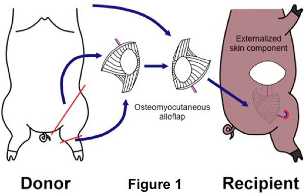
Figure 1. Schematic diagram of a swine heterotopic hind limb transplant. An osteomyocutaneous flap is harvested from the donor hind limb and transplanted to a subcutaneous pocket along the abdominal wall of the recipient.
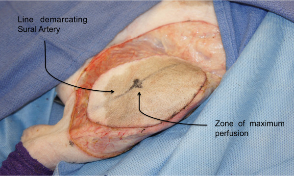
Figure 2: Osteomyocutaneous flap harvest: Donor skin paddle. Perforator zones in the anterolateral thigh are identified using laser angiography to demarcate the skin paddle of the osteomyocutaneous flap.
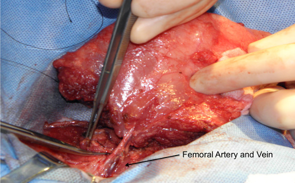
Figure 3: Osteomyocutaneous flap harvest: Graft on its vascular pedicle. Flap consisting of the distal femur, knee joint, proximal tibia, fibula, thigh muscles and skin paddle is harvested on a femoral vascular pedicle.
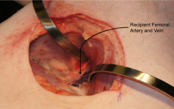
Figure 4: Recipient procedure: Creation of a subcutaneous abdominal pocket. Subcutaneous dissection is carried out to create an abdominal wall pocket extending from the groin to the dorsolateral abdominal wall.
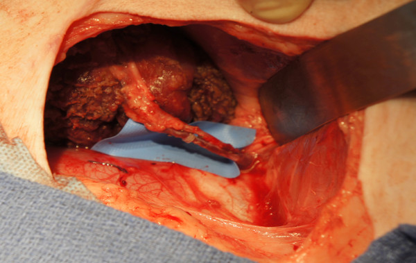
Figure 5: Recipient procedure: Graft inset and reperfusion following microvascular anastomosis. End-to-end femoral vessel anastomosis is performed after flap inset. The donor limb is used for the contralateral side of the recipient (i.e. the left donor limb for the right side of the recipient and the right donor limb for the left side of the recipient).
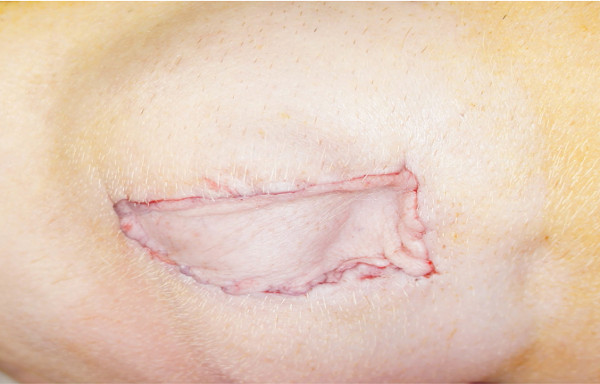
Figure 6: Skin component exteriorized to dorsolateral position for immune monitoring. During inset, the flap is positioned in a way that the skin paddle faces the dorsolateral abdominal wall where it is sutured to the recipient skin. This position allows easy monitoring of the flap.
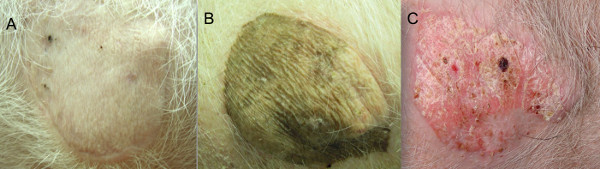
Figure 7: Representative images from transplanted animals: (A) Long term survivor (>150 days) with no clinical evidence of rejection. (B) Long term survivor (>150 days) with no clinical evidence of rejection. The allograft was obtained from a donor with dark color skin. Both (A) and (B) received co-stimulation blockade (CTLA4Ig) based immunomodulatory therapy. (C) Negative control (short-term tacrolimus therapy only) with de-epithelialization and advanced rejection as soon as tacrolimus was withdrawn (Day 30).
Historically, the heterotopic hind limb transplant protocol included exteriorization of a skin paddle to the ventral abdominal wall and the vessels were anastomosed in an end-to-side manner (Hettiarachty 2004). However, in our modified method, an inverted flap insetting and end-to-end anastomoses bring the skin paddle more laterally and hence facilitates immune monitoring in a standing position of the animal. The identification of the sural artery and the zones of maximum perfusion using near infrared laser angiography further improves the reliability of the skin paddle.
In our modified technique, we perform end-to-end anastomosis between the femoral vessels to minimize technical failures related to microvascular anastomosis. End-to-end anastomosis also enabled us to use a vascular coupling device (Synovis St. Paul MN) for venous anastomosis, which further decreased warm ischemia time. In our experience, ischemia time was more predictable with the use of a coupling device and since variation in ischemia time is directly correlated with immunologic outcome, this modification improved the reliability of this model. However, the use of a coupling device results in additional material costs but is still considered cost-effective based on the overall cost of the procedure and the avoidance of potential complications.
Our heterotopic hind limb transplants instantly produced circulating donor-derived bone marrow cells as evidenced by male donor derived SRY quantitative PCR analysis. Additionally, the viability of the bone marrow component of the allograft in our longterm survivors was confirmed using immunohistochemistry. This further improves reliability of our model as an investigative tool for unique immune privileged features of vascularized bone marrow, which is a key component of certain reconstructive transplants. This osteomyocutaneous flap also contains the knee joint with articular cartilage and a synovial membrane and serve as a reliable model to assess rejection of these specialized structures.
Despite the fact that this model seems to be ideal for translational studies investigating immunologic aspects of VCA and its components including skin, muscle, nerves, vessels and joints it still does not allow for the assessment of a functional outcome. Once the immunologic barrier is overcome, further modifications and/or additional translational models can be performed to investigate therapeutic strategies aimed at improving motor and sensory function of VCA.
Utilizing this model, our group demonstrated that high-dose bone marrow cell infusion combined with co-stimulatory blockade optimized induction therapy, reduced maintenance immunosuppression, and indefinitely prolonged graft survival16. Such targeted immunomodulatory protocols that combine bone marrow cell-based strategies and biologics might facilitate immune tolerance and eliminate the need for multidrug immunosuppression to maintain graft survival after VCA.
The authors have nothing to disclose.
We would like to acknowledge the following individuals for their contribution to this project: Kakali Sarkar, PhD, Joani Christensen, BS, Kate Buretta, BS, Nance Yuan, BS, William Lehao, MD, Johanna Grahammer, Georg Furtmüller, MD, Erin Rada MD, Mohammed Al-Rakan MD, Karim Sarhane MD, Saami Khalifian, BS, Mao Qi, MD, and Angelo Leto Barone MD, VCA Laboratory, Department of Plastic and Reconstructive Surgery, Johns Hopkins University School of Medicine, Janis Taube, MD, Mark Fischer, MD, Departments of Dermatology and Pathology, Johns Hopkins University School of Medicine, Sue Eller, Minimally Invasive Surgery Training Center, Johns Hopkins University School of Medicine and Cheng-Hung Lin, MD Chang Gung Memorial Hospital, Linkou, Taiwan.
Funding source: Armed Forces Institute of Regenerative Medicine (DoD W81XWH-08- 2-0032)
Name of the Reagent | Company | Catalogue Number | Comments (optional) |
REAGENTS | |||
HTK | Custodial | N/A | |
EQUIPMENT | |||
Electric Pen Drive | Synthes, Westchester PA | 05.001.011 | Reciprocating saw |
Vascular Coupling device | Synovis, Newtown PA | 21003B |