Here we describe an efficient and versatile protocol to induce, monitor and analyze novel glioblastomas (GBM) using transposon DNA injected into the ventricles of neonatal mice. Cells of the subventricular zone, which take up the plasmid, transform, proliferate and generate tumors with histo-pathological characteristics of human GBM.
An urgent need exists to test the contribution of new genes to the pathogenesis and progression of human glioblastomas (GBM), the most common primary brain tumor in adults with dismal prognosis. New potential therapies are rapidly emerging from the bench and require systematic testing in experimental models which closely reproduce the salient features of the human disease. Herein we describe in detail a method to induce new models of GBM with transposon-mediated integration of plasmid DNA into cells of the subventricular zone of neonatal mice. We present a simple way to clone new transposons amenable for genomic integration using the Sleeping Beauty transposon system and illustrate how to monitor plasmid uptake and disease progression using bioluminescence, histology and immuno-histochemistry. We also describe a method to create new primary GBM cell lines. Ideally, this report will allow further dissemination of the Sleeping Beauty transposon system among brain tumor researchers, leading to an in depth understanding of GBM pathogenesis and progression and to the timely design and testing of effective therapies for patients.
Glioblastoma multiforme (GBM) is the most common (60%) primary brain tumor in adults, with a median survival of 15-21 months when treated with surgery, radiation therapy, and chemotherapy1. Novel therapies for GBM are imperative. Experimental therapies require testing in animal models which adequately reproduce the salient features of the human disease. Strategies to induce GBM in rodents include chemical mutagenesis with alkylating agents, germline or somatic genetic alterations, or transplantation using glioma cell lines2. The most commonly used models employ the implantation of glioma cell lines, either orthotopically, into the brain or subcutaneously using syngeneic cells in animals with identical genotype; or xenogeneic cells, most commonly human GBM cell lines, implanted in immune compromised mice3. Xenografts offer many advantages for the study of intracranial tumors: convenience of reproducibility, standardized growth rates, time of death and tumor localization. However, these models have limitations due to the artificial, invasive surgical approach used for implantations and limited ability to accurately reproduce histological features characteristic of human GBM (WHO grade IV): pseudo-pallisading necrosis, nuclear atypias, diffuse invasion, micro-vascular proliferation and the formation of glomeruloid vascular abnormalities4-7. Induction of GBM by altering the genome of somatic cells with oncogenic DNA, either with viral vectors8-12, or with transposon-mediated integration13, reproduces more closely the etiology of human disease and recapitulates histo-pathological features of human GBM.
Historically, tumors of the CNS have been classified based on the perceived cell of origin, which it was observed, would be a predictive factor for survival14. GBMs are classified into primary and secondary. Emerging evidence today points to the highly heterogeneous nature of primary glioblastoma15 . Secondary GBM (15%), the result of malignant transformation of low grade astrocytomas (WHO grade I) and anaplasic astrocytomas (WHO grade II), are associated with earlier onset of the disease, better prognosis and a “proneural” pattern of gene expression, whereas primary GBM (85%) show a late onset, poor prognosis and glial (classical), neural or mesenchymal expression patterns. Whether these patterns of gene expression correlate with the actual cell of origin of the tumor is still being actively investigated. Accumulating data shows that the combination of genetic mutations associated with GBMs are predictive for survival. For example, loss of heterozygocity (LOH) of chromosomes 1p/19q, IDH1 mutations, PDGFRα amplifications, are associated with secondary GBMs, proneural expression pattern and better prognosis, whereas EGFR overexpression, Notch and Sonic hedgehog pathway activation, Nf1 and PTEN loss and mutations of p53 are correlated with neural, classical, or mesenchymal primary GBM and worse prognosis16,17. The advent of large scale sequencing projects and the accumulation of numerous patient specimens available for testing brings a wealth of new information with respect to genetic mutations and pathways implicated in GBM pathogenesis and progression and the possibility of individualized medicine, where therapies can be specifically tailored to the genetic abnormalities of the patient. Ultimately, to assess the predictive value of these mutations and pathways in a systematic way, and to test possible treatments in each case, requires animal models of GBM with pre-determined genetic alterations. Transposon mediated integration of genomic DNA offers a feasible approach.
The Sleeping Beauty transposon system, member of the Tc1/mariner class of transposons, was “awakened” (constructed) in a multi-step process of site specific mutagenesis from a salmonoid transposase gene, which became dormant more than 10 million years ago18,19. In essence, DNA transposons flanked by specific sequences (inverted repeats/direct repeats: IR/DR) can be integrated into the genome in a “cut and paste” manner by means of the activity of the Sleeping Beauty transposase. The transposase recognizes the ends of the IR sites, excises the transposon and integrates it randomly into another DNA site between the bases T and A, bases which are duplicated at each end of the transposon during transposition (Figure 1a). The Sleeping Beauty transposase is comprised of three domains, a transposon binding domain, a nuclear localization sequence and a catalytic domain. Four transposase molecules are required to bring the two ends of the transposon together and allow for transposition, however, if too many molecules of transposase are present, they can dimerize and tetramerize to inhibit the transposition reaction20. An efficient transposition reaction requires an optimal ratio of transposase to transposons. The DNA encoding the transposase can be delivered on the same plasmid with the transposon (in cis) or on a different plasmid (in trans). To ensure the optimal ratio between the transposase and the transposons, a promoter with adequate activity can be chosen for the expression of the transposase (for the “cis” model) or the ratio of the plasmids in the injection solution can be optimized (for the “trans” model). The Sleeping Beauty transposon system can be used successfully for functional genomics, insertional mutagenesis, transgenesis and somatic gene therapy21. Being a synthetic construct re-engineered to a functional molecule from a dormant salmonoid variant, the Sleeping Beauty transposase does not bind to other transposons in humans or other mammals20. Since its discovery, molecular engineering has enhanced the transposition efficacy of the SB transposon system through changes in the IR sequences and addition of TATA dinucleotides flanking the transposon, resulting in the pT2 transposons. These transposons have optimized binding of the SB to the IR site and increased efficacy of excision. The SB transposase also underwent significant improvement; the transposase used in experiments presented herein is the SB100X, a hyperactive transposase generated by a DNA shuffling strategy followed by a large-scale genetic screen in mammalian cells22.
In this report we present a rapid, versatile and reproducible method to induce intrinsic GBM in mice with non-viral, transposon-mediated integration of plasmid DNA into cells of the sub-ventricular zone of neonatal mice23. We present a simple way to create transposons with novel genes amenable for genomic integration using the Sleeping Beauty transposase and demonstrate how to monitor plasmid uptake and disease progression using bioluminescence. We also characterize histological and immuno-histochemical features of the GBM reproduced with this model. In addition, we present a quick method to generate primary GBM cell lines from these tumors. The Sleeping Beauty model, in which tumors are induced from cells original to the animal, allows the functional assessment of the role of candidate GBM genes in the induction and progression of tumors. This system is also well suited for the testing of novel GBM treatments, including immune therapies in immuno-competent mice, without the need of invasive inflammatory surgical procedures, which may alter the local microenvironment.
NOTE: All animal protocols have been approved by the University of Michigan Committee for the Use and Care of Animals (UCUCA).
1. Cloning of the Sequence of Interest into New Sleeping Beauty Transposons
NOTE: To insert genes or inhibitory elements (as shRNA) using the Sleeping Beauty transposase system, clone the sequence of interest into the backbone of a pKT or pT2 plasmid. Direct the cloning such that the regulatory elements, gene of interest and markers remain flanked by the inverted repeats (IR/DR). An example of cloning PDGFβ into a pKT backbone is detailed below (see also Figure 1b).
2. Intra-ventricular Neonatal Injections
3. Bioluminescence Monitoring of Tumor Formation and Progression
3.1) Monitoring Plasmid Uptake After Injection
3.2) Monitoring Tumor Formation and Progression
NOTE: Animals will start forming macroscopic tumors detectable by bioluminescence and histology within 2½-6 weeks, depending on the oncogenic plasmids injected.
4. Histological and Immunohistochemical Analysis of New GBMs
NOTE: When the tumors have reached the desired experimental time-point, animals can be sacrificed, brains perfused, fixed and analyzed. For survival analyses the moribund stage represents the endpoint of the experiment, when animals are humanely sacrificed at the first signs of tumor burden, as defined by the clinical stage when the animal becomes symptomatic showing impaired mobility, hunched posture and scruffy fur. A brief description of the standard histological and immuno-histochemical methods used is presented below.
4.1) Perfusion and Fixation
4.2) Hematoxylin-Eosin Staining of Paraffin Embedded Brains for Histo-pathological Analysis of Intrinsic GBMs
4.3) Immuno-histochemistry of Cryo-preserved Embedded Brains for Molecular and Cellular Characterization of De Novo Induced GBMs
5. Generation of Primary Tumor Cell Lines with Specific Genetic Alterations.
To characterize histo-pathological features of SB-induced glioblastomas, C57/ BL6 neonatal mice were injected at P1 with a plasmid encoding luciferase (pT2/SB100x-Luc) in combination with plasmids encoding transposons with oncogenic DNA, i.e., NRAS (pT/CAGGS-NRASV12) and SV40 LgT (pT/CMVSV40-LgT) (Figure 3c) or a plasmid encoding a short hairpin p53 with PDGFβ and a GFP reporter (pT2shp53/GFP4/mPDGFβ) in combination with NRAS (Figure 3d). Animals were monitored for bioluminescence the day after injection (Figure 2a) and periodically until reaching the moribund stage when they were euthanized. Moribund stage is defined as the clinical stage when the animal becomes symptomatic showing impaired mobility, hunched posture, scruffy fur and weight loss. Sometimes, animals develop seizures or abnormal patterns of movement, like walking in circles and sudden jumping. At the end of the experiment animals were anesthetized. The brains were perfused, embedded in paraffin, and processed for hematoxylin and eosin staining. Data show tumors displaying the hallmarks of human GBM (WHO grade IV) with hemorrhages (Figure 3c), pseudo-pallisading necrosis (Figure 3e) and perivascular and diffuse invasion into the brain parenchyma (Figure 3g, f). The formation of pseudopallisades is preceded by the rupture of large glomeruloid vessels with leaky endothelia (Figure 3i) which result in regions of hemorrhage with massive infiltration of mononuclear cells(Figure 3h). Atypical mitoses (Figure 3j) and gigantic multinucleated tumor cells (Figure 3k) are also pathognomonic of human GBM.
Glioblastomas generated using the Sleeping Beauty transposase system can be monitored throughout the progression of tumor growth with bioluminescence, if the plasmids injected encode luciferase. An example experiment is shown in Figure 4a. Animals usually succumb of tumor burden when luminescence reaches an intensity of 107-109 photons/s/cm2/sr. The median survival of animals is predictably dependent on the combinations of oncogenic transposons injected into the neonatal brain, as illustrated in the survival curves presented in Figure 4b. Note that the most aggressive tumors are induced with NRAS and SV40 LgT antigen (median survival of 30 days), whereas the median survival of animals with GBM induced with shp53 NRAS and PDGF is 62.5 days and of animals injected with shp53 and NRAS 83 days.
De novo formed tumors can be characterized immuno-histochemically by their expression of molecules characteristic of glial tumors. Figure 5 shows a nascent tumor (22 dpi, bioluminescence 2×10⁵ photons/s/cm2/sr), induced with shp53 and NRAS. The injected shp53 plasmid encodes for green fluorescent protein (GFP) to allow the identification of transfected cells and their progeny (pT2/shp53/GFP). GFP+ tumor cells express the neural stem cell marker nestin and some GFP+ cells also express glial fibrillary acidic protein (GFAP). Figure 6 illustrates a tumor from a moribund animal at 56 dpi, tumor which was also induced with shp53/GFP and NRAS. Numerous GFAP+ astrocytes are surrounding the tumor. GFP+ tumor cells express nestin, but not GFAP.
A great advantage when using this technique to induce GBMs is the ability to generate novel GBM cell lines with unique genetic alterations by means of the transposons injected. In addition, custom cell lines can be generated using transgenic animals with specific genetic makeup. These cell lines are instrumental in asking many mechanistic questions using biochemical assays. They are ideally suited for cytotoxicity studies with novel chemotherapeutic agents. Figure 7 illustrates a neurosphere from a Sleeping Beauty tumor induced with shp53, PDGF and NRAS, showing expression of GFP, which is encoded on one of the injected plasmids. Note that expression is not equally intense in all the cells, indicating the heterogeneous nature of these primary GBM cells.
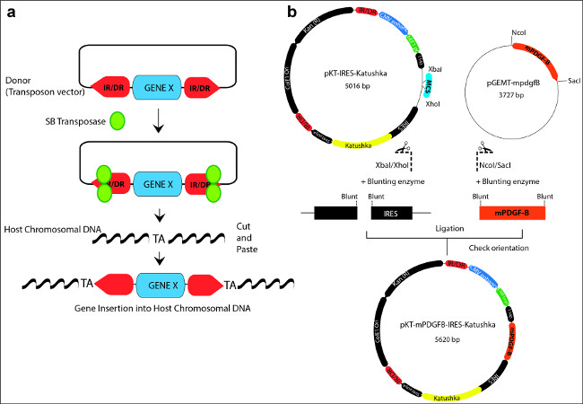
Figure 1: (a) Schematic representation illustrating the “cut and paste” mechanism used by the Sleeping Beauty transposase to integrate transposons into the host chromosomal DNA. A donor transposon plasmid is depicted with the gene of interest flanked by the inverted repeats/direct repeats (IR/DR; red arrow boxes) sequences. The SB transposase (green) binds to the IR/DR, excises the transposon and reintegrates it in between random TA dinucleotide base pairs on the host chromosomal DNA. (b) Example of cloning a gene of interest (mPDGFβ) into the backbone of a SB vector (pKT-IRES-Katushka). The SB vector contains two IR/DR repeat sequences (red) and a gene expression cassette which includes promoter and enhancer sequences, a multiple cloning site (MCS; blue), internal ribosomal entry sites (IRES), a fluorescent reporter marker (Katushka: yellow), and a polyadenilation site (polyA). The oncogene of interest (mPDGFβ; orange) is contained in a cloning vector pGEMT. The oncogene, flanked by specific restriction sites (NcoI/SacI) is released by enzymatic digestion, and blunted by the blunting enzyme. The vector is linearized and blunted as well. Finally, the mPDGFβ oncogene is inserted into the donor vector by means of a blunt-end ligation reaction to generate the new plasmid transposon vector: pKT-mPDGFβ-IRES-Katushka. Please click here to view a larger version of this figure.
Figure 2: Experimental Setup and guides for intra ventricular injections in neonatal mice (a) A stereotaxic frame (2) with micrometer dials is fitted with an automatic injector holding a 10 µl syringe (4). Inside the U frame, a neonatal adaptor frame is securely fastened (3). The control panel of the automatic injector (1) allows for precise selection of syringe, volume and rate of flow. (b) Photograph of a neonatal mouse (P1) with a needle inserted at the coordinates required for injections into the lateral ventricle: 1.5 mm ventral and 0.8 mm lateral to the lambda. (c) Illustration of a coronal section through the brain of a neonatal mouse (P1) highlighting the relative dimensions and position of the ventricles.
Figure 3: Tumors induced with the Sleeping Beauty transposon system show the histological hallmarks of human GBM (a) Bioluminescence image of a neonatal pup 24 hr after intraventricular injection with plasmids encoding NRAS, SV40 LgT. (b) Bioluminescence image of an adult animal (50 dpi) with a large tumor induced with shp53 NRAS and PDGF. (c) Hematoxilin and eosin stain of a coronal section from a brain of a moribund mouse with a tumor induced with NRAS and LgT (28 dpi). (d) Coronal section from a brain of a moribund mouse with a tumor induces with shp53 NRAS and PDGF. (e) Pseudopalisading necrosis, histological hallmark of human GBM is observed in de novo generated tumors. Arrow points to cells arranged in palisades migrating away from the central area of necrosis (N) (f) and (g) de novo generated tumors are highly invasive, showing invasion along blood vessels (g) and diffuse (f) into the normal brain parenchyma. (h) Region of hemorrhage with massive invasion of mononuclear cells (arrow), the initial stage of an area of pseudopalisading necrosis. (i) Large glomeruloid vessel with leaky endothelium (arrow) at the origin of diffusing hemorrhage inside a tumor induced with NRAS and SV40 –LgT. (j) Atypical mitosis in a tumor cells (arrowhead). (k) Gigantic tumor cell with multiple large nuclei (arrowhead).
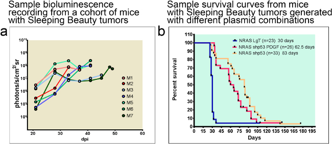
Figure 4: Tumor-bearing animals can be monitored with bioluminescence throughout the duration of disease progression. The median survival of animals is predicted by the combination of oncogenic DNA injected. (a) Example of bioluminescent traces from a cohort of 7 C57/BL6 mice. Note that mice succumb about a week after bioluminescence reaches 108 photons/s/cm2/sr. (b) Sample survival curves comparing median survival of animals with Sleeping Beauty tumors generated with different plasmid combinations. Median survival of tumors induced with NRAS and SV40 LgT is 30 dpi whereas of animals with tumors induced with shp53 NRAS and PDGF or shp53 and NRAS is 62.5 dpi or 83 dpi respectively. dpi: days post injection.
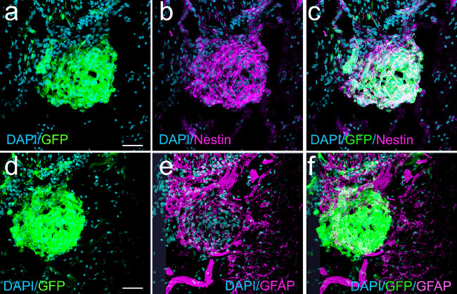
Figure 5: Nascent macroscopic GBM (22 dpi) induced with shp53 and NRAS show expression of nestin and GFAP in GFP+ tumor cells (confocal micrographs). Panel (a) shows a nascent tumor expressing GFP. Panel (b) represents the same field showing nestin expression. Panel (c) is an overlay of (a) and (b) illustrating co-localization of nestin and GFP. (d) GFP expression in tumor cells. (e) GFAP expression in some tumor cells and in cells surrounding the nascent tumor. (f) Overlay projection of (d) and (e) illustrating some tumor cells (white) co-expressing GFAP and GFP. Scale bars in (a) and (d) represent 75 µm.
Figure 6: GBM induced with shp53, NRAS and PDGF from a moribund animal (56 dpi) showing GFP and nestin expression in tumor cells and abundant staining for the mature glial marker GFAP surrounding the tumor. Panel (a) represents Nissl stain of acoronal section through the brain of a moribund animal with a tumor (intense blue staining from increased cellularity) induced with the Sleeping Beauty transposon system using shp53, PDGFΒ and NRAS. Panel (b), a coronal adjacent section to the one illustrated in panel (a) stained with the nuclear stain DAPI, showing expression of GFP in tumor cells. Panel (c) represents a confocal micrograph of the tumor border showing GFP expression in the tumor, identified by the high nuclear density with DAPI. Panel (d) illustrates the same field of view as in (a), showing intense GFAP expression in cells adjacent to the tumor. Panel (e) represents the overlay of panels (c) and (d). Panel (f) is a confocal micrograph of the tumor border, showing GFP expression in tumor cells. Panel (g) represents the same field of view as in (f), showing expression of nestin in tumor cells. Panel (h) is the overlay projection of panels (f) and (g). Scale bar in (c) represents 150 µm and in (f) represents 75 µm.
Figure 7: Neurospheres from a Sleeping Beauty tumor induced with shp53, PDGF and NRAS after 5 days in culture, passage 2. The cells are expressing GFP encoded on the shp53 PDGF plasmid (pT2shp53/GFP4/mPDGFβ). Panel (a) is a brightfield micrograph of a neurosphere. Panel (b) represents an epi-fluorescent micrograph of the same view as in (a) showing expression of GFP in the neurosphere cells. Panel (c) represents the overlay of panels (a) and (b).
In this article, we detail a versatile and reproducible method for generating new models of GBM using SB transposase- mediated integration of oncogenic plasmid DNA into cells surrounding the subventricular zone of neonatal mice. We present a protocol to generate transposon plasmids with new genes of interest, illustrate how to monitor the progression of the tumors in live animals, and how to characterize histo-pathological and immuno-histochemical features of these tumors.
As our lab (Figure 3) and others13 have shown, this model reliably creates tumors with the salient characteristics of human GBM including (1) pseudo-palisading necrosis, (2) vascular proliferation, (3) nuclear atypia, (4) abnormal mitoses and (5) perivascular and diffuse invasion. The use of neonatal mice is optimal from a logistical standpoint, providing a relatively easy technical procedure with minimal equipment and sedation/recovery time, allowing for rapid generation of large sample sizes of tumors with specific genetic alterations. These tumors cause animals to succumb to tumor burden with a predictable median survival dependent on the genetic alterations induced. Finally, the SB model has been used as a therapeutic intervention screen for treatment response24.
There are several important factors to consider when preparing the solutions used for the neonatal injections. DNA solutions need to be sterile, endotoxin free and concentrated (2-7 µg/µl) to allow for minimal volume of injections. The optimal ratio of nitrogen residues in the polyethyleneimine (PEI) to the phosphate residues in the DNA (N/P ratio) is 7. This ensures the formation of positively charged particle complexes which will bind anionic moieties on cell surfaces and will be endocytosed. Once in the cytoplasm, osmotic influx will cause the particles to burst and release the encapsulated plasmid DNA. The in vivo jet-PEI solution has a concentration of 150 mM expressed as nitrogen residues. Given that one µg DNA has 3 nmol of anionic phosphate, the amount of transfection reagent necessary can be calculated with the formula:
µl of in vivo jet-PEI = [(µg DNA x 3) x N/P ratio]/150.
For example, to prepare 40 µl of 0.5 µg/µl DNA solution with an N/P ratio of 7, 20 µg of DNA are needed. The volume of PEI required will be 20*3*7/150=2.8 µl.
Up to five different plasmids can be injected simultaneously. Experiments presented herein deliver the Sleeping Beauty transposase in trans on a plasmid that also encodes luciferase (SBLuc). As mentioned in the introduction, the ratio of transposase molecules to transposons is important to ensure optimal transposition. The optimal ratio (empirically established) of the SB100x plasmid to the transposon DNA plasmid is 1:4. Hence, if one uses two different transposon plasmids, T1 and T2 for example, the ratio of SBLuc:T1:T2 will be 1:2:2; or for a total of 20 µg DNA in a 40 µl solution 4 µg of SBLuc will be mixed with 8 µg of T1 and 8 µg of T2. For three different transposon plasmids the ratio will be SBLuc:T1:T2:T3 = 1:1.33:1.33:1.33 and for four transposons: 1:1:1:1:1.
It is useful to verify the uptake of the plasmid DNA by bioluminescence 24-72 hr after injection. As the animals grow, the increased optical density of the fur, skin, skull and brain, prevents monitoring of the tumor progression until the tumor has reached a threshold size, roughly 1-2 mm3 for darkly pigmented C57BL6 mice. Better transmission of the light is possible in these mice if their fur is shaved, or when using mice with white or no fur.
Several limitations need to be considered when using this model. First, the genetic material introduced with the injected plasmids can be integrated randomly in the host genome in different locations and number of copies. This may lead to variability between tumor cells and between tumors. Second, the DNA introduced is inserted primarily into intronic TA repeat sequences DNA25, however, the possibility of interference with coding host genomic DNA remains. Third, intraventricular injections may cause a small but persistent rate of hydrocephalus in young mice, which becomes clinically visible and symptomatic in the 3rd-4th week of life. Some strains of mice are more prone to hydrocephalus than others (i.e., C57/BL6 more than FVB). To minimize the risk of inducing hydrocephalus, it is recommended to inject as small a volume as possible (less than 1 µl in C57/BL6 mice), to ensure that the needles are sharp, the solutions have low salinity and to provide growing pups with enriched food, in order to allow for timely ossification of the skull bones. Finally, late-stage tumors often develop necrosis, which has to be considered when monitoring mice by bioluminescence. A lower reading may indicate an advanced tumor with large areas of necrosis and not necessarily a resolution of the tumor as a consequence of treatment. Ultimately, histological analysis remains the gold standard to assess tumor progression.
The advent of tumor whole genome sequencing in recent years has opened the door to exploration of the role of recurrently mutated genes in GBM. The Sleeping Beauty model is optimal for validating potential oncogenes or tumor suppressors. As in the example given in this paper with PDGFβ ligand, cloning the mouse cDNA sequence of a candidate oncogene into an established SB plasmid backbone will lead to constitutive expression within transfected cells. Evaluation of the role of tumor suppressors can be effectively performed by cloning into the backbone of the SB plasmid with candidate sequences, (available for example in the RNAi codex database26), to create robust expression of second-generation miR short-hairpins.
A current debate in the field of GBM research is related to the identity of the cell-of- origin. It has recently been shown that in mice, tumors induced by down-regulating p53 and Nf1 in neural stem cells, originate from oligodendrocyte precursor cells27. Using the Sleeping Beauty transposon model, similar studies can be designed to test whether other genetic alterations preferentially target other populations of stem cells/precursor cells to generate GBMs. Knowledge from such studies will greatly enhance our understanding of GBM etiology and provide avenues for novel therapeutic approaches.
The authors have nothing to disclose.
We thank Dr. John Ohlfest and Dr. Stacey Decker13 for the generous gift of plasmids and for the training provided to master this method. We thank Marta Dzaman for help with standardizing the Hematoxylin-Eosin staining procedure of paraffin-embedded sections. We also thank Molly Dahlgren for perusing this manuscript and providing helpful suggestions. This work is supported by NIH/NINDS grants to MGC and PRL, Leah’s Happy Hearts Foundation grant to MCG and PRL. Alex’s Lemonade Foundation young Investigator Award and the St. Baldrick’s Foundation Fellowship to CK.
Name of Material/ Equipment | Company | Catalog Number | Comments/Description |
Stoelting's Lab Standard with Rat and Mouse Adaptors | Stoelting | 51670 | Other companies produce similar frames, any of them with small mouse adaptors are suitable |
Quintessential Stereotaxic Injector (QSI) | Stoelting | 53311 | Injections can be made without it, ,but the automatic injector allows for increased reproducibility and convenience |
10 μL 700 series hand fitted MICROLITER syringe | Hamilton | Model 701 | This syringe model will deliver from 0.5 to 500 ul of solution reliably. Other syrnges (fro example 5 ul, Model 75) may also be used |
30 gauge gauge hypodermic needle (1.25", 15o bevel) |
Hamilton | S/O# 197462 | This needle is ideal to pierce the skin and the skull of a neonatal mouse without the need of other invasive procedures. If it gets dull (doesn't easily enter the skin), it needs to be replaced. |
in vivo-jetPEI | Polyplus Transfection | 201-10G | Aliquot in small volumes and keep at -20oC |
D-Luciferin, Potassium Salt | Goldbio.com | LuckK-1g | Other sources of firefly lucifierase are just as adequate |
Hematoxylin Solution, Harris Modified | Sigma Aldrich | HHS128-4L | |
Tissue-Tek O.C.T. Compound | Electron Microscopy Sciences | 62550-01 | for embedding brains for cryosectioning and immunohistovhemistry |
Advanced DMEM/F-12, no glutamine | Life Technologies | 12634-010 | for the culture of neurospheres |
B-27¨ Supplement (50X), serum free | Life Technologies | 17504-044 | serum free supplement for the culture of neurospheres |
N2 Supplement (100x) | Life Technologies | 17502-048 | serum free supplement for the culture of neurospheres |
Normocyn | InvivoGen | ant-nr-1 | anti-mycoplasma agent for the culture of neurospheres |
Recombinant Human EGF | PEPROTECH | AF-100-15 | growth factor supplement for the culture of neurospheres |
Recombinant Human FGF-basic | PEPROTECH | 100-18B | growth factor supplement for the culture of neurospheres |
HyClone HyQTase Cell Detachment Reagent | Thermo Scientific | SV3003001 | for the dissociation of neurospheres |
Kimble-Chase Kontes Pellet Pestle | Fisher Scientific | K749510-0590 | for the dissociation of freshly dissected tumors |
Falcon Cell Strainers mesh size 70um | Fisher Scientific | 08-771-2 | for generating single cell suspensions of dissociated neurospheres |
Xylenes Histological grade | Fisher Scientific | C8H10 | |
Protocol Harris Hematoxylin Mercury free ( acidified) | Fisher Scientific | 245-678 | |
Protocol Eosyn Y Solution ( intensified) | Fisher Scientific | 314-631 |