We demonstrate the effect of scleral crosslinking with riboflavin and UVA on an axial elongation rabbit eye. Axial elongation was induced in 13 day-old New Zealand rabbits (male and female) by suturing their right eye eyelids (tarsorrhaphy).
Myopic individuals, especially those with severe myopia, are at higher-than-normal risk of cataract, glaucoma, retinal detachment and chorioretinal abnormalities. In addition, pathological myopia is a common irreversible cause of visual impairment and blindness1-3. Our study demonstrates the effect of scleral crosslinking using riboflavin and ultraviolet-A radiation on the development of axial myopia in a rabbit model. The axial length of the eyeball was measured by A-scan ultrasound in New Zealand white rabbits aged 13 days (male and female). The eye then underwent 360° conjunctival peritomy with scleral crosslinking, followed by tarsorrhaphy. Axial elongation was induced in 13 day-old New Zealand rabbits by suturing their right eye eyelids (tarsorrhaphy). The eyes were divided into quadrants, and every quadrant had two scleral irradiation zones, each with an area of 0.2 cm² and a radius of 4 mm. Crosslinking was performed by dropping 0.1% dextran-free riboflavin-5-phosphate onto the irradiation zones 20 sec before ultraviolet-A irradiation and every 20 sec during the 200 sec irradiation time. UVA radiation (370 nm) was applied perpendicular to the sclera at 57 mW/cm² (total UVA light dose, 57 J/cm²). Tarsorrhaphies were removed on day 55, followed by repeated axial length measurements. This study demonstrates that scleral crosslinking with riboflavin and ultraviolet-A radiation effectively prevents occlusion-induced axial elongation in a rabbit model.
Myopia is the most common of the refractive disorders. The prevalence of myopia in the USA and Europe is reported to be around 30%, and in Asian countries it affects up to 60% of the general population1,2. Myopic progression occurs in up to 50% of myopes, usually at a rate of around -0.5 dioptres over a two-year interval3. The health costs imposed by myopia are considerable, including expenses for spectacles, contact lenses and refractive surgery and costs related to the increased health risks of glaucoma, cataract, retinal detachment and visual impairment4-6.
In animal studies of myopia, sight reduction is induced by eyelid suturing7-10, placement of an occluder at a short distance from the eye and corneal tattooing11. However, for artificial myopia to occur in these studies, the occlusion process has to be performed on very young animals, as no sight deprivation experiments carried out on adult specimens have proved successful.
One of the important features of severe myopia is a pathological change of the sclera with progressive thinning of the sclera, probably due to a disturbed feedback mechanism of emmetropization after visual deprivation12 or due to some metabolic disorder of the sclera, such as in Ehlers-Danlos syndrome13. Ultimately, both mechanisms lead to stretching and thinning of the sclera, retina and choroid due to structural abnormalities of the myopic sclera such as a decreased collagen fiber diameter14,15 and disturbances in fibrillogenesis16.
Several studies have shown that impaired collagen crosslinking is an important factor in the weakening process of the myopic sclera17-18. Wollensak et al.19-21 induced collagen crosslinking by applying the photosensitizer riboflavin and ultraviolet-A (UVA) irradiation (370 nm) and noted a significant, 157% increase in the rigidity of porcine and human sclera in vitro19 and a 465% increase in rabbit scleral rigidity in vivo (Young's modulus)20. Crosslinking also had a long-term effect on rabbit sclera in vivo: rigidity increased by 320.4% after 3 days, 277.6% after 4 months, and 502% after 8 months (Young's modulus)22.
Therapeutic attempts to arrest myopic progression have been published23-26 but the success of these methods is controversial. No efficient means of preventing progressive myopia has been found to date.
The etiology of myopia is still controversial, and its treatment poses a challenge. On the basis of these findings, it is hypothesized that scleral crosslinking can serve as a means for sclera-based treatment of myopic progression. The purpose of this study is to examine the scleral collagen crosslinking effect on the development of axial myopia induced by visual axis occlusion.
Animals were treated in accordance with the ARVO resolution on the use of animals in research. The study protocol was approved by the institutional Committee for Laboratory Animal Research (approval no. 022-4598-2; 021211).
1. Preparation for Surgery #1
2. Surgery #1 – Pre Cross Linking Step
3. Surgery #1 – Cross Linking
4. Surgery # 1 – Post Cross Linking Step
5. Surgery #1 – Post Operative Care
6. Surgery #2
Figures 1 and 2 graphically demonstrate the axial length measurements of two groups. Group 1 rabbits underwent scleral crosslinking and tarsorrhaphy on the right eye while the left eye was not operated on (Figure 1). Group 2 rabbits underwent only peritomy and tarsorrhaphy on the right eye while the left eye was not operated on (Figure 2).
In group 1, which underwent scleral crosslinking and tarsorrhaphy, the mean axial length in the right eye measured 10.68 ± 0.74 mm before eyelid suture and 14.29 ± 0.3 mm 55 days later, for a mean difference of 3.61 ± 0.76 mm. Corresponding values in the unoperated/unsutured left eye were 10.70 ± 0.79 mm and 15.14 ± 0.32 mm, for a mean difference of 4.44 ± 0.81 mm (Figure 1). Comparison of the axial lengths of the sutured and unsutured eyes at the end of the occlusion phase revealed a lesser net increase in the sutured eyes.
In group 2, which underwent only peritomy and tarsorrhaphies, mean axial length in the right eye measured 10.50 ± 0.67 mm before eyelid suture and 15.69 ± 0.39 mm 55 days later, for a mean difference of 5.19 ± 0.85 mm. Corresponding values in the unoperated/unsutured left eye were 10.54 ± 0.71 mm and 14.74 ± 0.38 mm, for a mean difference of 4.20 ± 0.67 mm (Figure 2). Comparison of the axial lengths of the sutured and unsutured eyes at the end of the occlusion phase revealed a greater net increase in the sutured eyes.
Comparison of the mean change in axial length of the right eyes between group 2 (5.19 ± 0.85 mm) and group 1 (3.61 ± 0.76 mm) yielded a significantly lower value at the end of the occlusion phase (55 days) in the eyes that underwent the crosslinking procedure (p <0.001, nonparametric Mann-Whitney test). The between-group difference in mean axial length in the left eyes (4.20 ± 0.67 mm vs. 4.44 ± 0.81 mm) was not statistically significant (p = 0.39, Mann-Whitney non-parametric test).
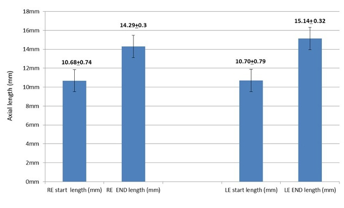
Figure 1. Right eye axial measurements before and after scleral crosslinking and tarsorrhaphy vs. left eye axial measurements. The mean axial length of the right eye before scleral crosslinking and tarsorrhaphy (RE start) and after removal of the tarsorrhaphy 55 days later (RE end). The mean axial length of the left eye at baseline (LE start) and 55 days later (LE end). The left eye was not operated on and was left open. The error bars indicates standard error of the mean. (Re-printed with permission from reference27). Please click here to view a larger version of this figure.
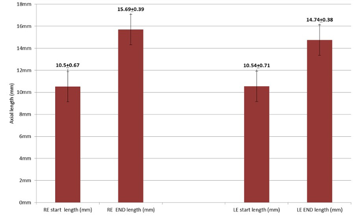
Figure 2. Right eye axial measurements before and after tarsorrhaphy vs. left eye axial measurements. The mean axial length of the right eye before tarsorrhaphy (RE start) and after removal of the tarsorrhaphy 55 days later (RE end). The mean axial length of the left eye at baseline (LE start) and 55 days later (LE end).The left eye was not operated on and was left open. The error bars indicates standard error of the mean. (Re-printed with permission from reference27). Please click here to view a larger version of this figure.
We present the first in vivo study of the prevention of axial myopia in a rabbit model using cross-linking technology with riboflavin and UVA irradiation. Although different laboratory animals can be used in this type of study, we chose rabbits mostly due to the size of the eyes and the need to perform crosslinking on the scleral surface.
We found that exposing the rabbit sclera and suturing the upper and lower eyelids to be challenging procedures. We recommend trimming the lid margins and using the sutures mentioned in our protocol to ensure that the eyelids are firmly closed for the given period without need for corrections.
One important thing to consider is the need to calibrate the irradiation device and the fiber optic for the required power energy.
H&E histological slides revealed no toxic changes to the retina on eyes that underwent the crosslinking procedure. Further investigations should be performed to address the potential toxicity of riboflavin and UVA irradiation on the retina, choroid and sclera, with a focus on position, amount of energy and exposure time.
Several limitations of this study warrant consideration. Only preoperative and postoperative axial lengths were measured. The axial length of the eyeball was measured by A-scan ultrasound. We did not evaluate biometric properties, refractive analysis or biomechanical properties of the sclera.
Taken together, we think that the effect of scleral crosslinking with riboflavin and UVA on the axial elongation of the rabbit eye will have a positive impact on myopia research.
The authors have nothing to disclose.
The authors are thankful to Ms. Dalia Sela and Mr. Emi Sharon for their professional and excellent technical work in the laboratory.
2-0 braided silk non-needled sutures | ETHICON | W193 | |
4-0 braided silk ivory color | ETHICON | W816 | |
0.1% dextran-free riboflavin-5-phosphate 1mg:1ml | Concept for Pharmacy Ltd | D2-5025 | |
UV A (370nm) light source | O/E LAND Inc | NCSU033B | |
Beveled down custom made fiber optic | Prizmatix Ltd | ||
26G lacrimal cannula | Beaver-visitec International Ltd. | REF581276 | |
25G tapered hydrodelineator [Blumenthal] | Beaver-visitec International Ltd. | REF585107 | |
13 days old rabbits | Harlan | 1NZWR40 | |
Ultrasonic biometer | Allergan-Humphrey | 820-519 | |
Skin marker | Devon | 4237101664X |