Summary

生成CRISPR / Cas9介导的单等位基因缺失来研究小鼠胚胎干细胞功能增强

Published: April 02, 2016
doi:

Summary

Experimental validation of enhancer activity is best approached by loss-of-function analysis. Presented here is an efficient protocol that uses CRISPR/Cas9 mediated deletion to study allele-specific regulation of gene transcription in F1 ES cells which contain a hybrid genome (Mus musculus129 x Mus castaneus).

Abstract

Enhancers control cell identity by regulating tissue-specific gene expression in a position and orientation independent manner. These enhancers are often located distally from the regulated gene in intergenic regions or even within the body of another gene. The position independent nature of enhancer activity makes it difficult to match enhancers with the genes they regulate. Deletion of an enhancer region provides direct evidence for enhancer activity and is the gold standard to reveal an enhancer’s role in endogenous gene transcription. Conventional homologous recombination based deletion methods have been surpassed by recent advances in genome editing technology which enable rapid and precisely located changes to the genomes of numerous model organisms. CRISPR/Cas9 mediated genome editing can be used to manipulate the genome in many cell types and organisms rapidly and cost effectively, due to the ease with which Cas9 can be targeted to the genome by a guide RNA from a bespoke expression plasmid. Homozygous deletion of essential gene regulatory elements might lead to lethality or alter cellular phenotype whereas monoallelic deletion of transcriptional enhancers allows for the study of cis-regulation of gene expression without this confounding issue. Presented here is a protocol for CRISPR/Cas9 mediated deletion in F1 mouse embryonic stem (ES) cells (Mus musculus129 x Mus castaneus). Monoallelic deletion, screening and expression analysis is facilitated by single nucleotide polymorphisms (SNP) between the two alleles which occur on average every 125 bp in these cells.

Introduction

转录调控元件由于异常的基因表达2是发展1和修改这些元素的过程中的基因表达的时空微调可导致疾病的关键。通过全基因组关联研究发现了许多疾病相关的区域是在非编码区,并有转录增强子3-4的功能。识别增强剂并将它们与它们调节是复杂的,因为它们通常位于从它们调节基因几千个碱基远并且可以以组织特异性方式5-6被激活的基因匹配。增强剂的预测通常是基于组蛋白修饰的标记,介体黏着复合物和细胞类型特异性转录结合因子7-10。预测增强剂的验证是最经常通过一个基于矢量的测定,其中所述增强剂激活报告基因11-12的表达进行。这些数据提供了v有关推测的增强子序列的调节潜力aluable信息,但不透露自己的功能其内生基因组范围内或识别它们调节的基因。基因组编辑充当一个强有力的工具来研究由失功能分析在它们的内源上下文转录调控元件的功能。

在基因组编辑,即CRISPR / Cas9基因组编辑系统的最新进展,有利于基因功能的研究。的CRISPR / Cas9系统易于使用和适应性对于许多生物系统。所述Cas9蛋白靶向于由导的RNA(gRNA)13中的基因组中的特定位点。所述SpCas9 / gRNA复杂扫描对其靶基因组序列的基因组中它必须是5'到protospacer相邻基序(PAM)的序列,NGG 14-15。的gRNA到其目标,一个20个核苷酸(nt)的序列与gRNA互补的碱基配对,激活导致域金字塔之戒SpCas9核酸酶活Ë链断裂(DSB)3碱基的序列PAM的上游。特异性是通过在gRNA种子区域完全碱基配对来实现,所述6-12 nt下邻近于PAM;相反地,不匹配5'种子的通常耐受16-17。引入的DSB可以修复或者由非同源末端连接(NHEJ)的DNA修复或同源性定向修复(HDR)mechanisms.NHEJ DNA修复往往造成在目标部位的几个碱基对,可以破坏的插入/缺失(插入缺失)的基因的开放阅读框(ORF)。以产生在基因组2 gRNAs,侧翼感兴趣的区域大的缺失,可以使用18-19。这种方法是对聚成基因座控制区或超增强剂它比常规增强剂9,18,20-22较大转录增强子的研究中特别有用的。

单等位基因缺失是研究转录 -regulation一个有价值的模型。观察到昌E在转录水平的增强子的单等位基因缺失之后关联到在基因调控该增强剂的不当两个等位基因的转录可能受影响的影响蜂窝健身时可能出现的混杂影响的作用。评估减少的表达是困难的但不区分野生型等位基因的删除的能力。此外,基因分型在每个等位基因缺失而不区分两个等位基因的能力是具有挑战性的,尤其是对大缺失> 10kb的至1兆23,其中它是难以通过PCR扩增整个野生型区域。使用通过杂交小家鼠 129小家鼠castaneus生成的F1 ES细胞的允许两个等位基因通过等位基因特异性PCR 18,24区别开来。在这些细胞中的基因组杂交便于等位基因特异缺失筛选和表达分析。上平均有一个SNP位这两个基因组之间的每一个125 bp的为表达和基因分型提供在引物设计的灵活性的分析。一种SNP的存在可以影响引物的熔化温度(T M)与靶实时定量PCR(qPCR的)扩增特异性允许两个等位基因25的歧视。此外,引物的3'末端中的一个错配极大地影响DNA聚合酶从引物防止不期望的等位基因靶26的扩增延伸的能力。描述在下面的协议是使用CRISPR / Cas9基因组编辑系统( 图1)大于1 kb的等位基因特异性增强缺失和随后的表达分析使用F1 ES细胞。
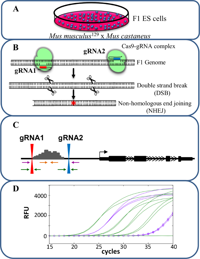
图1
图1.增强删除使用CRISPR / Cas9研究 -reg基因表达的ulation。(A)中由小家鼠 129小家鼠castaneus之间的交叉产生的F1 ES细胞用于允许等位基因特异性缺失。 (B)中的两个导向的RNA(gRNA)用于诱导增强子区的一个大Cas9介导的缺失。 (C)的引物组被用于识别大的单-和双等位基因缺失。橙色引物是内引物,紫色引物外侧的引物和绿色的引物的gRNA侧翼引物。 (D)基因表达的变化是使用等位基因特异性qPCR的监控。俄罗斯足协表示相对荧光单位。 请点击此处查看该图的放大版本。

Protocol

1.设计和建造的gRNA 删除转录增强子区域使用两个gRNAs一5'和一个3'的感兴趣区域的。使用由张实验室产生的鼠标UCSC基因组浏览器的轨道,以确定独特的gRNA序列(http://www.genome-engineering.org 15)。接下来检查这些gRNAs和他们的使用由桑格研究所(www.sanger.ac.uk/sanger/Mouse_SnpViewer/rel-1211)27-28提供在线工具SNP和插入缺失相邻的PAM。要定位具有同等效率两个等位基因,避免含有S…

Representative Results

这里所描述的协议使用的F1 ES细胞,研究基因表达的顺式 -regulation使用CRISPR / Cas9基因组编辑( 图1)产生的单等位基因增强剂删除的细胞。用于基因分型和基因表达的gRNA和等位基因特异性引物的设计是在该方法的关键因素。每个等位基因特异性引物组必须通过qPCR进行验证,以确认等位基因特异性。等位基因特异性引物仅扩增各自的基因组DNA靶标是理想?…

Discussion

CRISPR / Cas9介导的基因组编辑技术为基因改造一个简单,快捷,廉价的方法。这里详细生成和分析单等位基因缺失的增强功能性增强特性的方法发生在F1小鼠细胞单核苷酸多态性的优势的。这种类型的方法的优点是:1)单等位基因增强剂缺失不产生时的临界增强剂是从两个等位基因缺失, ,在规定的基因导致细胞杀伤力的蛋白水平大大降低的或改变的发生的混杂影响表型; 2)如果单等位…

Disclosures

The authors have nothing to disclose.

Acknowledgements

We would like to thank all the members of the Mitchell lab for helpful discussions. This work was supported by the Canadian Institutes of Health Research, the Canada Foundation for Innovation and the Ontario Ministry of Research and Innovation (operating and infrastructure grants held by JAM).

Materials

Phusion High-Fidelity DNA Polymerase NEB M0530S high fidelity DNA polymerase used in gRNA assembly
Gibson Assembly Master Mix NEB E2611L
gRNA_Cloning Vector Addgene 41824 A target sequence is cloned into this vector to create the gRNA plasmid
pCas9_GFP Addgene 44719 Codon-optimized SpCas9 and EGFP co-expression plasmid
AflII NEB R0520S
EcoRI NEB R3101S
Neon Transfection System 100 µL Kit Life Technologies MPK10096 Microporator transfection technology
prepGEM ZyGEM PT10500 genomic DNA extraction reagent
Nucleo Spin Gel & PCR Clean-up Macherey-Nagel 740609.5
High-Speed Plasmid Mini Kit Geneaid PD300
Maxi Plasmid Kit Endotoxin Free  Geneaid PME25
SYBR select mix for CFX Life Technologies 4472942 qPCR reagent
iScript cDNA synthesis kit Bio-rad 170-8891 Reverse transcription reagent
0.25% Trypsin with EDTA Life Technologies 25200072
PBS without Ca/Mg2+ Sigma D8537
0.5M EDTA Bioshop EDT111.500
HBSS Life Technologies 14175095
1M HEPES Life Technologies 13630080
BSA fraction V (7.5%) Life Technologies 15260037
Max Efficiency DH5α competent cells Invitrogen 18258012
FBS ES cell qualified FBS is subjected to a prior testing in mouse ES cells for pluripotency
DMSO Sigma D2650
Glutamax Invitrogen 35050
DMEM Life Technologies 11960069
Pencillin/Streptomycin Invitrogen 15140
Sodium pyruvate Invitrogen 11360
Non-essential aminoacid Invitrogen 11140
β-mercaptoethanol Sigma M7522
96-well plate Sarstedt 83.3924
Sealing tape Sarstedt 95.1994
CoolCell LX Biocision BCS-405 alcohol-free cell freezing container
CHIR99021 Biovision 1748-5 Inhibitor for F1 ES cell culture
PD0325901 Invivogen inh-pd32 Inhibitor for F1 ES cell culture
LIF Chemicon ESG1107 Inhibitor for F1 ES cell culture

References

  1. Sagai, T., Hosoya, M., Mizushina, Y., Tamura, M., Shiroishi, T. Elimination of a long-range cis-regulatory module causes complete loss of limb-specific Shh expression and truncation of the mouse limb. Development. 132 (4), 797-803 (2005).
  2. Kleinjan, D. A., Lettice, L. A. Long-range gene control and genetic disease. Adv Genet. 61, 339-388 (2008).
  3. Visel, A., Rubin, E. M., Pennacchio, L. A. Genomic views of distant-acting enhancers. Nature. 461 (7261), 199-205 (2009).
  4. Maurano, M. T., et al. Systematic localization of common disease-associated variation in regulatory DNA. Science. 337 (6099), 1190-1195 (2012).
  5. Heintzman, N. D., et al. Histone modifications at human enhancers reflect global cell-type-specific gene expression. Nature. 459 (7243), 108-112 (2009).
  6. Shen, Y., et al. A map of the cis-regulatory sequences in the mouse genome. Nature. 488 (7409), 116-120 (2012).
  7. Johnson, D. S., Mortazavi, A., Myers, R. M., Wold, B. Genome-wide mapping of in vivo protein-DNA interactions. Science. 316 (5830), 1497-1502 (2007).
  8. Rhee, H. S., Pugh, B. F. Comprehensive genome-wide protein-DNA interactions detected at single-nucleotide resolution. Cell. 147 (6), 1408-1419 (2011).
  9. Whyte, W. A., et al. Master transcription factors and mediator establish super-enhancers at key cell identity genes. Cell. 153 (2), 307-319 (2013).
  10. Chen, C. Y., Morris, Q., Mitchell, J. A. Enhancer identification in mouse embryonic stem cells using integrative modeling of chromatin and genomic features. BMC Genomics. 13 (1), 152 (2012).
  11. Patwardhan, R. P., et al. Massively parallel functional dissection of mammalian enhancers in vivo. Nat Biotechnol. 30 (3), 265-270 (2012).
  12. Melnikov, A., et al. Systematic dissection and optimization of inducible enhancers in human cells using a massively parallel reporter assay. Nat Biotechnol. 30 (3), 271-277 (2012).
  13. Jinek, M., et al. A programmable dual-RNA-guided DNA endonuclease in adaptive bacterial immunity. Science. 337 (6096), 816-821 (2012).
  14. Cong, L., et al. Multiplex genome engineering using CRISPR/Cas systems. Science. 339 (6121), 819-823 (2013).
  15. Mali, P., et al. RNA-guided human genome engineering via Cas9. Science. 339 (6121), 823-826 (2013).
  16. Hsu, P. D., et al. DNA targeting specificity of RNA-guided Cas9 nucleases. Nat Biotechnol. 31 (9), 827-832 (2013).
  17. Cho, S. W., et al. Analysis of off-target effects of CRISPR/Cas-derived RNA-guided endonucleases and nickases. Genome Res. 24 (1), 132-141 (2014).
  18. Zhou, H. Y., et al. A Sox2 distal enhancer cluster regulates embryonic stem cell differentiation potential. Genes Dev. 28 (24), 2699-2711 (2014).
  19. Fujii, W., Kawasaki, K., Sugiura, K., Naito, K. Efficient generation of large-scale genome-modified mice using gRNA and CAS9 endonuclease. Nucleic Acids Res. 41 (20), e187 (2013).
  20. Tuan, D. Y., Solomon, W. B., London, I. M., Lee, D. P. An erythroid-specific, developmental-stage-independent enhancer far upstream of the human ‘beta-like globin’ genes. Proc Natl Acad Sci U S A. 86 (8), 2554-2558 (1989).
  21. Amano, T., et al. Chromosomal dynamics at the Shh locus: limb bud-specific differential regulation of competence and active transcription. Dev Cell. 16 (1), 47-57 (2009).
  22. Li, Y., et al. CRISPR reveals a distal super-enhancer required for Sox2 expression in mouse embryonic stem cells. PLoS One. 9 (12), e114485 (2014).
  23. Canver, M. C., et al. Characterization of genomic deletion efficiency mediated by clustered regularly interspaced palindromic repeats (CRISPR)/Cas9 nuclease system in mammalian cells. J Biol Chem. 289 (31), 21312-21324 (2014).
  24. Mlynarczyk-Evans, S., et al. X chromosomes alternate between two states prior to random X-inactivation. PLoS Biol. 4 (6), e159 (2006).
  25. Lefever, S., Pattyn, F., Hellemans, J., Vandesompele, J. Single-nucleotide polymorphisms and other mismatches reduce performance of quantitative PCR assays. Clin Chem. 59 (10), 1470-1480 (2013).
  26. Huang, M. M., Arnheim, N., Goodman, M. F. Extension of base mispairs by Taq DNA polymerase: implications for single nucleotide discrimination in PCR. Nucleic Acids Res. 20 (17), 4567-4573 (1992).
  27. Keane, T. M., et al. Mouse genomic variation and its effect on phenotypes and gene regulation. Nature. 477 (7364), 289-294 (2011).
  28. Yalcin, B., et al. Sequence-based characterization of structural variation in the mouse genome. Nature. 477 (7364), 326-329 (2011).
  29. Gibson, D. G., et al. Enzymatic assembly of DNA molecules up to several hundred kilobases. Nat Methods. 6 (5), 343-345 (2009).
  30. Gibson, D. G., Smith, H. O., Hutchison, C. A., Venter, J. C., Merryman, C. Chemical synthesis of the mouse mitochondrial genome. Nat Methods. 7 (11), 901-903 (2010).
  31. Ding, Q., et al. Enhanced efficiency of human pluripotent stem cell genome editing through replacing TALENs with CRISPRs. Cell Stem Cell. 12 (4), 393-394 (2013).
  32. Basu, S., Campbell, H. M., Dittel, B. N., Ray, A. Purification of specific cell population by fluorescence activated cell sorting (FACS). J Vis Exp. (41), (2010).
  33. Forlenza, M., Kaiser, T., Savelkoul, H. F., Wiegertjes, G. F. The use of real-time quantitative PCR for the analysis of cytokine mRNA levels. Methods Mol Biol. 820, 7-23 (2012).
  34. Wu, J. H., Hong, P. Y., Liu, W. T. Quantitative effects of position and type of single mismatch on single base primer extension. J Microbiol Methods. 77 (3), 267-275 (2009).
  35. Sanyal, A., Lajoie, B. R., Jain, G., Dekker, J. The long-range interaction landscape of gene promoters. Nature. 489 (7414), 109-113 (2012).
check_url/53552?article_type=t

Play Video

Cite This Article
Moorthy, S. D., Mitchell, J. A. Generating CRISPR/Cas9 Mediated Monoallelic Deletions to Study Enhancer Function in Mouse Embryonic Stem Cells. J. Vis. Exp. (110), e53552, doi:10.3791/53552 (2016).

View Video