Summary

इमेजिंग झिल्ली फ्लोरोसेंट वोल्टेज सेंसर आनुवंशिक रूप से इनकोडिंग के दो प्रकार के साथ संभावित

Published: February 04, 2016
doi:

Summary

A method for imaging changes in membrane potential using genetically encoded voltage indicators is described.

Abstract

Genetically encoded voltage indicators (GEVIs) have improved to the point where they are beginning to be useful for in vivo recordings. While the ultimate goal is to image neuronal activity in vivo, one must be able to image activity of a single cell to ensure successful in vivo preparations. This procedure will describe how to image membrane potential in a single cell to provide a foundation to eventually image in vivo. Here we describe methods for imaging GEVIs consisting of a voltage-sensing domain fused to either a single fluorescent protein (FP) or two fluorescent proteins capable of Förster resonance energy transfer (FRET) in vitro. Using an image splitter enables the projection of images created by two different wavelengths onto the same charge-coupled device (CCD) camera simultaneously. The image splitter positions a second filter cube in the light path. This second filter cube consists of a dichroic and two emission filters to separate the donor and acceptor fluorescent wavelengths depending on the FPs of the GEVI. This setup enables the simultaneous recording of both the acceptor and donor fluorescent partners while the membrane potential is manipulated via whole cell patch clamp configuration. When using a GEVI consisting of a single FP, the second filter cube can be removed allowing the mirrors in the image splitter to project a single image onto the CCD camera.

Introduction

इस पत्र के प्रमुख ध्यान केंद्रित आनुवंशिक रूप से इनकोडिंग फ्लोरोसेंट प्रोटीन का उपयोग कर इन विट्रो में झिल्ली क्षमता में परिवर्तन के ऑप्टिकल इमेजिंग का प्रदर्शन है। झिल्ली क्षमता में परिवर्तन इमेजिंग neuronal सर्किट की गतिविधि का अध्ययन करने के रोमांचक संभावना प्रदान करता है। एक प्रतिदीप्ति तीव्रता परिवर्तन में झिल्ली क्षमता परिणाम में परिवर्तन, कैमरे के प्रत्येक पिक्सेल एक किराए की इलेक्ट्रोड neuronal गतिविधि के nonintrusive माप सक्षम हो जाता है। चालीस से अधिक वर्षों के लिए, कार्बनिक वोल्टेज के प्रति संवेदनशील रंगों झिल्ली क्षमता 1-4 में परिवर्तन के अवलोकन के लिए उपयोगी हो गया है। हालांकि, इन रंगों सेलुलर विशिष्टता की कमी है। इसके अलावा, कुछ प्रकार की कोशिकाओं के दाग के लिए मुश्किल हैं। आनुवंशिक रूप से इनकोडिंग वोल्टेज संकेतक (GEVIs) कोशिकाओं विशेष रूप से अध्ययन किया जा फ्लोरोसेंट वोल्टेज के प्रति संवेदनशील जांच अभिव्यक्त होने से इन सीमाओं को पार।

वहाँ GEVIs के तीन वर्गों रहे हैं। GEVI के प्रथम श्रेणी VO का उपयोग करता हैया तो एक फ्लोरोसेंट प्रोटीन (एफपी) 5-9 या एक Förster प्रतिध्वनि ऊर्जा हस्तांतरण (झल्लाहट) की जोड़ी को 10-12 के साथ ltage संवेदन वोल्टेज संवेदन फॉस्फेट से डोमेन। सेंसर के द्वितीय श्रेणी के एक फ्लोरोसेंट सूचक सीधे 13-15 के रूप में या electrochromic झल्लाहट 16,17 के माध्यम से माइक्रोबियल rhodopsin उपयोग करता है। तृतीय श्रेणी के दो घटकों, आनुवंशिक घटक एक झिल्ली लंगर एफपी और एक दूसरे घटक एक झिल्ली बाध्य शमन डाई 18-20 जा रहा जा रहा है इस्तेमाल करता है। दूसरी और तीसरी कक्षाओं विट्रो और टुकड़ा प्रयोगों में 19,20 के लिए उपयोगी होते हैं, केवल सेंसर के प्रथम श्रेणी वर्तमान में विवो 6 विश्लेषण के लिए उपयोगी होते हैं।

इस रिपोर्ट में हम इन विट्रो में (चित्रा 1) GEVIs के प्रथम श्रेणी का उपयोग कर झिल्ली क्षमता की इमेजिंग प्रदर्शन करेंगे। वोल्टेज सेंसर की यह प्रथम श्रेणी के लिए सबसे आसान vivo इमेजिंग के लिए संक्रमण है। GEVIs यू के बाद सेएक वोल्टेज संवेदन डोमेन एक एफपी के लिए जुड़े हुए हैं के बारे में tilizing सेंसर की rhodopsin वर्ग की तुलना में उज्जवल 50 गुना, वे बजाय चाप दीपक रोशनी का उपयोग कर एक अत्यंत शक्तिशाली लेजर की आवश्यकता होती है imaged किया जा सकता है। चमक में असमानता का एक और परिणाम है कि GEVIs के प्रथम श्रेणी आसानी से मस्तिष्क की ऑटो प्रतिदीप्ति अधिक हो सकती है। rhodopsin आधारित जांच नहीं कर सकते। सेंसर के तृतीय श्रेणी के बस के रूप में प्रथम श्रेणी के रूप में उज्ज्वल है, लेकिन एक रासायनिक पीने की वस्तु है जो विवो में प्रशासन के लिए मुश्किल है के अलावा आवश्यकता है।

हम होगा, इसलिए, एक एकल एफपी (Bongwoori) 8 और एक झल्लाहट जोड़ी से मिलकर एक जांच (नबी 2) 12 के साथ एक जांच के अधिग्रहण प्रदर्शित करता है। 11 एक हरी फ्लोरोसेंट दाता, तिपतिया घास से मिलकर, और एक लाल फ्लोरोसेंट स्वीकर्ता, mRuby2, नबी २.२४२ और नबी २.२४४ नाम – झल्लाहट इस रिपोर्ट में constructs VSFP-सीआर (तिपतिया घास-mRuby2 वोल्टेज के प्रति संवेदनशील फ्लोरोसेंट प्रोटीन) की तितली संस्करण हैं <s> 12 अप। रिकॉर्डिंग के इन प्रकार के परिचय के शोधकर्ताओं जानकारी GEVIs के प्रकार प्रदान कर सकते हैं की एक बेहतर समझ देना चाहिए।

आकृति 1
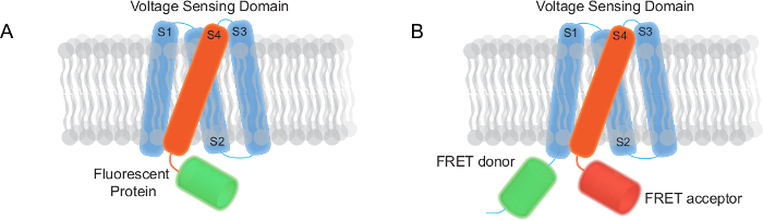
चित्रा 1. वोल्टेज संकेतक आनुवंशिक रूप से इनकोडिंग (GEVIs) यह रिपोर्ट (ए) में imaged एक मोनो आधारित GEVI एक पार झिल्ली वोल्टेज संवेदन डोमेन और एक फ्लोरोसेंट प्रोटीन होने एफपी की दो प्रकार। (बी) के एक झल्लाहट आधारित एक पार झिल्ली वोल्टेज संवेदन डोमेन, एक झल्लाहट दाता और स्वीकर्ता के शामिल GEVI। यह आंकड़ा का एक बड़ा संस्करण देखने के लिए यहां क्लिक करें।

Protocol

आचार कथन: पशु प्रयोग प्रोटोकॉल केआईएसटी पशु प्रोटोकॉल 2014-001 में संस्थागत पशु की देखभाल और उपयोग समिति द्वारा अनुमोदित किया गया था। 1. उपकरण सेटअप इमेजिंग सेटअप एक कंपन अलगाव की मेज पर ए…

Representative Results

क्षणिक ट्रांसफ़ेक्ट कोशिकाओं प्रतिदीप्ति तीव्रता में महत्वपूर्ण परिवर्तन और प्लाज्मा झिल्ली अभिव्यक्ति की डिग्री प्रदर्शन कर सकते हैं। भले ही coverslip पर कुछ कोशिकाओं आंतरिक प्रतिदीप्ति ?…

Discussion

तंत्रिका तंत्र कई अलग अलग तरीकों में वोल्टेज, अवरोध एक मामूली hyperpolarization, synaptic इनपुट एक अपेक्षाकृत बड़े वोल्टेज परिवर्तन में एक मामूली विध्रुवण और एक संभावित कार्रवाई के परिणाम का कारण बनता है का कारण बनता …

Disclosures

The authors have nothing to disclose.

Acknowledgements

This work was supported by the World Class Institute (WCI) program of the National Research Foundation of Korea funded by Ministry of Education, Science, and Technology of Korea Grant WCI 2009-003 and Korea Institute of Science and Technology Institutional Program Project 2E24210. Sungmoo Lee was supported by Global Ph.D. Fellowship program (NRF-2013H1A2A1033344) of the National Research Foundation (NRF) under the Ministry of Education (MOE, Korea).

Materials

Inverted Microscope Olympus IX71
60X objective lens (numerical aperture = 1.35) Olympus UPLSAPO 60XO
Excitation filter Semrock  FF02-472/30 For voltage imaging of super ecliptic pHluorin in Bongwoori
Dichroic mirror Semrock FF495-Di03-25×36
Emission filter Semrock FF01-497/LP
75W Xenon arc lamp CAIRN OptoSource Illuminator LEDs and lasers are also effective light sources
Slow speed CCD camera Hitachi KP-D20BU
Dual port camera adaptor Olympus U-DPCAD
High speed CCD camera RedShirtImaging, LLC NeuroCCD-SM
Image splitter CAIRN Optosplit 2
Excitation filter Semrock FF01-475/23-25 For voltage imaging of FRET pair based GEVI consisting of Clover and mRuby2)
Dichroic mirror Semrock FF495-Di03-25×36
Emission filter Chroma ET520/40
Dichroic mirror Semrock FF560-FDi01-25X36
Emission filter Chroma ET645/75
Vibration isolation system Kinetic systems 250BM-IC, 5702E-3036-31
Patching chamber Warner instruments RC-26G, 64-0235
#0 Micro Coverglass (22x40mm) Electron Microscopy Sciences 72198-20
Temperature controller Warner instruments TC-344B
#0 (0.08~0.13mm) – 10mm diameter glass coverslip Ted Pella 260366
Lipofection agent Life Technologies 11668-027
Calcium phosphate reagent Clontech – Takara 631312
Patch clamp amplifier HEKA EPC 10 USB amplifier
Multi-channel data acquisition software HEKA Patchmaster
Image acquisition and analysis software RedShirtImaging Neuroplex
Spreadsheet application software Microsoft Microsoft Excel 2010
Data analysis software OriginLab OriginPro 8.6.0
Demagnifier Qioptiq LINOS Optem standard camera coupler 0.38x SC38 J clamp
Confocal microscope Nikon Nikon A1R confocal microscope
Anti-fade reagent Life Technologies P36930

References

  1. Salzberg, B. M., Davila, H. V., Cohen, L. B. Optical recording of impulses in individual neurones of an invertebrate central nervous system. Nature. 246, 508-509 (1973).
  2. Cohen, L. B., et al. Changes in axon fluorescence during activity: molecular probes of membrane potential. J. Membrane Biol. 19, 1-36 (1974).
  3. Tasaki, I., Warashina, A. Dye-membrane interaction and its changes during nerve excitation. Photochem Photobiol. 24, 191-207 (1976).
  4. Grinvald, A., Hildesheim, R. VSDI: a new era in functional imaging of cortical dynamics. Nat Rev Neurosci. 5, 874-885 (2004).
  5. Jin, L., Han, Z., Platisa, J., Wooltorton, J. R., Cohen, L. B., Pieribone, V. A. Single action potentials and subthreshold electrical events imaged in neurons with a fluorescent protein voltage probe. Neuron. 75, 779-785 (2012).
  6. Cao, G., Platisa, J., Pieribone, V. A., Raccuglia, D., Kunst, M., Nitabach, M. N. Genetically targeted optical electrophysiology in intact neural circuits. Cell. 154, 904-913 (2013).
  7. St-Pierre, F., Marshall, J. D., Yang, Y., Gong, Y., Schnitzer, M. J., Lin, M. Z. High-fidelity optical reporting of neuronal electrical activity with an ultrafast fluorescent voltage sensor. Nat Neurosci. 17, 884-889 (2014).
  8. Piao, H. H., Rajakumar, D., Kang, B. E., Kim, E. H., Baker, B. J. Combinatorial mutagenesis of the voltage-sensing domain enables the optical resolution of action potentials firing at 60 Hz by a genetically encoded fluorescent sensor of membrane potential. J Neurosci. 35, 372-385 (2015).
  9. Jung, A., Garcia, J. E., Kim, E., Yoon, B. J., Baker, B. J. Linker length and fusion site composition improve the optical signal of genetically encoded fluorescent voltage sensors. Neurophoton. 2, 021012 (2015).
  10. Dimitrov, D., et al. Engineering and characterization of an enhanced fluorescent protein voltage sensor. PLoS One. 2, e440 (2007).
  11. Lam, A. J., et al. Improving FRET dynamic range with bright green and red fluorescent proteins. Nat Methods. 9, 1005-1012 (2012).
  12. Sung, U., et al. Developing fast fluorescent protein voltage sensors by optimizing FRET interactions. PLoS One. 10, e0141585 (2015).
  13. Kralj, J. M., Douglass, A. D., Hochbaum, D. R., Maclaurin, D., Cohen, A. E. Optical recording of action potentials in mammalian neurons using a microbial rhodopsin. Nat Methods. 9, 90-95 (2012).
  14. Flytzanis, N. C., et al. Archaerhodopsin variants with enhanced voltage-sensitive fluorescence in mammalian and Caenorhabditis elegans neurons. Nat Commun. 5, 4894 (2014).
  15. Hochbaum, D. R., et al. All-optical electrophysiology in mammalian neurons using engineered microbial rhodopsins. Nat Methods. 11, 825-833 (2014).
  16. Gong, Y., Wagner, M. J., Zhong Li, J., Schnitzer, M. J. Imaging neural spiking in brain tissue using FRET-opsin protein voltage sensors. Nat Commun. 5, 3674 (2014).
  17. Zou, P., et al. Bright and fast multicoloured voltage reporters via electrochromic FRET. Nat Commun. 5, 4625 (2014).
  18. Chanda, B., Blunck, R., Faria, L. C., Schweizer, F. E., Mody, I., Bezanilla, F. A hybrid approach to measuring electrical activity in genetically specified neurons. Nat Neurosci. 8, 1619-1626 (2005).
  19. Wang, D., McMahon, S., Zhang, Z., Jackson, M. B. Hybrid voltage sensor imaging of electrical activity from neurons in hippocampal slices from transgenic mice. J Neurophysiol. 108, 3147-3160 (2012).
  20. Weigel, S., Flisikowska, T., Schnieke, A., Luksch, H. Hybrid voltage sensor imaging of eGFP-F expressing neurons in chicken midbrain slices. J Neurosci Methods. 233, 28-33 (2014).
  21. Waters, J. C., Sluder, G., Wolf, D. E. Live-Cell Fluorescence Imaging. Methods in Cell Biology Volume 81. , 115-140 (2007).
  22. Kaech, S., Banker, G. Culturing hippocampal neurons. Nat Protoc. 1, 2406-2415 (2006).
  23. Beaudoin, G. M., et al. Culturing pyramidal neurons from the early postnatal mouse hippocampus and cortex. Nat Protoc. 7, 1741-1754 (2012).
  24. Jiang, M., Chen, G. High Ca2+-phosphate transfection efficiency in low-density neuronal cultures. Nat Protoc. 1, 695-700 (2006).
  25. Molleman, A. . Patch clamping: an introductory guide to patch clamp electrophysiology. , 101-102 (2003).
  26. Osorio, N., Delmas, P. Patch clamp recording from enteric neurons in situ. Nat Protoc. 6, 15-27 (2010).
  27. Schroder, M., Kaufman, R. J. The mammalian unfolded protein response. Annu Rev Biochem. 74, 739-789 (2005).
  28. Wilt, B. A., Fitzgerald, J. E., Schnitzer, M. J. Photon shot noise limits on optical detection of neuronal spikes and estimation of spike timing. Biophys J. 104, 51-62 (2013).
  29. Lundby, A., Mutoh, H., Dimitrov, D., Akemann, W., Knopfel, T. Engineering of a genetically encodable fluorescent voltage sensor exploiting fast Ci-VSP voltage-sensing movements. PLoS One. 3, e2514 (2008).
  30. Peterka, D. S., Takahashi, H., Yuste, R. Imaging voltage in neurons. Neuron. 69, 9-21 (2011).
check_url/53566?article_type=t&slug=imaging-membrane-potential-with-two-types-genetically-encoded

Play Video

Cite This Article
Lee, S., Piao, H. H., Sepheri-Rad, M., Jung, A., Sung, U., Song, Y., Baker, B. J. Imaging Membrane Potential with Two Types of Genetically Encoded Fluorescent Voltage Sensors. J. Vis. Exp. (108), e53566, doi:10.3791/53566 (2016).

View Video