Summary

背マウス皮膚から毛包幹細胞と表皮角化細胞の単離

Published: April 29, 2016
doi:

Summary

An ideal model for studying adult stem cell biology is the mouse hair follicle. Here we present a protocol for isolating different populations of hair follicles stem cells and epidermal keratinocytes, employing enzymatic digestion of mouse dorsal skin followed by FACS analysis.

Abstract

The hair follicle (HF) is an ideal system for studying the biology and regulation of adult stem cells (SCs). This dynamic mini organ is replenished by distinct pools of SCs, which are located in the permanent portion of the HF, a region known as the bulge. These multipotent bulge SCs were initially identified as slow cycling label retaining cells; however, their isolation has been made feasible after identification of specific cell markers, such as CD34 and keratin 15 (K15). Here, we describe a robust method for isolating bulge SCs and epidermal keratinocytes from mouse HFs utilizing fluorescence activated cell-sorting (FACS) technology. Isolated hair follicle SCs (HFSCs) can be utilized in various in vivo grafting models and are a valuable in vitro model for studying the mechanisms that govern multipotency, quiescence and activation.

Introduction

成体幹細胞(SCS)は、死細胞を置き換えることにより、組織の恒常性を維持し、けがの際に損傷した組織を修復するために不可欠です。これらのSCは、継続的な自己再生を受けると、様々な細胞系列1-3に分化する能力によって定義されます。その補充のための成人のSCに依存している最も研究システムは、造血系、腸及び皮膚1,2,4を含みます。

胚発生の間、皮膚は、表皮細胞の単層として始まります。間葉系細胞は、皮膚を移植し、下層のコラーゲン性真皮5を形成する場合、毛包(HF)の形態形成が開始されます。下向き6を成長し始める毛プラコードを形成するために、後に、真皮乳頭(DP)を構成する表皮層の直下に整理し、上皮を刺激する間葉系細胞を、専門に。高いHFの底部に位置するマトリックス細胞を、増殖、内層が毛幹(HS)と周囲の内毛根鞘(IRS)2,3を形成するために同心円筒に分化し始める一方で、これらの間葉系細胞を包むと毛球を形成します。

毛包間表皮(IFE)、皮脂腺(SG)およびHF:出生後の生活の中で、皮膚の表皮には3つの区画で構成されています。恒常性の一定の状態にあるIFEとSGとは対照的に、HFは、持続的な成長のサイクル(成長期)、破壊(退行期)、残り(休止期)4,7を受ける動的なミニ臓器です。毛包幹細胞(HFSCs)、燃料、この永久サイクルは、バルジ4として知られているHF内に特殊なニッチに存在します。 HFSCsが膨らみを終了する成長期の間に、DPからの起動信号以下、増殖し始めると下向きの下降従って、外毛根鞘として知られている細胞の長い直線状の軌跡を作成する(ORS)8-10。マトリックス細胞、すなわちHF、急速サイクルの基部にDPを囲むので、HSとIRS 10( 図1)を生成する最終分化を受けて上方へ移行します。成長期の持続時間は、毛髪の長さを決定し、マトリクスのセル6の増殖および分化能力に依存します。 HFが退行期に入ると、バルブ止めにおけるトランジット増幅マトリックス細胞は、増殖、アポトーシスを起こし、それがHF 8,11の非循環部に達するまで上方DPを引きながら完全に退行します。この後退の間、HFは退行期の特徴である上皮鎖として知られる一時的な構造を形成し、多くのアポトーシス細胞が含まれています。マウスでは、退行期は3-4間日間持続し、非常に最初の毛周期に同期されます。 HFは休止期に達すると、すべてのHFSCsが静止状態になります。 HFサイクルの異なる段階はまたメートルのため、マウスの皮膚の色の変化によって特徴付けられますelanin生産。退行期中の濃い灰色に成長期の間に黒から皮膚の変化は、休止期6,7,12,13の間にピンクに。

図1
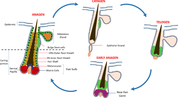
図1:毛包サイクル 。 HFは永久上部と急速な成長(成長期)、破壊(退行期)および相対的静穏期や休符(休止期)の連続的なサイクルを経る下絶えず改造、サイクリング部で構成されている。 大きい方を表示するには、こちらをクリックしてください。この図のバージョン。

HFを維持するのSCは、最初だけSG 14の下方HFの恒久的な地域に住んでスローサイクリングラベル保持細胞(LRC)の人口を明らかにしたトリチウム化チミジンで、チェイス実験を用いて同定しました。 HFSCの進歩特徴付けは、HFのニッチ15からの特定のSCを同定および単離するために使用することができるマーカーの数が少ないことが明らかになりました。おそらくHFSCsの濃縮のための最良のマーカーは、CD34、また、ヒト16中の造血SCマーカーとして同定された細胞表面マーカーです。このCD34 +集団内の2つの異なる集団はまた、α6インテグリンの発現2に基づいて単離されています。他のマーカーは、CD34発現と共局在し、K15プロモーターは、標的化およびトランスジェニック動物15,17-19にHFSCsを単離するために使用され、ケラチン高いバルジ領域で発現される15(K15)です。過去10年間でHFSCsおよび前駆細胞の他のいくつかの異なる集団はまた、HF 17,20-27内に存在することが報告されています。

HFSCsの追加の刺激的な特徴は、皮膚修復への貢献です。通常の条件下ではHFSCsは、HFを補充し、IFEの恒常性に関与しません。ハウ版、創傷に応じて、これらの細胞はIFE 9を再増殖にそのSCのニッチや援助を終了します。我々は最近、マウスがプロアポトーシスSept4 / ARTS遺伝子の表示のために、アポトーシスに対する耐性を実証CD34、K15およびSox9を+ HFSCs、数の増加を削除することを実証しました。 HFSCsはSept4 / ARTSから単離した– / –背部皮は蛍光活性化細胞選別(FACS)を利用し、CD34 +およびK15 + HFSCsの数の2倍を超える増加がありました。これらのSept4 / ARTS – / – HFSCsが、 インビトロで拡大していなかっただけでそれ以上のコロニーを生じたが、対照28と比較しても過酷な条件に耐えることができました。

HFSCs数が増加した結果、Sept4 / ARTS – / –マウスは、皮膚切除損傷に応答して有意に速く治癒します。驚くべきことに、Sept4 / ARTS – / –マウスdisplayeダ創傷床から再生されたHFSの数が多い、とかなり小さい傷跡。さらに、XIAP(アポトーシスのX連鎖阻害剤)、ARTSの生化学的目標のために削除されたマウスは、治癒障害28を実証しました。

我々の結果と他の研究室で行われた作業はHFSCsは大人のSCの生物学と機能を研究するための理想的なモデルとして役立つことが示されています。ここでは、4つのマーカーの発現に基づいてHFSCsと表皮角化細胞の濃縮及び単離するための方法論について説明します。インテグリンα6。インテグリンβ1; SCA-1(表皮角化細胞のマーカー)およびCD34。 K15 + HFSCsの同様の分離はまた、K15-GFPレポーターマウス19を用いて行うことができます。

Protocol

この研究は健康のイスラエルの省の実験動物の管理と使用に関するガイドに記載勧告に厳密に従って行きました。動物のすべてが承認された制度的動物管理プロトコルイスラエル工科大学のIL-02302から2015に従って取り扱われました。 1.実験の準備キレート化されたウシ胎児血清の調製注:カルシウムに対するこれらの細胞の露出を制御することが重要ですの?…

Representative Results

このプロトコルは、詳細に濃縮し、集団の2つのタイプの分離について説明します。バルジのSC及び表皮角化細胞を2プロトコルの主要なステップを示しています。バック8週齢のマウスの背部摘出皮膚を用いて、我々は唯一のHFSCsで表現されているCD34マーカーを使用して、バルジのSCを豊かに。およびSCA-1、表皮ケラチノサイトにラベルを付けま…

Discussion

ここで説明するプロトコルは、よく成体マウスの背部皮膚からHFSCsを単離するための確立されていますが、同じように、マーカー2,16,23,28,29の選択に基づいて、HF構造内の他の集団の単離のために適用することができます。特定の細胞型は、不均一な細胞集団の混合物から選択され、収穫することができるという点で、この方法は、組織解離、細胞単離の他の方法、上特に有利です。さ…

Disclosures

The authors have nothing to disclose.

Acknowledgements

This work was supported in part by NIH grant RO1GM60124 (to H.S.). H.S. is an Investigator with the Howard Hughes Medical Institute. Y.F. is supported by the Deloro Career Advancement Chair and The German Israeli Foundation (I-2381-412.13/2015). D.S. is supported by the Coleman-Cohen post-doctoral fellowship.

Materials

Isoflurane  Primal Critical Care  66794-017-10
Carbon dioxide
Electro Shaver Oster  Golden A5 Shaver from any other company could be used
70% ethanol Gadot Lab 830000051 96% ehtanol diluted with distilled water 
Dissection mat Dissection tools from any provider can be used
Forceps Dumont 11251-10 Foreceps from any other company could be used
Scissors  Dumont 14094-11 Scissors from any other company could be used
Needles/Pins
Scalpel Albion 10 Ensure that the scalpel has a blunt end
Tissue culture dish 60mm x 15mm Sigma-Aldrich CLS430166
PBS In-house PBS without Calcium and Magnesium
0.25% Trypsin/EDTA Biological Industries 03-050-1A Trypsin obtained from a different company might have a different activity and duration of the trypsin digest has to be adjusted accordingly
Pipettes 10ml Sigma-Aldrich Corning, 4488
Ice
50 ml sterie centrifuge tubes Minplast Ein-shemer 35050-43
70µM Cell strainer Fisher 22362548
40µM Cell strainer Fisher 22362549
Staining buffer
Centrifuge Eppendorf 5804 R 5805 000.017
FACS tubes with Cell strainer caps  Falcon  352235
FACS tubes  Falcon  352063
Integrin β1 eBioscience 25-0291 1:400
Integrin α6 eBioscience 15-0495 1:600
Sca I eBioscience 11-5981 1:200
CD34 eBioscience 9011-0349 1:300
DAPI Sigma-Aldrich D9542 50ng/ml
Dry Chelex BioRad 142-2842
Beaker  Pyrex
Distilled H2O 
Stir bar
NHCl BioLab 1903059
Fetal bovine serum (FBS) Beit Haemek Biological Industries 400718 FBS obtained from a different company can be used
1L glass bottle Ilmabor Boro 3.3
Bottle top filter Autofil 1102-RLS

References

  1. Wagers, A. J., Weissman, I. L. Plasticity of adult stem cells. Cell. 116, 639-648 (2004).
  2. Blanpain, C., Lowry, W. E., Geoghegan, A., Polak, L., Fuchs, E. Self-renewal, multipotency, and the existence of two cell populations within an epithelial stem cell niche. Cell. 118, 635-648 (2004).
  3. Fuchs, E. Scratching the surface of skin development. Nature. 445, 834-842 (2007).
  4. Hsu, Y. C., Fuchs, E. A family business: stem cell progeny join the niche to regulate homeostasis. Nat Rev Mol Cell Biol. 13, 103-114 (2012).
  5. Blanpain, C., Fuchs, E. Epidermal homeostasis: a balancing act of stem cells in the skin. Nat Rev Mol Cell Biol. 10, 207-217 (2009).
  6. Alonso, L., Fuchs, E. The hair cycle. J Cell Sci. 119, 391-393 (2006).
  7. Muller-Rover, S., et al. A comprehensive guide for the accurate classification of murine hair follicles in distinct hair cycle stages. J Invest Dermatol. 117, 3-15 (2001).
  8. Zhang, Y. V., Cheong, J., Ciapurin, N., McDermitt, D. J., Tumbar, T. Distinct self-renewal and differentiation phases in the niche of infrequently dividing hair follicle stem cells. Cell Stem Cell. 5, 267-278 (2009).
  9. Ito, M., et al. Stem cells in the hair follicle bulge contribute to wound repair but not to homeostasis of the epidermis. Nat Med. 11, 1351-1354 (2005).
  10. Hsu, Y. C., Pasolli, H. A., Fuchs, E. Dynamics between stem cells, niche, and progeny in the hair follicle. Cell. 144, 92-105 (2011).
  11. Fuchs, E. The tortoise and the hair: slow-cycling cells in the stem cell race. Cell. 137, 811-819 (2009).
  12. Plikus, M. V., Chuong, C. M. Complex hair cycle domain patterns and regenerative hair waves in living rodents. J Invest Dermatol. 128, 1071-1080 (2008).
  13. Ito, M., Kizawa, K., Hamada, K., Cotsarelis, G. Hair follicle stem cells in the lower bulge form the secondary germ, a biochemically distinct but functionally equivalent progenitor cell population, at the termination of catagen. Differentiation. 72, 548-557 (2004).
  14. Cotsarelis, G., Sun, T. T., Lavker, R. M. Label-retaining cells reside in the bulge area of pilosebaceous unit: implications for follicular stem cells, hair cycle, and skin carcinogenesis. Cell. 61, 1329-1337 (1990).
  15. Cotsarelis, G. Epithelial stem cells: a folliculocentric view. J Invest Dermatol. 126, 1459-1468 (2006).
  16. Trempus, C. S., et al. Enrichment for living murine keratinocytes from the hair follicle bulge with the cell surface marker CD34. J Invest Dermatol. 120, 501-511 (2003).
  17. Morris, R. J., et al. Capturing and profiling adult hair follicle stem cells. Nat Biotechnol. 22, 411-417 (2004).
  18. Lyle, S., et al. The C8/144B monoclonal antibody recognizes cytokeratin 15 and defines the location of human hair follicle stem cells. J Cell Sci. 111 (Pt 21), 3179-3188 (1998).
  19. Liu, Y., Lyle, S., Yang, Z., Cotsarelis, G. Keratin 15 promoter targets putative epithelial stem cells in the hair follicle bulge. J Invest Dermatol. 121, 963-968 (2003).
  20. Vidal, V. P., et al. Sox9 is essential for outer root sheath differentiation and the formation of the hair stem cell compartment. Curr Biol. 15, 1340-1351 (2005).
  21. Snippert, H. J., et al. Lgr6 marks stem cells in the hair follicle that generate all cell lineages of the skin. Science. 327, 1385-1389 (2010).
  22. Nijhof, J. G., et al. The cell-surface marker MTS24 identifies a novel population of follicular keratinocytes with characteristics of progenitor cells. Development. 133, 3027-3037 (2006).
  23. Jensen, U. B., et al. A distinct population of clonogenic and multipotent murine follicular keratinocytes residing in the upper isthmus. J Cell Sci. 121, 609-617 (2008).
  24. Jensen, K. B., et al. Lrig1 expression defines a distinct multipotent stem cell population in mammalian epidermis. Cell Stem Cell. 4, 427-439 (2009).
  25. Jaks, V., et al. Lgr5 marks cycling, yet long-lived, hair follicle stem cells. Nat Genet. 40, 1291-1299 (2008).
  26. Horsley, V., et al. Blimp1 defines a progenitor population that governs cellular input to the sebaceous gland. Cell. 126, 597-609 (2006).
  27. Goldstein, J., Horsley, V. Home sweet home: skin stem cell niches. Cell Mol Life Sci. 69, 2573-2582 (2012).
  28. Fuchs, Y., et al. Sept4/ARTS regulates stem cell apoptosis and skin regeneration. Science. 341, 286-289 (2013).
  29. Nowak, J. A., Fuchs, E. Isolation and culture of epithelial stem cells. Methods Mol Biol. 482, 215-232 (2009).
check_url/53931?article_type=t

Play Video

Cite This Article
Soteriou, D., Kostic, L., Sedov, E., Yosefzon, Y., Steller, H., Fuchs, Y. Isolating Hair Follicle Stem Cells and Epidermal Keratinocytes from Dorsal Mouse Skin. J. Vis. Exp. (110), e53931, doi:10.3791/53931 (2016).

View Video