Reliable and accurate outcome assessment is the key for translation of preclinical therapies into clinical treatment. The current paper describes how to assess three clinically relevant primary outcome parameters of cardiac performance and damage in a pig acute myocardial infarction model.
Method Article
Primary Outcome Assessment in a Pig Model of Acute Myocardial Infarction
In This Article
Summary
Abstract
Mortality after acute myocardial infarction remains substantial and is associated with significant morbidity, like heart failure. Novel therapeutics are therefore required to confine cardiac damage, promote survival and reduce the disease burden of heart failure. Large animal experiments are an essential part in the translational process from experimental to clinical therapies. To optimize clinical translation, robust and representative outcome measures are mandatory. The present manuscript aims to address this need by describing the assessment of three clinically relevant outcome modalities in a pig acute myocardial infarction (AMI) model: infarct size in relation to area at risk (IS/AAR) staining, 3-dimensional transesophageal echocardiography (TEE) and admittance-based pressure-volume (PV) loops. Infarct size is the main determinant driving the transition from AMI to heart failure and can be quantified by IS/AAR staining. Echocardiography is a reliable and robust tool in the assessment of global and regional cardiac function in clinical cardiology. Here, a method for three-dimensional transesophageal echocardiography (3D-TEE) in pigs is provided. Extensive insight into cardiac performance can be obtained by admittance-based pressure-volume (PV) loops, including intrinsic parameters of myocardial function that are pre- and afterload independent. Combined with a clinically feasible experimental study protocol, these outcome measures provide researchers with essential information to determine whether novel therapeutic strategies could yield promising targets for future testing in clinical studies.
Introduction
Heart failure with reduced ejection fraction (HFrEF) accounts for about 50% of all heart failure cases, affecting an estimated 1 - 2% of people in the western world1. Its most prevalent cause is acute myocardial infarction (AMI). As acute mortality after AMI has declined significantly due to increased awareness and improved treatment options, emphasis has shifted towards its chronic sequelae; the most prominent being HFrEF2,3. Together with increasing health care costs4, the growing epidemic of heart failure stresses the need for novel diagnostics and therapies, which can be studied in a highly translational porcine model of adverse remodeling after AMI as previously described5.
Both, determinants (e.g., infarct size) and functional assessments (e.g., echocardiography) of adverse remodeling are often used for efficacy testing of new therapeutics, indicating the need for reliable and relatively inexpensive methods. The aim of the current paper is to address this need by introducing important and reliable outcome measures for efficacy testing in a pig model of acute myocardial infarction. These include infarct size (IS) in relation to area at risk (AAR), 3D transesophageal echocardiography (3D-TEE) and detailed admittance-based pressure-volume (PV) loop acquisition.
Infarct size is the main determinant of adverse remodeling and survival after AMI6. Although timely reperfusion of ischemic myocardium may salvage reversibly injured cardiomyocytes and limit infarct size, reperfusion itself causes additional damage through the generation of oxidative stress and a disproportionate inflammatory response (ischemia-reperfusion injury (IRI))7. Hence, IRI has been identified as a promising therapeutic target. The ability of novel therapeutics to decrease infarct size is quantified by assessing infarct size in relation to the area at risk (AAR). AAR quantification is mandatory to correct for inter-individual variability in coronary anatomy of animal models, as a larger AAR leads to a larger absolute infarct size. Since infarct size is directly related to cardiac performance and myocardial contractility, variations in AAR can influence study outcome measures irrespective of treatment modalities8.
Three-dimensional transesophageal echocardiography (3D-TEE) is a safe, reliable and, most importantly, clinically applicable inexpensive method to measure cardiac function non-invasively. Whereas transthoracic echocardiography (TTE) images are limited to 2D parasternal long- and short-axis views in pigs9, 3D-TEE can be used to obtain complete 3-dimensional images of the left ventricle. Therefore, it does not require mathematical approximations of left ventricular (LV) volumes such as the modified Simpson's rule10. The latter falls short of correctly estimating LV volumes after LV remodeling due to the lack of cylindrical geometry11. Moreover, 3D-TEE is preferable over epicardial echocardiography as it does not require surgical interventions, which have been observed to exert cardioprotective effects in the present model12. Although the use of 2D-TEE for the assessment of myocardial function has been described before13,14, limitations regarding ventricular geometry are similar to those observed in 2D-TTE and depend on the extent of LV remodeling. Hence, the larger the infarct (and thus the higher the probability of heart failure), the more likely 2D measurements become flawed by incorrect geometrical assumptions and the higher the need for 3D techniques.
Nonetheless, most imaging modalities are limited in their ability to assess intrinsic functional properties of the myocardium. PV loops provide such relevant additional information and their acquisition is therefore described in detail below.
Protocol
All animal experiments were approved by the Ethical Committee on Animal Experimentation of the University Medical Center Utrecht (Utrecht, the Netherlands) and conform to the 'Guide for the care and use of laboratory animals'.
NOTE: The protocol to perform a closed-chest balloon occlusion is not part of the current manuscript and is described in detail elsewhere5. In short, pigs (60 - 70 kg) are subjected to 75 min transluminal balloon occlusion of the midportion of the left anterior descending artery (LAD).
Both, three-dimensional transesophageal echocardiography (3D-TEE) and pressure-volume (PV) loop measurements can be performed at baseline, short-term and long-term follow-up. Note that these measurements are considered unreliable in the first hours after myocardial infarction due to frequent arrhythmias in this phase. Infarct size (IS) and area-at-risk (AAR) measurements are preferably assessed at short-term follow-up (24 - 72 hr)15,16, since changes in microvasculature and secondary myocardial scar thinning culminate in less reliable results. Infarct size staining is performed using 2,3,5-triphenyltetrazolium chloride (TTC) (CAUTION, irritant), which is considered highly reproducible and relatively inexpensive. TTC is a white powder that colorlessly dissolves in saline. Upon contact with various dehydrogenases, it is converted to a brick red color. Thereby, it discriminates between viable (red) and dead myocardial tissue (white). For an overview on both invasive and non-invasive infarct size determination, readers are directed to a comprehensive review on this subject17.
Figure 1 shows the timeline including anesthesia, surgical preparation and primary outcome measurements used in this study.
1. Medication and Anesthesia
- Ensure that the animal does not eat or drink for at least 5 hours prior to the procedure. Pre-treatment, anesthesia and post-operative pain treatment protocols have been described in detail elsewhere5.
- In short, the day prior to surgery a buprenorfine patch (5 µg/hr) is applied to the skin that is active for seven days to limit post-operative pain. On the day of surgery, sedate pigs by intramuscular injection of 0.4 mg/kg midazolam, 10 mg/kg ketamine and 0.014 mg/kg atropine. Wait for approximately 10 - 15 min. Insert an 18 G cannula in one of the ear veins and administer 5 mg/kg sodium thiopental to induce anesthesia.
- Intubate the pig using an endotracheal tube (size 8.5 for pigs of 60 - 70 kg). If necessary, perform balloon-ventilation (frequency 12/min) and transport the pig to the operating theater.
- Upon arrival in the operation theater, start mechanical positive pressure ventilation with FiO2 0.50, 10 ml/kg tidal volume and a frequency of 12/min using continuous capnography recording.
- Start balanced anesthesia by continuous intravenous infusion of a combination of midazolam (0.5 mg/kg/hr), sufentanil (2.5 μg/kg/hr) and pancuronium (0.1 mg/kg/hr).
- Confirm anesthesia by testing the corneal reflex and monitoring the breathing pattern (e.g., spontaneous breathing in combination with mechanical ventilation indicates incomplete anesthesia). Use vet ointment on the eyes to prevent dryness while the animal is under anesthesia.
2. 3D Transesophageal Echocardiography (TEE)
- To allow for heart rate monitoring and data acquisition, connect the animal to the 5 leads ECG on the echocardiography machine.
- Place the animal in the right lateral position. Make sure the probe is straight and flexible at the tip by unlocking the operating piece.
- Open the pig's mouth and carefully insert the echo probe in the esophagus. If necessary, use a laryngoscope for visualization. Be careful to avoid ending up in the normal anatomic pharyngeal pouch, resembling a Zenker's diverticulum18.
- Insert the probe for 50 - 60 cm (measure from the tip of the snout). Slowly rotate the probe and flex the head to a left anterolateral position to visualize the heart (Figure 2A - B). Make sure all walls are clearly visible.
- Use the "3D full volume" option on the display of the echocardiography machine to display two perpendicular images of the left ventricle as shown in Figure 2C - D. Then maximize the sector width that is being acquired by selecting "FV Opt Volume". Pause ventilation by temporarily switching off mechanical ventilation and press "Acquire" to obtain full volume measurements.
- After echo acquisition, make sure the tip is flexible by unlocking the operating piece. Then slowly remove the probe from the animal.
NOTE: Do not leave the animal unattended until it has regained sufficient consciousness to maintain sternal recumbency. Do not return an animal that has undergone surgery to the company of other animals until fully recovered. - Perform offline analysis with validated software as described previously19.
3. Admittance-based Pressure-volume Loop Acquisition
- Pre-soak the sensing tips of the 7 F tetra-polar admittance catheter in 0.9% saline (room temperature to 37 °C) for a minimum of 20 min to ensure proper hydration and minimal baseline pressure drift during the experiment20.
- Administer medication and anesthesia as described in section 1.
- Perform surgical preparation and obtain vascular access as described previously5.
- In short, shave and clean the neck. Disinfect the surgical area with iodine 2% and cover the non-sterile parts of the pigs with sterile surgical drapes.
- Make a medial incision in the neck to expose the carotid artery and internal jugular vein. Insert an 8 F sheath into the carotid artery and a 9 F sheath into the jugular vein.
- Insert a Swan-Ganz (SG) catheter through the 9 F sheath in the jugular vein and wedge it in a small pulmonary artery by inflating the balloon at the tip of the catheter. After adequate placement in the peripheral part of the lung, deflate the balloon. Connect the SG to an external cardiac output device.
- Attach a 20 ml syringe containing 0.9% sterile saline to the injection port that connects to the lumen with the most proximal debouchment. Measure cardiac output by rapid infusion of 5 ml 0.9% saline (room temperature) and obtain heart rates to calculate stroke volume (SV). Repeat this procedure three times and calculate the average SV.
NOTE: Cardiac output is (automatically) calculated using the Stewart-Hamilton thermodilution equation and is based on temperature changes in the pulmonary artery upon infusion of room temperature saline21. - Remove the SG catheter. Insert an 8 F Fogarty catheter through the 9 F sheath in the jugular vein and position it in the inferior vena cava.
- Calibrate the pressure signal of the PV loop catheter using the "Course" and "Fine" button, while the tip remains in 0.9% saline. Then input the measured SV into the system.
- Advance the PV loop catheter through the 8 F sheath in the carotid artery and center the tip in the left ventricle (LV) under fluoroscopy.
- Select the largest adequately placed-segment by plotting the raw conductance signal against the pressure signal. Ensure that the pressure-conductance loops are of rectangle shape. Phase signal is expected to show a sinus trace with values between 3 and 5 degrees. Pause ventilation and perform a baseline scan to convert Conductance to Volume.
- Accept the baseline data by pressing "Continue" when the signals are stable (no arrhythmias), heart rate is equal to ECG or pressure derived heart rates and end-systolic (ES) / end-diastolic (ED) conductance are adequately sensed by the system20.
NOTE: The latter can be verified by plotting the raw conductance signal against the pressure signal and comparing ES / ED conductance values derived from the baseline scan to real-time conductance. If any of the requirements above is not met, repeat the procedure.
- Accept the baseline data by pressing "Continue" when the signals are stable (no arrhythmias), heart rate is equal to ECG or pressure derived heart rates and end-systolic (ES) / end-diastolic (ED) conductance are adequately sensed by the system20.
- Acquire baseline pressure-volume loops by recording 10 - 12 consecutive beats during apnea by pausing ventilation.
- Inflate the Fogarty catheter under fluoroscopic guidance to reduce preload and record 10 - 12 consecutive beats as described above. Make sure systolic blood pressure remains >60 mmHg and no arrhythmias interfere with the measurements.
- Remove the Fogarty and PV loop catheters. Keep recording arterial pressure before and during removal of the PV loop catheter to enable correcting for pressure drift (i.e., ex vivo pre- and post-procedural baseline pressure difference).
NOTE: Do not leave the animal unattended until it has regained sufficient consciousness to maintain sternal recumbency. Do not return an animal that has undergone surgery to the company of other animals until fully recovered. - Perform offline analysis of geometrical measurements and functional parameters with validated software22.
4. Area At Risk (AAR) and Infarct Size (IS) Quantification
- Dissolve 1.00 g Evans blue (CAUTION23, toxic) in 50 ml 0.9% saline, fill two 50 ml Luer lock syringes with 20 ml and 30 ml of 2% Evans blue solution respectively and keep at room temperature.
NOTE: Work in a fume hood and wear a dust mask to limit exposure to hazardous dusts and use gloves and protective glasses to prevent contact from skin and eyes. - Taking similar precautions, dissolve 1% 2,3,5-triphenyl-tetrazoliumchloride (TTC) (CAUTION, irritant) in 37 °C 0.9% saline and keep at 37 °C.
- Surgically prepare the animal to obtain vascular access to both carotid arteries. Perform a sternotomy to allow for direct visualization of the effect of in vivo Evans blue infusion5.
- Insert a 7 F and an 8 F introducer sheath in the respective carotid artery. Alternatively, insert both introducer sheaths in one single carotid artery or use one of the femoral arteries for one of both guiding catheters.
- Connect two standard Y-connectors to a 7 F JL4 and an 8 F JL4 guiding catheter respectively. For a femoral approach, use a JR4 for the right coronary artery (RCA) and a JL4 for the left main coronary artery (LCMA). Connect an additional three-way tap with 10 cm extension to both Y-connectors.
- Administer 100 IU/kg heparin. Position the 8 F JL4 guiding catheter in the ostium of the LMCA via one of two introducer sheaths.
- Using a 0.014" guidewire, advance a coronary dilatation catheter through the LCMA catheter and position the balloon at the site where coronary occlusion was performed during MI induction. Do not inflate yet.
- Position the second 8 F JL4 guiding catheter in the ostium of the RCA via the second introducer sheath.
- Perform a coronary angiography (CAG) by infusing contrast agent under fluoroscopy to confirm correct positioning of both guiding catheters and the balloon in the coronary arteries, using anteroposterior and LAO 30° views.
- Attach the two 50 ml syringes containing 30 ml (LCMA) and 20 ml (RCA) 2% Evans blue to the respective three-way taps attached to the Y-connectors on the guiding catheters.
- Inflate the balloon and confirm occlusion of the coronary artery by CAG. Only when the balloon completely blocks the passage of any contrast agent, inject Evans blue dye through both guiding catheters (5 ml/s) while the balloon is inflated.
- Directly after the completion of Evans blue infusion, induce ventricular fibrillation by placing a 9 V battery on the non-infarcted part of the heart.
- Incise the caval vein to release pressure and make sure a suction unit is available to allow for drainage of blood.
- Deflate the balloon, retract it together with both guiding catheters and explant the heart by dissecting surrounding membranes. A transverse cut through the large vessels (i.e., aorta, pulmonary artery/veins) allows for complete explantation. Rapidly wash off blood and superfluous dye on the exterior surface and in the cardiac cavities using 0.9% saline.
- Carefully dissect the left ventricle and make cuts in 5 equal 10 mm thick sections from apex to base, in a plane parallel to the atrioventricular (AV) groove.
- Photograph both sides of all five slices separately under ambient light conditions, as a possible Evans blue washout may occur in the subsequent step. For calibration, make sure a ruler is present in the image.
- Incubate for 10 min in 1% TTC solution at 37 °C, turning the sections around after 5 min for equal staining.
- Again, photograph both sides of all five slices separately under ambient light conditions and make sure a ruler is visualized in the image for calibration.
- Weigh all slices. Use software suitable for the analyses5. When using ImageJ (version 1.47), click the "Straight line" button. Now, draw a straight line with a known distance using the ruler in the image (e.g., 5 cm). Click "Analyze" -> "Set scale" and enter the distance in the box "Known distance". This procedure allows for calibration of distance in pixels to SI units of length.
- Using the "Polygon selections"button, select the total area that corresponds to the LV myocardium in the present image, click "Analyze" -> "Measure" to acquire measurements. Perform this procedure for both sides of each slice of myocardium, and average per slice.
- Multiply by the weight of the slice proportional to the total weight of all five slices and average these measurements for all slices.
- Perform similar measurements for area at risk (AAR) and infarct size (IS). Divide IS/AAR, AAR/LV and IS/LV and multiply by 100% to obtain respective outcome measurements5.
Results
3D Transesophageal Echocardiography
3D transesophageal echocardiography (3D-TEE) can be used for the assessment of global cardiac function. After AMI, global cardiac function differs from healthy baseline values. In particular, left ventricular ejection fraction (LVEF) decreases from 59 ± 4% to 37 ± 6% after one week of reperfusion (n = 10) (GPJ van Hout, 2015). An increase in end-systolic volume (51 ± 7 to 82 ± 13 ml) and decrease in stroke volume (74 ± 11 to 47 ± 8 ml) is also observed, whereas end-diastolic volume does not differ between both time points (125 ± 14 to 129 ± 13 ml). Representative images one week after myocardial infarction (ischemia-reperfusion) are displayed in Figure 3. In our ample experience, we have not encountered any complications related to TEE.
Admittance-based Pressure-volume Loops
Pressure-volume (PV) loops can be used both to assess global cardiac function and specific intrinsic myocardial muscle properties. Outcome measurements of the former can be easily calculated from the graphs in Figure 4A and include EDV (lower right corner), ESV (upper left corner) and LVEF ((EDV - ESV)/EDV x 100%). Both, ESV and EDV provide important information on left ventricular geometry and LVEF is an important measure for determining left ventricular pump function. A previous study compared admittance-based PV loops to gold-standard cardiac magnetic resonance imaging (CMRI) in a pig model of AMI24. After eight weeks, PV loop measurements significantly overestimated both ESV and EDV. With regard to LVEF however, no significant difference was observed between PV loops and CMRI. In addition, both techniques showed a fairly good correlation of EDV and LVEF.
For intrinsic cardiac performance, different measurements can be derived from PV loops, such as end-systolic and end-diastolic pressure-volume relationship (ESPVR; EDPVR)25. Representative PV loop images with preload reduction and some examples of systolic and diastolic functional parameters are shown in Figure 4B. The ESPVR slope decreases, indicating decreased contractility. Additional valuable functional parameters that can be derived from PV loops are presented in Table 1.
Infarct Size/Area At Risk Quantification
In female Dalland landrace pigs (6 months; 60 - 70 kg), occlusion of the left anterior descending artery (LAD) directly distal to the first septal and first diagonal branch during 75 minutes leads to an area at risk (AAR) of 22 ± 2% of the left ventricle (LV) (n = 5) (GHJM Ellenbroek, 2015). Infarct size constitutes 16 ± 2% of the left ventricle and 73 ± 7% of the AAR. This fairly large IS/AAR has been chosen for as patients with a larger infarct size are more prone to the development of heart failure than patients with a smaller infarct size. In pigs, the greatest therapeutic benefit can therefore be gained when applying 75 minutes of ischemia. Moreover, due to greater infarct size, cardiac function deteriorates, which allows for functional improvement as well. When a shorter period of index ischemia is applied, cardiac infarct size is lower and function is only mildly impaired, which allows for only a very small window of functional improvement. Figure 5 shows a representative example of a TTC and Evans blue staining that allows clear identification of the 3 areas: 1) remote myocardium, 2) AAR, and 3) infarcted myocardium.

Figure 1. Timeline of the Experimental Protocol. This timeline provides an overview of the most important experimental steps in the used pig AMI model. Adequate induction of anesthesia is required prior to each measurement. Time indications can be observed under each proceeding. Infarct size is preferably assessed after 24 - 72 hours. 3D-TEE and PV loop data acquisition can be performed at baseline and at short- and long-term follow-up. The first hours after AMI, arrhythmias are frequent and can greatly interfere with cardiac hemodynamics and therefore prevent reliable data acquisition. AMI: acute myocardial infarction; 3D-TEE: three-dimensional transesophageal echocardiography; PV loop: pressure-volume loop. Please click here to view a larger version of this figure.
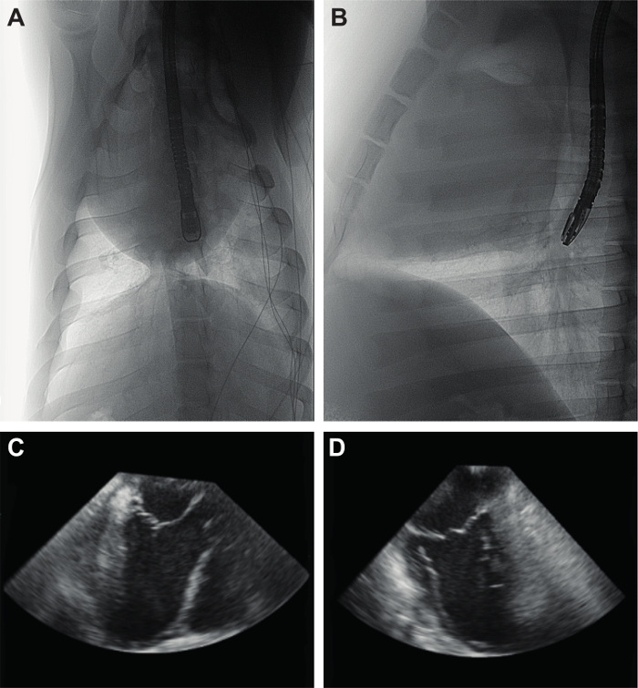
Figure 2. Positioning and Acquisition of 3D TEE Images. Anteroposterior (A) and mediolateral (B) X-ray images of the 3D-TEE probe positioning in the esophagus. Image acquisition follows upon correct visualization of the left atrium, left ventricle and aorta (C) and a perpendicular image of both left atrium and left ventricle (D). 3D-TEE: three-dimensional transesophageal echocardiography. Please click here to view a larger version of this figure.
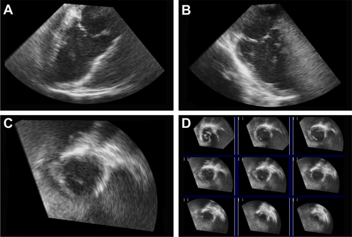
Figure 3. Full Volume 3D-TEE Images of the Left Ventricle. 3D-TEE recordings of the left ventricle one week after acute myocardial infarction (75 minutes) and reperfusion. Perpendicular long-axis images of the left atrium and left ventricle can be observed in the upper half panel (A, B). A magnified example (C) of multiple cross-sectional images (D) of the left ventricle is displayed in the lower half panel. 3D-TEE: three-dimensional transesophageal echocardiography. Please click here to view a larger version of this figure.
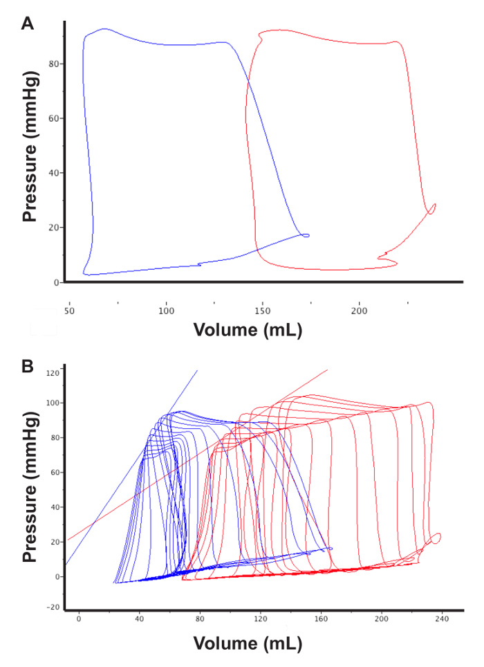
Figure 4. Pressure-volume Loop Images at Baseline and After Myocardial Infarction. Representative PV loop images during apnea (i.e., pausing ventilation) at baseline (blue) and eight week after AMI (red) (A). Increases in EDV and ESV and a decrease in SV can be observed, indicating a decrease in LVEF (%). PV loop images with preload reduction are used to assess intrinsic myocardial function parameters (B). Compared to baseline, infarcted myocardium shows a decrease in contractility as derived from the ESPVR (straight blue and red lines). PV loop: pressure-volume loop; AMI: acute myocardial infarction; EDV: end-diastolic volume; SV: stroke volume; LVEF: left ventricular ejection fraction; ESPVR: end-systolic pressure-volume relationship. Please click here to view a larger version of this figure.
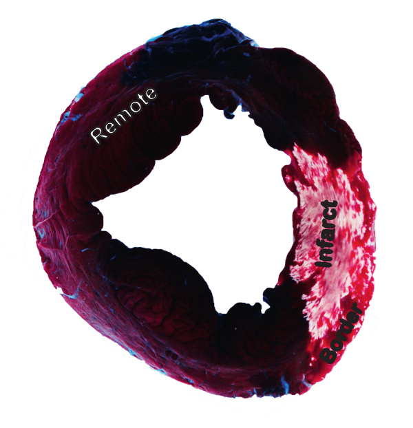
Figure 5. Infarct Size and Area-at-risk Staining. Representative image of infarct size and area-at-risk staining of the left ventricle after acute myocardial infarction (75 minutes) and subsequent reperfusion for three days. (Hemorrhagic) infarct tissue can be observed in rosy brown and gray-white, whereas the border zone is stained red. Surrounding blue-stained areas indicate remote myocardium. Please click here to view a larger version of this figure.
| Volume parameters | Pressure parameters | Loading-independent parameters | Other | |||
| Systolic | Diastolic | Systolic | Diastolic | Systolic | Diastolic | Other |
| ESV | EDV | ESP | EDP | ESPVR | EDPVR | HR |
| dP/dT | dP/dT | Ees | PRSW | SW | ||
| Derivatives | τ (tau) | ESV100 | PRSW | |||
| LVEF, SV, CO | PHT | Ea | dP/dV | |||
Table 1. Valuable Functional Parameters that can be Derived from Pressure-volume Loops. Categorized into volume, pressure and loading-independent parameters, this table describes the most commonly used (systolic and diastolic) parameters derived from PV loops. PV loops: pressure-volume loops; ESV: end-systolic volume; EDV: end-diastolic volume; LVEF: left ventricular ejection fraction; SV: stroke volume; CO: cardiac output; ESP: end-systolic pressure; dP/dT: derivative of pressure; τ (tau): isovolumic relaxation constant; PHT: pressure half-time; ESPVR: end-systolic pressure-volume relationship; Ees: end-systolic elastance; ESV100: end-systolic volume corrected for pressure (100 mmHg); Ea: arterial elastance; EDPVR: end-diastolic pressure-volume relationship; PRSW: preload recruitable stroke work; HR: heart rate; SW: stroke work; dP/dV: EDPVR slope (chamber stiffness).
Discussion
Cardiac remodeling is largely depending on myocardial infarct size and the quality of myocardial infarct repair6,26. To assess the former in a standardized manner, the present manuscript provides an elegant method of in vivo infusion of Evans blue combined with ex vivo TTC staining, which has been validated and extensively used8,16,27,28. This method allows for quantification of the area at risk (AAR) and infarct size in relation to AAR16. The current approach reduces the risk of dye diffusion into the AAR, the infarct region or — with malpositioning — papillary muscle, as it does not require myocardial puncture. Moreover, there is no need for external ligation of the coronary artery, which may be imprecise, inaccurate and occasionally damage the myocardium. An alternative method, combining catheter-based Evans blue infusion into the LV and cross-clamping of the ascending aorta29, is undesirable for different reasons. Clamping occludes the left ventricular guiding catheter, hampering Evans blue infusion into the LV. Besides, compression and traction forces may lead to LCMA catheter and intracoronary balloon malpositioning and inaccurate AAR measurements. Moreover, since balloon occlusion of the LAD requires guiding catheter positioning in the LCMA, coronary filling from the LV is limited, preventing Evans blue entry from the LV into the coronary artery.
However, though superior to myocardial puncture and cross-clamping of the ascending aorta, the technique presented in this manuscript requires some precautions. Complete occlusion of (one of the) coronary arteries through an obstructing guiding catheter needs to be prevented. This can be controlled by monitoring wash-out rates and pressure, and can usually be avoided by slightly retracting the guiding catheter from of the coronary ostium. If inevitable, shorten the time the guiding catheter is positioned in the coronary artery as much as possible by preparation of other parts of the protocol. In addition, make sure the balloon completely occludes the target vessel before Evans blue infusion.
When Evans blue infusion is completed, induce VF and incise the caval vein to release blood pressure before balloon deflation and catheter withdrawal in order to prevent Evans blue diffusion into the AAR. Care should be taken to gently but firmly position the guiding catheter in the coronary ostium, allowing for diffusion of Evans blue in both the LAD and LCx. In addition, Evans blue infusion rates should not be too high since limited flow into the coronary arteries may lead to Evans blue wash-out into the systemic circulation. Although selectively infused into the coronary arteries, Evans blue diffusion into the systemic circulation cannot be completely prevented. Therefore, histologic analysis of other non-cardiac tissue (e.g., spleen, kidney) may still be problematic. Simultaneous TTC co-infusion into the AAR has been described before, but is undesirable in our opinion, as TTC does not reach the part of the AAR obstructed by the balloon. Moreover, previous analyses show that TTC might react with residual intravasal blood in the infarct area and overlap with the red color in the non-infarct AAR30. Future applications of this technique could be to preserve non-cardiac tissues by obstructing blood flow into the systemic circulation. This could be achieved by balloon obstruction of the descending thoracic aorta through a femoral approach.
Echocardiography to date remains a cornerstone for cardiac function assessment in both clinical care and various animal models in cardiovascular research. However, due to the thoracic shape of landrace pigs, transthoracic echocardiography (TTE) is limited to 2-dimensional long- and short-axis views of the LV9. Therefore, cardiac volume and LVEF have to be estimated by mathematical approximations such as the modified Simpson's rule, which assumes a cylindrical left ventricular morphology10. As a result of LV remodeling after MI however, cardiac dimensions change. Therefore, this particular geometric assumption cannot be made, reducing the accuracy and reliability of such measurements31.
This problem can be solved by using 3D echocardiography to obtain 3D images of the complete left ventricle. In pigs, LVEF assessment by epicardial 3D echocardiography shows excellent correlation with gold-standard CMRI24,32. However, this requires surgery prior to AMI induction for baseline measurements. Regardless of the approach, i.e., open chest vs. subxiphoidal approach, invasive surgery for epicardial echocardiography has been shown to be cardioprotective12,33,34. Concomitant adhesions hinder resternotomy, which renders epicardial echocardiography undesirable for baseline measurements in a closed-chest AMI model.To avoid these drawbacks, 3D images of the heart can be obtained through 3D transesophageal echocardiography (3D-TEE). This technique is portable, widely available and allows for serial measurements and visualization of the entire left ventricular volume. Moreover, it is reliable, relatively inexpensive and safe.
Note that it is important to gently insert the TEE probe into the mouth and esophagus, since ending up in the Zenker's diverticulum and applying too much pressure may lead to esophageal rupture. Moreover, since the anatomical relationship between stomach and heart differs from man, 3D-TEE in pigs does not allow for regional measurements (e.g., strain, tissue Doppler imaging) and is restricted to volume measurements. In the data presented in the manuscript, no increase of EDV 7 days after AMI was observed. A longer follow-up period is needed to extensively drive adverse remodeling, leading to an increased EDV at several weeks follow-up11.
In contrast to conventional echocardiography, admittance-based PV loops moderately overestimate LV volumes, both at baseline35 and after 8 weeks of follow-up24. Still, fairly good correlations and high degrees of agreement with CMRI have been found. Although PV loop measurements several weeks after AMI are less precise compared to baseline, LV dimensions and derivatives hereof (LVEF) are useful for the global assessment of cardiac function35.
In addition, PV loops provide specific information about intrinsic myocardial properties, such as ESPVR. Since regional functional measurements in TTE and TEE are limited and epicardial echocardiography is undesirable at baseline, PV loops provide an elegant and safe technique for the assessment of intrinsic myocardial function. Both, the decline in ESPVR slope and the typical shift in V0 can be used to compare different therapeutics. These classical features are validated in ex vivo canine heart suffering from pan-ischemia. Hence, in regional ischemia models, like AMI models, these specific characteristics are not always present, which can be attributed to many factors, of which ventricular remodeling and regional ischemia are the most important ones25,36,37.
For adequate data acquisition, it is critical to make sure no arrhythmias are present when converting conductance to volume and when acquiring PV loops. If arrhythmias are present, reposition the PV loop catheter so it does not irritate the myocardium. The administration of anti-arrhythmic drugs (e.g., 150 - 300 mg amiodarone) may also help. However, note that PV loop acquisition within several hours after acute myocardial infarction is not reliable due to frequent arrhythmias (e.g., premature ventricular complexes, bigemini).
Slightly advancing or retracting the PV loop catheter into the LV or from the muscular wall may also help to improve the shape of the PV loops. After changing PV loop catheter positioning, always double-check that the largest adequately placed segment is selected.
In conclusion, the current paper introduces three methods for cardiac assessment in a previously described pig AMI model with additional value for the evaluation of new therapeutics to decrease the burden of the ongoing heart failure epidemic.
Disclosures
The authors have nothing to disclose.
Acknowledgements
The authors gratefully acknowledge Marlijn Jansen, Joyce Visser, Grace Croft, Martijn van Nieuwburg, Danny Elbersen and Evelyn Velema for their excellent technical support during the animal experiments.
Materials
| Name | Company | Catalog Number | Comments |
|---|---|---|---|
| 3-dimensional Transesophageal Echocardiography | |||
| iE33 ultrasound device | Philips | - | |
| X7-2t transducer | Philips | - | |
| Aquasonic® 100 ultrasound transmission gel | Parker Laboratories Inc. | 01-34 | Alternative product can be used |
| Battery handle type C (laryngoscope handle) | Riester | 12303 | |
| Ri-Standard Miller blade MIL 4 (laryngoscope blade) | Riester | 12225 | |
| Qlab 10.0 (3DQ Advanced) analysis software | Philips | - | |
| Name | Company | Catalog Number | Comments |
| Pressure-volume loop acquisition | |||
| Cardiac defibrillator | Philips | ||
| 0.9% Saline | Braun | ||
| 8 F Percutaneous Sheath Introducer Set | Arrow | CP-08803 | Alternative product can be used |
| 9 F Radifocus® Introducer II Standard Kit | Terumo | RS*A90K10SQ | Alternative product can be used |
| 8 F Fogarty catheter | Edward Life Sciences | 62080814F | Alternative product can be used |
| 7 F Criticath™ SP5107H TD catheter (Swan-Ganz) | Becton Dickinson (BD) | 680078 | Alternative product can be used |
| Ultraview SL Patient Monitor and Invasive Command Module (external cardiac output device) | Spacelabs Healthcare | 91387 | Alternative product can be used |
| ADVantage system™ | Transonic SciSense | - | |
| 7 F Tetra-polar admittance catheter (7.0 VSL Pigtail / no lumen) | Transonic SciSense | - | |
| Multi-channel acquisition system (Iworx 404) | Iworx | - | |
| Labscribe V2.0 analysis software | Iworx | - | Alternative product can be used |
| Name | Company | Catalog Number | Comments |
| Infarct size / area-at-risk quantification | |||
| Diathermy | - | Alternative product can be used | |
| Lebsch knife | - | Alternative product can be used | |
| Hammer | - | Alternative product can be used | |
| Bone marrow wax | Syneture | Alternative product can be used | |
| Klinkenberg scissors | - | Alternative product can be used | |
| Retractor | - | Alternative product can be used | |
| Surgical scissors | - | ||
| 7 F Percutaneous Sheath Introducer Set | Arrow | CP-08703 | Alternative product can be used |
| 8 F Percutaneous Sheath Introducer Set | Arrow | CP-08803 | Alternative product can be used |
| 7 F JL4 guiding catheter | Boston Scientific | H749 34357-662 | Alternative product can be used |
| 8 F JL4 guiding catheter | Boston Scientific | H749 34358-662 | Alternative product can be used |
| COPILOT Bleedback Control Valves | Abbott Vascular | 1003331 | Alternative product can be used |
| BD Connecta™ | Franklin Lakes | 394995 | Alternative product can be used |
| Contrast agent | Telebrix | ||
| Persuader 9 Steerable Guidewire 9 (0.014", 180 cm, straight tip), hydrophilic coating | Medtronic Inc. | 9PSDR180HS | Alternative product can be used |
| SAPPHIRE™ Coronary Dilatation Catheter (PTCA balloon suitable for the size of the particular coronary artery (2.75 - 3.25 mm)) | OrbusNeich | 103-3015 | Alternative product can be used |
| Evans Blue | Sigma-Aldrich | E2129-100G | Toxic. Alternative product can be used |
| 2,3,5-triphenyl-tetrazolium chloride (TTC) | Sigma-Aldrich | T8877-100G | Irritant. Alternative product can be used |
| 9 V Battery | - | - | |
| Ruler | - | - | |
| Photocamera | Sony | - | |
| ImageJ | National Institutes of Health | - | Alternative product can be used |
References
- Mosterd, A., Hoes, A. W. Clinical epidemiology of heart failure. Heart. 93 (9), 1137-1146 (2007).
- Nichols, M., et al. European Cardiovascular Disease Statistics. , Brussels. (2012).
- Krumholz, H. M., et al. Reduction in Acute Myocardial Infarction Mortality in the United States. JAMA. 302 (7), 767-773 (2010).
- Go, A. S., et al. Heart disease and stroke statistics - 2013 update: A Report from the American Heart Association. Circulation. 127 (1), (2013).
- Koudstaal, S., et al. Myocardial infarction and functional outcome assessment in pigs. J. Vis. Exp. (86), e51269(2014).
- Chareonthaitawee, P., Christian, T. F., Hirose, K., Gibbons, R. J., Rumberger, J. A. Relation of initial infarct size to extent of left ventricular remodeling in the year after acute myocardial infarction. J. Am. Coll. Cardiol. 25 (3), 567-573 (1995).
- Yellon, D. M., Hausenloy, D. J. Myocardial reperfusion injury. N. Engl. J. Med. 357 (11), 1221-1235 (2007).
- Suzuki, Y., Lyons, J. K., Yeung, A. C., Ikeno, F. In vivo porcine model of reperfused myocardial infarction: In situ double staining to measure precise infarct area/area at risk. Catheter Cardiovasc. Interv. 71 (1), 100-107 (2008).
- Weidemann, F., et al. Myocardial function defined by strain rate and strain during alterations in inotropic states and heart rate. Am. J. Physiol. Heart Circ. Physiol. 283 (2), H792-H799 (2002).
- Mercier, J. C., et al. Two-dimensional echocardiographic assessment of left ventricular volumes and ejection fraction in children. Circulation. 65 (5), 962-969 (1982).
- De Jong, R., et al. Cardiac Function in a Long-Term Follow-Up Study of Moderate and Severe Porcine Model of Chronic Myocardial Infarction. Biomed. Res. Int. 2015, 1-11 (2015).
- Van Hout, G. P. J., et al. Invasive surgery reduces infarct size and preserves cardiac function in a porcine model of myocardial infarction. J. Cell. Mol. Med. , 2655-2663 (2015).
- Meybohm, P., et al. Assessment of left ventricular systolic function during acute myocardial ischemia: A comparison of transpulmonary thermodilution and transesophageal echocardiography. Minerva Anestesiol. 77 (2), 132-141 (2011).
- Gruenewald, M., et al. Visual evaluation of left ventricular performance predicts volume responsiveness early after resuscitation from cardiac arrest. Resuscitation. 82 (12), 1553-1557 (2011).
- Bolli, R., Becker, L., Gross, G., Mentzer, R., Balshaw, D., Lathrop, D. A. Myocardial protection at a crossroads: The need for translation into clinical therapy. Circ. Res. 95 (2), 125-134 (2004).
- Timmers, L., et al. Exenatide reduces infarct size and improves cardiac function in a porcine model of ischemia and reperfusion injury. J. Am. Coll. Cardiol. 53 (6), 501-510 (2009).
- Csonka, C., et al. Measurement of myocardial infarct size in preclinical studies. J. Pharmacol. Toxicol. Methods. 61 (2), 163-170 (2010).
- Law, R., Katzka, D. A., Baron, T. H. Zenker's Diverticulum. Clin. Gastroenterol. Hepatol. 12 (11), 1773-1782 (2014).
- Philips Healthcare. QLAB 10.0 Quick Card: 3DQ and 3DQ Adv measurements guide. , (2013).
- Transonic. ADV500 Pressure-Volume Measurement System Use and Care Manual, version 5. , (2006).
- Schramm, W. Is the cardiac output obtained from a Swan-Ganz catheter always zero? J. Clin. Monit. Comput. 22 (6), 431-433 (2008).
- iWorx. LabScribe 3: Software Manual for Pressure-Volume Analyses. , (2014).
- Hueper, W. C., Ichniowski, C. T. Toxicopathologic studies on the dye T-1824. Arch. Surg. 48 (1), 17-26 (1944).
- Van Hout, G. P. J., et al. Admittance-based pressure-volume loops versus gold standard cardiac magnetic resonance imaging in a porcine model of myocardial infarction. Physiol. Rep. 2 (4), 1-9 (2014).
- Burkhoff, D., Mirsky, I., Suga, H. Assessment of systolic and diastolic ventricular properties via pressure-volume analysis: a guide for clinical, translational, and basic researchers. Am. J. Physiol. Heart Circ. Physiol. Heart Circ. Physiol. 289 (2), H501-H512 (2005).
- Frangogiannis, N. G. The inflammatory response in myocardial injury, repair, and remodelling. Nat. Rev. Cardiol. 11 (5), 255-265 (2014).
- Fishbein, M., et al. Early phase acute myocardial infarct size quantification: validation of the triphenyl tetrazolium chloride tissue enzyme staining technique. Am. Heart. J. 101 (5), 593-600 (1981).
- Arslan, F., et al. Treatment with OPN-305, a humanized anti-toll-like receptor-2 antibody, reduces myocardial ischemia/reperfusion injury in pigs. Circ. Cardiovasc. Interv. 5 (2), 279-287 (2012).
- Meyns, B., Stolinski, J., Leunens, V., Verbeken, E., Flameng, W. Left ventricular support by Catheter-Mountedaxial flow pump reduces infarct size. J. Am. Coll. Cardiol. 41 (7), 1087-1095 (2003).
- Khalil, P. N., et al. Histochemical assessment of early myocardial infarction using 2,3,5-triphenyltetrazolium chloride in blood-perfused porcine hearts. J. Pharmacol. Toxicol. Methods. 54 (3), 307-312 (2006).
- Gardner, B. I., Bingham, S. E., Allen, M. R., Blatter, D. D., Anderson, J. L. Cardiac magnetic resonance versus transthoracic echocardiography for the assessment of cardiac volumes and regional function after myocardial infarction: an intrasubject comparison using simultaneous intrasubject recordings. Cardiovasc. Ultrasound. 7, 38(2009).
- Santos-Gallego, C., et al. 3D-Echocardiography Demonstrates Excellent Correlation With Cardiac Magnetic Resonance for Assessment of Left Ventricular Function and Volumes in a Model of Myocardial Infarction. J. Am. Coll. Cardiol. 59 (13), E1564(2012).
- Keith Jones,, W,, et al. Peripheral nociception associated with surgical incision elicits remote nonischemic cardioprotection via neurogenic activation of protein kinase C signaling. Circulation. 120, Suppl 1. S1-S9 (2009).
- Gross, G. J., Baker, J. E., Moore, J., Falck, J. R., Nithipatikom, K. Abdominal Surgical Incision Induces Remote Preconditioning of Trauma (RPCT) via Activation of Bradykinin Receptors (BK2R) and the Cytochrome P450 Epoxygenase Pathway in Canine Hearts. Cardiovasc. Drugs Ther. 25 (6), 517-522 (2011).
- Van Hout, G. P. J., de Jong, R., Vrijenhoek, J. E. P., Timmers, L., Duckers, H. J., Hoefer, I. E. Admittance-based pressure-volume loop measurements in a porcine model of chronic myocardial infarction. Exp. Physiol. 98 (11), 1565-1575 (2013).
- Sunagawa, K., Maughan, W. L., Burkhoff, D., Sagawa, K. Left ventricular interaction with arterial load studied in isolated canine ventricle. Am. J. Physiol. 245 (5 Pt 1), H773-H780 (1983).
- Steendijk, P., Baan, J., Der Velde, E. T. V. an, Baan, J. Effects of critical coronary stenosis on global systolic left ventricular function quantified by pressure-volume relations during dobutamine stress in the canine heart. J. Am. Coll. Cardiol. 32 (3), 816-826 (1998).
Reprints and Permissions
