Summary

Kinematisk analyse av celledeling og Expansion: Kvantifisering Cellular Grunnlag for Vekst og prøvetaking Utviklingssoner i<em> Zea mays</em> Blader

Published: December 02, 2016
doi:

Summary

Quantifying cell division and expansion is of crucial importance to the understanding of whole-plant growth. Here, we present a protocol to calculate cellular parameters determining maize leaf growth rates and highlight the use of these data for investigating molecular growth regulatory mechanisms by directing developmental stage-specific sampling strategies.

Abstract

Growth analyses are often used in plant science to investigate contrasting genotypes and the effect of environmental conditions. The cellular aspect of these analyses is of crucial importance, because growth is driven by cell division and cell elongation. Kinematic analysis represents a methodology to quantify these two processes. Moreover, this technique is easy to use in non-specialized laboratories. Here, we present a protocol for performing a kinematic analysis in monocotyledonous maize (Zea mays) leaves. Two aspects are presented: (1) the quantification of cell division and expansion parameters, and (2) the determination of the location of the developmental zones. This could serve as a basis for sampling design and/or could be useful for data interpretation of biochemical and molecular measurements with high spatial resolution in the leaf growth zone. The growth zone of maize leaves is harvested during steady-state growth. Individual leaves are used for meristem length determination using a DAPI stain and cell-length profiles using DIC microscopy. The protocol is suited for emerged monocotyledonous leaves harvested during steady-state growth, with growth zones spanning at least several centimeters. To improve the understanding of plant growth regulation, data on growth and molecular studies must be combined. Therefore, an important advantage of kinematic analysis is the possibility to correlate changes at the molecular level to well-defined stages of cellular development. Furthermore, it allows for a more focused sampling of specified developmental stages, which is useful in case of limited budget or time.

Introduction

Vekst analyse avhenger av et sett med verktøy som ofte brukes av planteforskere til å beskrive genotype bestemt vekstforskjeller og / eller fenotypiske responser på miljøfaktorer. De inkluderer størrelse og vekt målinger av hele anlegget eller et orgel og beregninger av vekstrater for å utforske de underliggende mekanismene for vekst. Organ vekst bestemmes av celledeling og ekspansjon på cellenivå. Derfor, inkludert kvantifisering av disse to prosessene i vekst analyser er nøkkelen til å forstå forskjeller i hel-organ vekst en. Følgelig er det avgjørende å ha en passende metode for å bestemme cellevekstparametre som er forholdsvis lett å bruke av ikke-spesialiserte laboratorier.

Er allerede etablert kinematisk analyse som en tilnærming som gir et kraftig rammeverk for utvikling av organvekstmodeller 2. Teknikken har blitt optimalisert for lineære systemer,slik som Arabidopsis thaliana røtter og enfrøbladede blader, men også for ikke-lineære systemer, slik som tofrøbladede blader 3. I dag er denne metodikken i økende grad blir brukt til å studere hvordan genetiske, hormonelle, utviklingsmessige, og miljøfaktorer påvirker celledeling og ekspansjon i ulike organer (tabell 1). Dessuten gir det også et rammeverk for å knytte cellulære prosesser til sine underliggende biokjemiske, molekylære og fysiologiske bestemmelser (Tabell 2), selv om begrensningene kan pålegges av orgel størrelse og romlige organiseringen for teknikker som krever større mengder plantemateriale (f.eks metabolitten målinger, proteomikk, etc.).

Enfrøbladede blader, som for eksempel mais (Zea mays) blad, representere lineære systemer i hvilke celler beveger seg fra undersiden av bladet mot spissen, sekvensielt passerer gjennom meristem og forlengelse sone for å oppnå den modnesone. Dette gjør det til et ideelt modellsystem for kvantitative studier av romlige mønstre av vekst fire. Videre mais blader har store vekstsonene (meristem og forlengelse sone som strekker seg over flere centimeter 5) og gi muligheter for studier på andre nivåer i organisasjonen. Dette gir mulighet for undersøkelse av (antatte) reguleringsmekanismer som styrer celledeling og vekst, kvantifisert ved kinematisk analyse gjennom en rekke molekylære teknikker, fysiologiske målinger og cellebiologi tilnærminger (tabell 2).

Her gir vi en protokoll for å utføre en kinematisk analyse i monocot blader. Først forklarer vi hvordan du kan gjennomføre en skikkelig analyse av både celledeling og celleforlengelse som en funksjon av posisjon langs bladet aksen og hvordan man regner kinematiske parametre. For det andre viser vi også hvordan denne kan brukes som et grunnlag for prøvetaking utforming. Her diskuterer vi to tilfeller: høyoppløselig sampling end fokusert prøvetaking, slik at bedre data tolkning og lagring av tid / penger, henholdsvis.

Tabell 1. Oversikt over kinematiske analyser fremgangsmåter for kvantifisering av celledeling og vekst i forskjellige organer.

organ henvisning
monocotyledonous blader 16, 20, 21, 22
root tips 2, 23, 24, 25, 26, 27, 28, 29
dicotyledonous blader 21, 30, 31
skyte apikale meristem 32

Tabell 1. Oversikt over kinematiske analyser fremgangsmåter for kvantifisering av celledeling og vekst i forskjellige organer.

<p class="jove_content" fo:keep-together.within-page = "1"> Figur 3

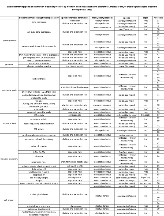
Tabell 2. Kobling mellom celleprosesser kvantifisert ved den kinematiske analysen til deres regulering på molekylnivå. Referanser til ulike studier som forbinder kvantifisering av cellulære prosesser til resultatene fra biokjemiske og molekylære assays i forskjellige arter og organer. Xyloglucan endotransglucosylase (XET), malondialdehyde (MDA), cyclin-avhengige kinaser (CDK). Klikk her for å se en større versjon av denne tabellen.

Protocol

MERK: Følgende protokoll for kinematisk analyse er kun gyldig for blader under steady-state vekst. Dette innebærer en stabil blad forlengelseshastighet og romlige mønster av cellelengden og ekspansjon i et blad i løpet av en periode på flere dager 6. 1. Plantevekst og Målinger av Leaf Forlengelse Rate (LER) Velg et blad i steady-state vekst og utviklingsstadiet av interesse. MERK: Det er en forskjell mellom steady-state vekst og repeterende vekst, …

Representative Results

Her viser vi en sammenligning mellom godt vannes planter (kontroll, 54% jord vanninnhold, (SWC)) og planter utsatt for tørkestress forhold (tørke, 34% SWC) i form av deres blad vekst. Alle plantene ble dyrket i et vekstkammer under kontrollerte forhold (16 timer dag / 8 timer natt, 25 ° C / 18 ° C dag / natt, 300-400 μEm -2 sek -1 fotosyntetisk aktiv stråling (PAR). De tørke ble etablert av forskuddstrekk vann til riktig SWC ble nådd, og deretter videre oppr…

Discussion

En full kinematisk analyse på mais bladene muliggjør bestemmelse av den cellulære basis av bladveksten og gir mulighet for utforming av effektive strategier sampling. Selv om protokollen er relativt enkelt, er en viss forsiktighet anbefales i følgende kritiske trinn: (1) Det er viktig å koble de yngre, lukkede blader (trinn 2.3) uten å skade meristem, siden meristem lengde bestemmelse (trinn 3) krever fullstendig meristem å være til stede. Noen praksis på forhånd kan være nødvendig. (2) Meristematic lengde b…

Disclosures

The authors have nothing to disclose.

Acknowledgements

Dette arbeidet ble støttet av en doktorgrad fellesskap fra Universitetet i Antwerpen til VA; PhD fellesskap fra den flamske Science Foundation (FWO, 11ZI916N) til KS; prosjektstøtte fra FWO (G0D0514N); en felles forskningsvirksomhet (GOA) forskningsstipend, "A Systems Biology tilnærming av Leaf morphogenesis" fra Forskningsrådet ved Universitetet i Antwerpen; og Interuniversity attraksjon polakker (IUAP VII / 29, MARS), "Mais og Arabidopsis Root og skyt Growth" fra den belgiske Federal Science Policy kontor (BELSPO) til GTSB Han Asard, Bulelani L. Sizani og Hamada AbdElgawad alle bidratt til video .

Materials

Pots Any Any We use pots with the following measueres, but can be different depending on the treatment/study : bottom diameter: 11cm, opening diameter: 15 cm, height: 12 cm. We grow one maize plant per pot.
Planting substrate Any Any We use potting medium (Jiffy, The Netherlands), but other substrates can be used, depending on treatment/study.
Ruler Any Any An extension ruler that covers at least 1,5 meters is needed to measure the final leaf length of the plants.
Seeds  Any NA Seeds can be ordered from a breeder.
Scalpel Any Any The scalpel is used during leaf harvesting to detach the leaf of interest from its surrounding leaves and right after harvesting to cut a proper sample for cell length and meristem length measurements. 
15 ml falcon tubes Any Any The 15 ml falcon tubes are used for storing samples used for cell length measurements during sample clearing with absolute ethanol and lactic acid.
Eppendorf tubes Any Any The eppendorf tubes are used for storing samples used for meristem length measurements in ethanol:acetic acid 3:1 (v:v) solution.
Gloves Any Any Latex gloves, which protect against corrosive reagents.
Acetic acid Any Any CAUTION: Corrosive to metals, category 1 Skin corrosion, categories 1A,1B,1C Serious eye damage, category 1; Flammable liquids, categories 1,2,3
Absolute ethanol Any Any CAUTION: Hazardous in case of skin contact (irritant), of eye contact (irritant), of inhalation. Slightly hazardous in case of skin contact (permeator), of ingestion
Lactic acid >98% Any Any CAUTION: Corrosive to metals, category 1 Skin corrosion, categories 1A,1B,1C Serious eye damage, category 1
Sodium chloride (NaCl) Any Any
Ethylenediaminetetraacetic acid (EDTA) Any Any CAUTION: Acute toxicity (oral, dermal, inhalation), category 4 Skin irritation, category 2 Eye irritation, category 2 Skin sensitisation, category 1 Specific Target Organ Toxicity – Single exposure, category 3
Tris(hydroxymethyl)aminomethane hydrochloride (Tris-HCl) Any Any This material can be an irritant, contact with eyes and skin should be avoided. Inhalation of dust may be irritating to the respiratory tract.
4′,6-Diamidine-2′-phenylindole dihydrochloride (DAPI) Any Any Cell permeable fluorescent minor groove-binding probe for DNA. Causes skin irritation. May cause an allergic skin reaction. May cause respiratory irritation.
Ice Any NA The DAPI solution has to be kept on ice.
Fluorescent microscope AxioScope A1, Axiocam ICm1 from Zeiss or other Any fluorescent microscope can be used for determining meristem length.
Microscopic slide Any Any
Cover glass Any Any
Tweezers Any Any Tweezers are needed for unfolding the rolled maize leaf right after harvesting in order to cut a proper sample for cell length and meristem length measurements. 
Image-analysis software Axiovision (Release 4.8) from Zeiss NA The software can be downloaded at: http://www.zeiss.com/microscopy/en_de/downloads/axiovision.html. Other softwares such as ImageJ (https://imagej.nih.gov/ij/) could be used as well.
Microscope equipped with DIC AxioScope A1, Axiocam ICm1 from Zeiss or other Any  microscope, equipped with differential interference contrast (DIC) can be used to measure cell lengths.
R statistical analysis software R Foundation for Statistical Computing NA Open source; Could be downloaded at https://www.r-project.org/
R script NA NA We use the kernel smoothing function locpoly of the Kern Smooth package (Wand MP, Jones MC.  Kernel Smoothing: Chapman & Hall/CRC (1995)). The script is available for Mac and Windows upon inquire with the corresponding author. We have versions for Mac and Windows.

References

  1. Fiorani, F., Beemster, G. T. S. Quantitative analyses of cell division in plants. Plant Mol. Biol. 60, 963-979 (2006).
  2. Silk, W. K., Erickson, R. O. Kinematics of Plant-Growth. J. Theor. Biol. 76, 481-501 (1979).
  3. Rymen, B., Coppens, F., Dhondt, S., Fiorani, F., Beemster, G. T. S., Hennig, L., Köhler, C. Kinematic Analysis of Cell Division and Expansion. Plant Developmental Biology. , (2010).
  4. Avramova, V., Sprangers, K., Beemster, G. T. S. The Maize Leaf: Another Perspective on Growth Regulation. Trends Plant Sci. 20, 787-797 (2015).
  5. Rymen, B., et al. Cold nights impair leaf growth and cell cycle progression in maize through transcriptional changes of cell cycle genes. Plant Physiol. 143, 1429-1438 (2007).
  6. Muller, B., Reymond, M., Tardieu, F. The elongation rate at the base of a maize leaf shows an invariant pattern during both the steady-state elongation and the establishment of the elongation zone. J. Exp. Bot. 52, 1259-1268 (2001).
  7. Beemster, G. T. S., Masle, J., Williamson, R. E., Farquhar, G. D. Effects of soil resistance to root penetration on leaf expansion in wheat (Triticum aestivum L): Kinematic analysis of leaf elongation. J. Exp. Bot. 47, 1663-1678 (1996).
  8. Bernstein, N., Silk, W. K., Lauchli, A. Growth and Development of Sorghum Leaves under Conditions of Nacl Stress – Spatial and Temporal Aspects of Leaf Growth-Inhibition. Planta. 191, 433-439 (1993).
  9. Sylvester, A. W., Smith, L. G., Bennetzen, J. L., Hake, S. C. Cell Biology of Maize Leaf Development. Handbook of maize: It’s Biology. , (2009).
  10. Nelissen, H., et al. A Local Maximum in Gibberellin Levels Regulates Maize Leaf Growth by Spatial Control of Cell Division. Curr. Biol. 22, 1183-1187 (2012).
  11. Avramova, V., et al. Drought Induces Distinct Growth Response, Protection, and Recovery Mechanisms in the Maize Leaf Growth Zone. Plant Physiol. 169, 1382-1396 (2015).
  12. Picaud, J. C., et al. Total malondialdehyde (MDA) concentrations as a marker of lipid peroxidation in all-in-one parenteral nutrition admixtures (APA) used in newborn infants. Pediatr. Res. 53, 406 (2003).
  13. Basu, P., Pal, A., Lynch, J. P., Brown, K. M. A novel image-analysis technique for kinematic study of growth and curvature. Plant Physiol. 145, 305-316 (2007).
  14. Vander Weele, C. M., et al. A new algorithm for computational image analysis of deformable motion at high spatial and temporal resolution applied to root growth. Roughly uniform elongation in the meristem and also, after an abrupt acceleration, in the elongation zone. Plant Physiol. 132, 1138-1148 (2003).
  15. Nelissen, H., Rymen, B., Coppens, F., Dhondt, S., Fiorani, F., Beemster, G. T. S., DeSmet, I. . Plant Organogenesis. , (2013).
  16. Ben-Haj-Salah, H., Tardieu, F. Temperature Affects Expansion Rate of Maize Leaves without Change in Spatial-Distribution of Cell Length – Analysis of the Coordination between Cell-Division and Cell Expansion. Plant Physiol. 109, 861-870 (1995).
  17. Fiorani, F., Beemster, G. T. S., Bultynck, L., Lambers, H. Can meristematic activity determine variation in leaf size and elongation rate among four Poa species? A kinematic study. Plant Physiol. 124, 845-855 (2000).
  18. Pettko-Szandtner, A., et al. Core cell cycle regulatory genes in rice and their expression profiles across the growth zone of the leaf. J. Plant Res. 128, 953-974 (2015).
  19. Poorter, H., Remkes, C. Leaf-Area Ratio and Net Assimilation Rate of 24 Wild-Species Differing in Relative Growth-Rate. Oecologia. 83, 553-559 (1990).
  20. Macadam, J. W., Volenec, J. J., Nelson, C. J. Effects of Nitrogen on Mesophyll Cell-Division and Epidermal-Cell Elongation in Tall Fescue Leaf Blades. Plant Physiol. 89, 549-556 (1989).
  21. Tardieu, F., Granier, C. Quantitative analysis of cell division in leaves: methods, developmental patterns and effects of environmental conditions. Plant Mol. Biol. 43, 555-567 (2000).
  22. Bernstein, N., Silk, W. K., Lauchli, A. Growth and Development of Sorghum Leaves under Conditions of Nacl Stress – Possible Role of Some Mineral Elements in Growth-Inhibition. Planta. 196, 699-705 (1995).
  23. Erickson, R. O., Sax, K. B. Rates of Cell-Division and Cell Elongation in the Growth of the Primary Root of Zea-Mays. P. Am. Philos. Soc. 100, 499-514 (1956).
  24. Beemster, G. T. S., Baskin, T. I. Analysis of cell division and elongation underlying the developmental acceleration of root growth in Arabidopsis thaliana. Plant Physiol. 116, 1515-1526 (1998).
  25. Goodwin, R. H., Stepka, W. Growth and differentiation in the root tip of Phleum pratense. Am. J. Bot. 32, 36-46 (1945).
  26. Hejnowicz, Z. Growth and Cell Division in the Apical Meristem of Wheat Roots. Physiologia Plantarum. 12, 124-138 (1959).
  27. Gandar, P. W. Growth in Root Apices .1. The Kinematic Description of Growth. Bot. Gaz. 144, 1-10 (1983).
  28. Baskin, T. I., Cork, A., Williamson, R. E., Gorst, J. R. Stunted-Plant-1, a Gene Required for Expansion in Rapidly Elongating but Not in Dividing Cells and Mediating Root-Growth Responses to Applied Cytokinin. Plant Physiol. 107, 233-243 (1995).
  29. Sacks, M. M., Silk, W. K., Burman, P. Effect of water stress on cortical cell division rates within the apical meristem of primary roots of maize. Plant Physiol. 114, 519-527 (1997).
  30. Granier, C., Tardieu, F. Spatial and temporal analyses of expansion and cell cycle in sunflower leaves – A common pattern of development for all zones of a leaf and different leaves of a plant. Plant Physiol. 116, 991-1001 (1998).
  31. De Veylder, L., et al. Functional analysis of cyclin-dependent kinase inhibitors of Arabidopsis. Plant Cell. 13, 1653-1667 (2001).
  32. Kwiatkowska, D. Surface growth at the reproductive shoot apex of Arabidopsis thaliana pin-formed 1 and wild type. J. Exp. Bot. 55, 1021-1032 (2004).
  33. Kutschmar, A., et al. PSK-alpha promotes root growth in Arabidopsis. New Phytol. 181, 820-831 (2009).
  34. Vanneste, S., et al. Plant CYCA2s are G2/M regulators that are transcriptionally repressed during differentiation. Embo J. 30, 3430-3441 (2011).
  35. Eloy, N. B., et al. Functional Analysis of the anaphase-Promoting Complex Subunit 10. Plant J. 68, 553-563 (2011).
  36. Eloy, N. B., et al. SAMBA, a plant-specific anaphase-promoting complex/cyclosome regulator is involved in early development and A-type cyclin stabilization. P. Natl. Acad. Sci. USA. 109, 13853-13858 (2012).
  37. Dhondt, S., et al. SHORT-ROOT and SCARECROW Regulate Leaf Growth in Arabidopsis by Stimulating S-Phase Progression of the Cell Cycle. Plant Physiol. 154, 1183-1195 (2010).
  38. Baute, J., et al. Correlation analysis of the transcriptome of growing leaves with mature leaf parameters in a maize RIL population. Genome Biol. 16, (2015).
  39. Andriankaja, M., et al. Exit from Proliferation during Leaf Development in Arabidopsis thaliana: A Not-So-Gradual Process. Dev. Cell. 22, 64-78 (2012).
  40. Beemster, G. T. S., et al. Genome-wide analysis of gene expression profiles associated with cell cycle transitions in growing organs of Arabidopsis. Plant Physiol. 138, 734-743 (2005).
  41. Spollen, W. G., et al. Spatial distribution of transcript changes in the maize primary root elongation zone at low water potential. Bmc Plant Biol. 8, (2008).
  42. Candaele, J., et al. Differential Methylation during Maize Leaf Growth Targets Developmentally Regulated Genes. Plant Physiol. 164, 1350-1364 (2014).
  43. West, G., Inze, D., Beemster, G. T. S. Cell cycle modulation in the response of the primary root of Arabidopsis to salt stress. Plant Physiol. 135, 1050-1058 (2004).
  44. Zhang, Z., Voothuluru, P., Yamaguchi, M., Sharp, R. E., Peck, S. C. Developmental distribution of the plasma membrane-enriched proteome in the maize primary root growth zone. Front. Plant Sci. 4, (2013).
  45. Bonhomme, L., Valot, B., Tardieu, F., Zivy, M. Phosphoproteome Dynamics Upon Changes in Plant Water Status Reveal Early Events Associated With Rapid Growth Adjustment in Maize Leaves. Mol. Cell Proteomics. 11, 957-972 (2012).
  46. Schnyder, H., Nelson, C. J. Growth-Rates and Assimilate Partitioning in the Elongation Zone of Tall Fescue Leaf Blades at High and Low Irradiance. Plant Physiol. 90, 1201-1206 (1989).
  47. Schnyder, H., Nelson, C. J., Spollen, W. G. Diurnal Growth of Tall Fescue Leaf Blades .2. Dry-Matter Partitioning and Carbohydrate-Metabolism in the Elongation Zone and Adjacent Expanded Tissue. Plant Physiol. 86, 1077-1083 (1988).
  48. Schnyder, H., Nelson, C. J. Growth-Rates and Carbohydrate Fluxes within the Elongation Zone of Tall Fescue Leaf Blades. Plant Physiol. 85, 548-553 (1987).
  49. Vassey, T. L., Shnyder, H. S., Spollen, W. G., Nelson, C. J. Cellular Characterisation and Fructan Profiles in Expanding Tall Fescue. Curr. T. Pl. B. 4, 227-229 (1985).
  50. Allard, G., Nelson, C. J. Photosynthate Partitioning in Basal Zones of Tall Fescue Leaf Blades. Plant Physiol. 95, 663-668 (1991).
  51. Spollen, W. G., Nelson, C. J. Response of Fructan to Water-Deficit in Growing Leaves of Tall Fescue. Plant Physiol. 106, 329-336 (1994).
  52. Volenec, J. J., Nelson, C. J. Carbohydrate-Metabolism in Leaf Meristems of Tall Fescue .1. Relationship to Genetically Altered Leaf Elongation Rates. Plant Physiol. 74, 590-594 (1984).
  53. Volenec, J. J., Nelson, C. J. Carbohydrate-Metabolism in Leaf Meristems of Tall Fescue .2. Relationship to Leaf Elongation Rates Modified by Nitrogen-Fertilization. Plant Physiol. 74, 595-600 (1984).
  54. Silk, W. K., Walker, R. C., Labavitch, J. Uronide Deposition Rates in the Primary Root of Zea-Mays. Plant Physiol. 74, 721-726 (1984).
  55. Granier, C., Inze, D., Tardieu, F. Spatial distribution of cell division rate can be deduced from that of p34(cdc2) kinase activity in maize leaves grown at contrasting temperatures and soil water conditions. Plant Physiol. 124, 1393-1402 (2000).
  56. Voothuluru, P., Sharp, R. E. Apoplastic hydrogen peroxide in the growth zone of the maize primary root under water stress.1. Increased levels are specific to the apical region of growth maintenance. J. Exp. Bot. 64, 1223-1233 (2012).
  57. Wu, Y. J., Jeong, B. R., Fry, S. C., Boyer, J. S. Change in XET activities, cell wall extensibility and hypocotyl elongation of soybean seedlings at low water potential. Planta. 220, 593-601 (2005).
  58. Macadam, J. W., Nelson, C. J., Sharp, R. E. Peroxidase-Activity in the Leaf Elongation Zone of Tall Fescue .1. Spatial-Distribution of Ionically Bound Peroxidase-Activity in Genotypes Differing in Length of the Elongation Zone. Plant Physiol. 99, 872-878 (1992).
  59. Macadam, J. W., Sharp, R. E., Nelson, C. J. Peroxidase-Activity in the Leaf Elongation Zone of Tall Fescue .2. Spatial-Distribution of Apoplastic Peroxidase-Activity in Genotypes Differing in Length of the Elongation Zone. Plant Physiol. 99, 879-885 (1992).
  60. Beemster, G. T. S., De Vusser, K., De Tavernier, E., De Bock, K., Inze, D. Variation in growth rate between Arabidopsis ecotypes is correlated with cell division and A-type cyclin-dependent kinase activity. Plant Physiol. 129, 854-864 (2002).
  61. Kavanova, M., Lattanzi, F. A., Schnyder, H. Nitrogen deficiency inhibits leaf blade growth in Lolium perenne by increasing cell cycle duration and decreasing mitotic and post-mitotic growth rates. Plant Cell Environ. 31, 727-737 (2008).
  62. Macadam, J. W., Nelson, C. J. Secondary cell wall deposition causes radial growth of fibre cells in the maturation zone of elongating tall fescue leaf blades. Ann. Bot-London. 89, 89-96 (2002).
  63. Schnyder, H., Nelson, C. J. Diurnal Growth of Tall Fescue Leaf Blades .1. Spatial-Distribution of Growth, Deposition of Water, and Assimilate Import in the Elongation Zone. Plant Physiol. 86, 1070-1076 (1988).
  64. Gastal, F., Nelson, C. J. Nitrogen Use within the Growing Leaf Blade of Tall Fescue. Plant Physiol. 105, 191-197 (1994).
  65. Vanvolkenburgh, E., Boyer, J. S. Inhibitory Effects of Water Deficit on Maize Leaf Elongation. Plant Physiol. 77, 190-194 (1985).
  66. Silk, W. K., Hsiao, T. C., Diedenhofen, U., Matson, C. Spatial Distributions of Potassium, Solutes, and Their Deposition Rates in the Growth Zone of the Primary Corn Root. Plant Physiol. 82, 853-858 (1986).
  67. Meiri, A., Silk, W. K., Lauchli, A. Growth and Deposition of Inorganic Nutrient Elements in Developing Leaves of Zea-Mays L. Plant Physiol. 99, 972-978 (1992).
  68. Neves-Piestun, B. G., Bernstein, N. Salinity-induced inhibition of leaf elongation in maize is not mediated by changes in cell wall acidification capacity. Plant Physiol. 125, 1419-1428 (2001).
  69. Bouchabke, O., Tardieu, F., Simonneau, T. Leaf growth and turgor in growing cells of maize (Zea mays L.) respond to evaporative demand under moderate irrigation but not in water-saturated soil. Plant Cell Environ. 29, 1138-1148 (2006).
  70. Westgate, M. E., Boyer, J. S. Transpiration-Induced and Growth-Induced Water Potentials in Maize. Plant Physiol. 74, 882-889 (1984).
  71. Horiguchi, G., Gonzalez, N., Beemster, G. T. S., Inze, D., Tsukaya, H. Impact of segmental chromosomal duplications on leaf size in the grandifolia-D mutants of Arabidopsis thaliana. Plant J. 60, 122-133 (2009).
  72. Fleury, D., et al. The Arabidopsis thaliana homolog of yeast BRE1 has a function in cell cycle regulation during early leaf and root growth. Plant Cell. 19, 417-432 (2007).
  73. Vlieghe, K., et al. The DP-E2F-like gene DEL1 controls the endocycle in Arabidopsis thaliana. Curr. Biol. 15, 59-63 (2005).
  74. Boudolf, V., et al. The plant-specific cyclin-dependent kinase CDKB1;1 and transcription factor E2Fa-DPa control the balance of mitotically dividing and endoreduplicating cells in Arabidopsis. Plant Cell. 16, 2683-2692 (2004).
  75. Baskin, T. I., Beemster, G. T. S., Judy-March, J. E., Marga, F. Disorganization of cortical microtubules stimulates tangential expansion and reduces the uniformity of cellulose microfibril alignment among cells in the root of Arabidopsis. Plant Physiol. 135, 2279-2290 (2004).
check_url/54887?article_type=t

Play Video

Cite This Article
Sprangers, K., Avramova, V., Beemster, G. T. S. Kinematic Analysis of Cell Division and Expansion: Quantifying the Cellular Basis of Growth and Sampling Developmental Zones in Zea mays Leaves. J. Vis. Exp. (118), e54887, doi:10.3791/54887 (2016).

View Video