A liquid crystal nanoparticle (LCNP) nanocarrier is exploited as a vehicle for the controlled delivery of a hydrophobic cargo to the plasma membrane of living cells.
The controlled delivery of drug/imaging agents to cells is critical for the development of therapeutics and for the study of cellular signaling processes. Recently, nanoparticles (NPs) have shown significant promise in the development of such delivery systems. Here, a liquid crystal NP (LCNP)-based delivery system has been employed for the controlled delivery of a water-insoluble dye, 3,3′-dioctadecyloxacarbocyanine perchlorate (DiO), from within the NP core to the hydrophobic region of a plasma membrane bilayer. During the synthesis of the NPs, the dye was efficiently incorporated into the hydrophobic LCNP core, as confirmed by multiple spectroscopic analyses. Conjugation of a PEGylated cholesterol derivative to the NP surface (DiO-LCNP-PEG-Chol) enabled the binding of the dye-loaded NPs to the plasma membrane in HEK 293T/17 cells. Time-resolved laser scanning confocal microscopy and Förster resonance energy transfer (FRET) imaging confirmed the passive efflux of DiO from the LCNP core and its insertion into the plasma membrane bilayer. Finally, the delivery of DiO as a LCNP-PEG-Chol attenuated the cytotoxicity of DiO; the NP form of DiO exhibited ~30-40% less toxicity compared to DiOfree delivered from bulk solution. This approach demonstrates the utility of the LCNP platform as an efficient modality for the membrane-specific delivery and modulation of hydrophobic molecular cargos.
Since the advent of interfacing nanomaterials (materials ≤100 nm in at least one dimension) with living cells, a continuing goal has been to take advantage of the unique size-dependent properties of nanoparticles (NPs) for various applications. These applications include cell and tissue labeling/imaging (both in vitro and in vivo), real-time sensing, and the controlled delivery of drugs and other cargos1. Examples of such relevant NP properties include the size-dependent emission of semiconductor nanocrystals (quantum dots, QDs); the photothermal properties of gold nanoparticles; the large loading capacity of the aqueous core of liposomes; and the ballistic conductivity of carbon allotropes, such as single-wall carbon nanotubes and graphene.
More recently, significant interest has arisen in the use of NPs for the controlled modulation of drugs and other cargos, such as contrast/imaging agents. Here, the rationale is to significantly enhance/optimize the overall solubility, delivered dose, circulation time, and eventual clearance of the drug cargo by delivering it as an NP formulation. This has come to be known as NP-mediated drug delivery (NMDD), and there are currently seven FDA-approved NP drug formulations for use in the clinic to treat various cancers and hundreds more in various stages of clinical trials. In essence, the goal is to "achieve more with less;" that is, to use the NP as a scaffold to deliver more drug with fewer dosing administrations by taking advantage of the large surface area:volume (e.g., hard particles, such as QDs and metal oxides) of NPs or their large interior volume for loading large cargo payloads (e.g., liposomes or micelles). The purpose here is to reduce the necessity for multiple systemically delivered dosing regimens while at the same time promoting aqueous stability and enhanced circulation, particularly for challenging hydrophobic drug cargos that, while highly effective, are sparingly soluble in aqueous media.
Thus, the goal of the work described herein was to determine the viability of using a novel NP scaffold for the specific and controlled delivery of hydrophobic cargos to the lipophilic plasma membrane bilayer. The motivation for the work was the inherent limited solubility and difficulty in the delivery of hydrophobic molecules to cells from aqueous media. Typically, the delivery of such hydrophobic molecules requires the use of organic solvents (e.g., DMSO) or amphiphilic surfactants (e.g., Poloxamers), which can be toxic and compromise cell and tissue viability2, or micelle carriers, which can have limited internal loading capacities. The NP carrier chosen here was a novel liquid crystal NP (LCNP) formulation developed previously3 and that had been shown previously to achieve a ~40-fold improvement in the efficacy of the anticancer drug doxorubicin in cultured cells4.
In the work described herein, the representative cargo selected was the potentiometric membrane dye, 3,3'-dioctadecyloxacarbocyanine perchlorate (DiO). DiO is a water-insoluble dye that has been used for anterograde and retrograde tracing in living and fixed neurons, membrane potential measurements, and for general membrane labeling5,6,7,8,9. Due to its hydrophobic nature, DiO is typically added directly to cell monolayers or tissues in a crystalline form10, or it is incubated at very high concentrations (~1-20 µM) after dilution from a concentration stock solution11,12.
Here, the approach was use to the LCNP platform, a multifunctional NP whose inner core is completely hydrophobic and whose surface is simultaneously hydrophilic and amenable to bioconjugation, as a delivery vehicle for DiO. DiO is incorporated into the LCNP core during synthesis, and the NP surface is then functionalized with a PEGylated cholesterol moiety to promote the membrane binding of the DiO-LCNP ensemble to the plasma membrane. This approach resulted in a delivery system that partitioned the DiO into the plasma membrane with greater fidelity and membrane residence time than the free form of DiO delivered from bulk solution (DiOfree). Further, this method showed that the LCNP-mediated delivery of DiO substantially modulates and drives the rate of specific partitioning of the dye into the lipophilic plasma membrane bilayer. This is achieved while concomitantly reducing the cytotoxicity of the free drug by ~40% by delivering it as an LCNP formulation.
It is anticipated that the methodology described herein will be a powerful enabling technique for researchers whose work involves or requires the cellular delivery of highly hydrophobic cargos that are sparingly soluble or completely insoluble in aqueous solution.
1. Preparation of DiO-LCNP and DiO-LCNP-PEG-Chol
2. Characterization of DiO-LCNP and DiO-LCNP-PEG-Chol
3. Preparation of Cell Culture Dishes for Delivery Experiments and Imaging
NOTE: DiO-LCNP labeling is performed on HEK 293T/17 human embryonic kidney cells (between passages 5 and 15) that are cultured as described previously4. Perform the delivery experiments and the subsequent cell imaging as described below.
4. Cellular Delivery of DiO and DiO-LCNPs and Imaging of Fixed Cells
5. Cellular Delivery of DiO and DiO-LCNPs and FRET Imaging in Live Cells
NOTE: In this method, cells are colabeled with 6 µM each DiO-LCNP-PEG-Chol (where DiO is a FRET donor) and 1,1'-dioctadecyl-3,3,3',3'-tetramethylindocarbocyanine perchlorate (DiIfree, where DiI is a FRET acceptor). The release of DiO from DiO-LCNP-PEG-Chol and its incorporation into the plasma membrane is confirmed by an observed increase in energy transfer from the DiO donor to the DiI acceptor.
6. Cytotoxicity Assay of DiO and DiO-LCNPs to the HEK 293T/17 Cells
NOTE: The cytotoxicity of the DiO-LCNP materials is assessed using a tetrazolium dye-based proliferation assay17. Cells are cultured in a multiwell plate in the presence of varying concentrations of the materials under conditions that emulate delivery/labeling. The cells are then cultured for 72 hr to allow for proliferation. A dye (MTS, (3-(4,5-dimethylthiazol-2-yl)-5-(3-carboxymethoxyphenyl)-2-(4-sulfophenyl)-2H-tetrazolium) is then added to the wells, and metabolically active cells convert the dye into a blue formazan product. The amount of color formation is directly proportional to the number of viable cells.
7. Data Analysis
LCNPs were prepared in which the hydrophobic core of the NP was loaded with a representative membrane-labeling probe to demonstrate the utility of the LCNP as an efficient delivery vehicle for hydrophobic cargos. For this purpose, the cargo chosen was the highly water-insoluble potentiometric membrane-labeling dye, DiO. DiO-loaded LCNPs (DiO-LCNPs) were synthesized using a two-phase mini-emulsion technique with the chemical components DACTP11, AC10COONa, and DiO, as shown in Figure 118. In this NP system, the covalently linked polymeric network of the crystalline cross-linking agent DACTP11 provides a stable hydrophobic core where the DiO resides within the interstitial spaces in the crosslinked network. The carboxylate groups on the NP surface provide colloidal stability to the particle in aqueous media while also serving as a functional group "handle" for the attachment of cell-targeting ligands (and other biologicals). To tether the DiO-LCNPs to the plasma membrane, an amine-terminated PEGylated cholesterol moiety (PEG-Chol) was covalently attached to the LCNP surface via EDC coupling (Figure 1). After NP synthesis and the confirmation of successful bioconjugation, the ability of the DiO-LCNPs to label the plasma membrane of living cells and to deliver the embedded DiO cargo to the lipophilic portion of the plasma membrane bilayer with improved specificity and kinetics compared to the free form (DiOfree) delivered from bulk solution was assessed.
As shown in Figure 2A, DiOfree (6.0 µM) robustly stained the plasma membrane with high efficiency (nearly 100% of cells labeled) after 15 min of incubation with HEK 293T/17 cells. However, significant cellular internalization of DiOfree was observed upon only a modest increase in the incubation time to 30 min (Figure 2A and C). The extent of DiOfree internalization was determined by time-resolved colocalization experiments in which cells were first incubated with 6.0 µM DiOfree (30 min). The DiOfree-containing incubation medium was then removed, and the cells were subsequently cultured for up to 4 hr. The plasma membranes of the cells were counterstained with a dye-labeled membrane phospholipid (phosphoethanolamine conjugated to Rhodamine B; PE-Rhoda). As shown in Figure 2C, after 15 min of incubation, almost 100% of DiOfree was colocalized with the PE-Rhoda marker (Pearson's colocalization coefficient; PCC = 0.99 ± 0.01). However, after 30 min of incubation ~80% of DiOfree remained bound to the membrane (~20% internalized). The degree of DiOfree internalization increased steadily as the incubation time was extended to as long as 4 hr, and this was reflected in the concomitant decrease in the PCC between the DiO and Rhoda-PE (Figure 2C). A fit of the data to a one-phase exponential decay equation revealed that the DiOfree internalization reached a maximum of ~50% at 1 hr, with an internalization rate (k = 0.045 min-1) and half-life (15 min) that reflected the rapid and efficient cellular uptake of DiOfree. These data demonstrate the rapid time-dependent transition of DiOfree from the plasma membrane to the cytosol, where it remained largely excluded from the nucleus over the 4 hr time period examined.
Given the uncontrolled cellular uptake of DiOfree, the goal became to modulate this behavior through the delivery of DiO as an LCNP-PEG-Chol NP formulation. When delivered as an LCNP-PEG-Chol bioconjugate, a more persistent membrane residence time of the DiO compared to DiOfree was noted (Figure 2B). After 15 min of incubation of the DiO-LCNP-PEG-Chol with the cells, nearly 100% of the DiO signal was located at the plasma membrane, where it was colocalized with the PE-Rhoda membrane marker, a result that was comparable to that observed for DiOfree. It was noted, however, that while the DiOfree labeling of the membrane was quite uniform and contiguous, the staining pattern of the DiO-LCNP-PEG-Chol after 15 min of incubation was more punctate and not nearly as uniform in nature (Figure 2B). This result indicated that the DiO-LCNP-PEG-Chol NPs were collecting in discrete regions within the plasma membrane. When the incubation time was increased to 30 min, ~94% of the DiO signal remained at the membrane (compared to ~80% for DiOfree at this same time point) (Figure 2B and 2C). This trend became even more pronounced as the cells were cultured with the DiO-LCNP-PEG-Chol NPs for increasingly longer times after the initial 30 min incubation. For example, after culture for 1 hr after the initial incubation, nearly 80% of the DiO-LCNP-PEG-Chol NP signal remained membranous and colocalized with the PE-Rhoda marker (compared to ~55% for DiOfree). Notably, the cellular internalization of the DiO-LCNP-PEG-Chol NPs reached a maximum of 30% at 2 hr (70% membrane retention). This corresponds to a cellular uptake rate (k = 0.024 min-1; half-life = 29 min) that is exactly one half of that observed for the internalization of DiOfree.
Next, the ability of the NP-embedded DiO cargo to efficiently efflux out of the LCNP core and enter the lipophilic environment of the plasma membrane bilayer in a controlled and time-dependent manner was determined. To assess this, a FRET-based strategy was devised wherein the DiO serves as a FRET donor engaged in energy transfer to the acceptor dye, DiI. As expected, upon initial labeling (t = 0 min), the emission signal was dominated by the DiO donor contained within the membrane-tethered NPs (Figure 3B). FRET imaging of this same field 4 hr later (t = 240 min), however, revealed a significant increase in the emission signal of the DiI acceptor, providing strong evidence of the transition of the DiO donor from the LCNP core into the plasma membrane bilayer (Figure 3C). Examination of the time-resolved nature of this transition showed a steady decrease in DiO donor emission coupled with a corresponding increase in DiI acceptor emission over the 4 hr experimental window (Figure 3D). Notably, the FRET efficiency during this transition reached its maximum at ~180 min post-initial labeling, suggesting that the efflux and membrane partitioning of the DiO donor had reached its maximum at this time point (Figure 3E). These data provided strong evidence of the time-resolved partitioning of the DiO from the NP to the plasma membrane bilayer that reached its maximum ~3 hr post-initial labeling with the DiO-LCNP-PEG-Chol NPs.
Finally, comparative cytotoxicity analysis was performed for DiOfree versus DiO-LCNP-PEG-Chol. When incubated with HEK 293T/17 cells (at a DiO concentration of 6 µM in both cases), it was clear that the encapsulation of the DiO within the LCNP core attenuated its cytotoxicity. While DiOfree elicited cellular viabilities of ~50%, cells incubated with DiO-LCNP-PEG-Chol exhibited cellular viabilities ~90%. (Figure 4). Cumulatively, the cell labeling data coupled with the cellular toxicity data demonstrate enhancements in both the efficiency of DiO-based membrane labeling and the modulation of the cytotoxicity of DiO.
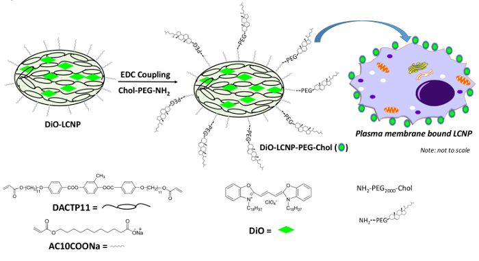
Figure 1: Schematic representation of membrane-binding DiO-LCNP-PEG-Chol. DiO-LCNP is composed of an acrylate liquid crystal cross-linking agent (DACTP11); a carboxyl-terminated polymerizable surfactant (AC10COONa); and a lipophilic dye, DiO. Cholesterol-terminated poly(ethylene glycol) (PEG-Chol) was conjugated to DiO-LCNP via EDC coupling. Addition of Chol to the DiO-LCNP surface mediates preferential binding of the NP to the plasma membrane. Please click here to view a larger version of this figure.
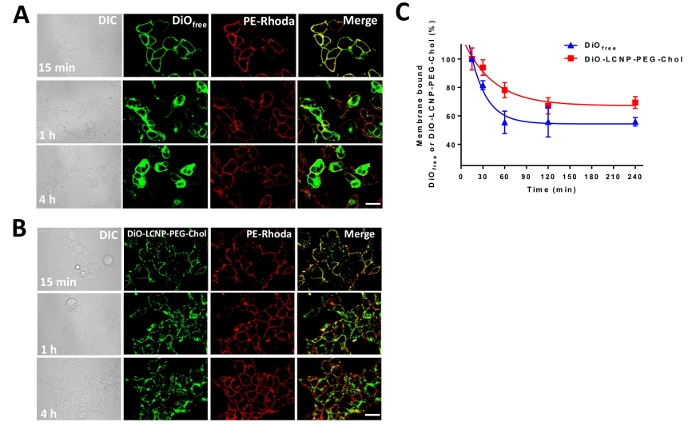
Figure 2: Time-resolved cellular uptake of DiOfree in HEK293T/17 cells. DiOfree (6.0 µM, panel (A) or DiO-LCNP-PEG-Chol ([DiO] = 6.0 µM, panel (B) was incubated on cell monolayers for 15 min, removed, and replaced with growth medium; the cells were cultured for the times indicated (up to 4 hr). Cells were subsequently stained with PE-Rhoda (2.0 µM) and fixed. The samples were imaged for DiO (green) and membrane-bound PE-Rhoda (red) using CLSM. (C) Time-resolved plot of the percent of membrane-bound DiOfree or DiO-LCNP-PEG-Chol signal as a function of increasing incubation time. The data was obtained from the Pearson's colocalization coefficient (PCC, n = 3 ± standard deviation) of the green (DiO) and red (PE-Rhoda) channels, and is expressed as a percentage (± SEM) after normalization to the PCC corresponding to 15 min of incubation. Scale bar = 20 µm. Reprinted with permission from reference18. Please click here to view a larger version of this figure.
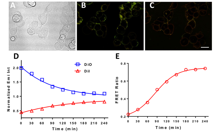
Figure 3: Time-resolved FRET confirms the efflux of DiO from DiO-LCNP-PEG-Chol to the plasma membrane. HEK293T/17 cells were costained with DiO-LCNP-PEG-Chol and DiI, where the DiO (green emitting) and DiI (red emitting) dyes act as the FRET donor and acceptor, respectively. (A) DIC image of the live cells costained with DiO-LCNP-PEG-Chol ([DiO] = 6.0 µM) and DiI (6.0 µM). The sample was imaged to monitor the change in FRET signal over the period of 4 hr. (B) and (C) are the FRET images of the live cell on the same focal plane at t = 0 min and t = 240 min, respectively. (D) Normalized, time-resolved emission intensity of the DiO donor and DiI acceptor from cells stained with DiO-LCNP-PEG-Chol and DiI and imaged in FRET excitation mode. (E) Time-resolved FRET ratio (DiIemi/DiOemi) for cells labeled with DiO-LCNP-PEG-Chol and DiI and imaged in FRET configuration. Scale bar = 20 µm. Reprinted with permission from reference18. Please click here to view a larger version of this figure.
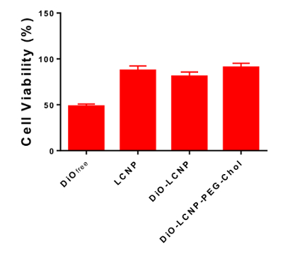
Figure 4: Quantification of cytotoxicity. DiOfree, LCNP, DiO-LCNPs, or DiO-LCNP-PEG-Chol were incubated on HEK 293T/17 cell monolayers for 15 min and then removed. Cells were washed and cultured in growth medium for 72 hr prior to MTS assay. The bar graph represents a comparison of the cell viability (n = 5 ± SEM) of DiOfree, LCNP, DiO-LCNP, and DiO-LCNP-PEG-Chol at [DiO] = 6.0 µM. The difference between DiOfree and LCNP, DiO-LCNP, or DiO-LCNP-PEG-Chol are significant (p < 0.001) at 72 hr of incubation. Reprinted with permission from reference18. The data was analyzed using the univariate analysis of variance (ANOVA); the Bonferroni's post hoc test was used for multiple comparisons. Please click here to view a larger version of this figure.
A continuing goal of NMDD is the controlled targeting and delivery of drug formulations to cells and tissues, combined with simultaneous improved drug efficacy. One specific class of drug molecules for which this has posed a significant challenge is hydrophobic drugs/imaging agents that have sparingly to no solubility in aqueous media. This problem has plagued the transition of potent drugs from in vitro cell culture systems to the clinical setting and has resulted in a number of promising drug molecules being “shelved” or abandoned and not pursued further in the clinical setting. Wortmannin (Wtmn), for example, is a potent inhibitor of phosphatidylinositol 3′ kinases and phosphatidylinositol 3′ kinase-related kinases and has great potential as an anticancer agent, but its lack of water solubility has hampered its progression in clinical trials.
Recently, Karve et al. showed improved water solubility of Wtmn when formulated as a lipid-polymer NP assembly19. One arena that could greatly benefit from this type of improved, controlled delivery as an NP formulation is the delivery of hydrophobic drugs and imaging agents to the plasma membrane. Thus, the goal here was to address this technical hurdle and to develop a methodology to overcome this technological hurdle.
In developing this methodology, a model membrane-targeting dye, DiO, was selected that has historically been plagued by poor solubility in aqueous media. This imparts significant challenges in the specific delivery of the dye to the plasma membrane. Among these challenges are the need to add the crystalline form of the dye directly to cells or tissues10 or to incubate cells with very high concentrations of DiO after dilution from stock solutions containing solvents such as DMSO11,12. The approach here was two-fold: 1) incorporate the DiO into the hydrophobic core of LCNPs during NP synthesis and 2) covalently modify the as-synthesized DiO-LCNPs with a PEGylated cholesterol conjugate (DiO-LCNP-PEG-Chol) to facilitate the direct interaction of the DiO-loaded NPs with the plasma membrane. Indeed, this approach effectively improved a number of aspects of the delivery of DiO to the plasma membrane.
First, the membrane residence time of DiO was effectively doubled when delivered as a LCNP formulation compared to when DiO was delivered free from bulk solution. This extended membrane residence time was attributed to the localization of the DiO-LCNP-PEG-Chol conjugates to the lipid raft microdomains of the plasma membrane, as confirmed by co-staining experiments. Second, the partitioning of the DiO cargo into the lipid bilayer was confirmed by FRET experiments in which the DiO served as the donor to a membrane-resident DiI acceptor. Finally, the slow, sustained release of the DiO cargo into the membrane bilayer effectively attenuated the cytotoxicity of the DiO by ~40%.
It is important to highlight several points in the protocol that are critical for success. First, the percent loading of the DiO into the LCNP core must be empirically optimized in trial synthesis runs to obtain a balance between the generation of high-quality DiO-LCNP-PEG-Chol NPs and the desired level of fluorescence signal in cellular delivery experiments. Likewise, the bioconjugation of the Chol-PEG-NH2 moiety to the DiO-LCNP surface must be optimized to obtain the desired level of cellular labeling. Finally, the number of cells applied to the fibronectin-coated culture dishes is critical for success, and care must be taken to seed the cells at a density of ~8 x 10⁴ cells/ml (~2.4 x 10⁵ cells per dish). When the cells are seeded too sparsely, it results in inefficient membrane labeling, while seeding the cells too densely results in the acquisition of poor images that do not clearly show membranous labeling.
One potential limitation of the as-described protocol is that the efficiency of incorporation of various other membrane-targeting dyes and drugs will vary depending on the hydrophobicity of the dye/drug cargo. In these instances, the LCNP synthesis protocol will have to be tested empirically to determine the optimum synthesis conditions for ideal dye/drug cargo incorporation.
In summary, a method has been developed for the LCNP-based controlled delivery of a hydrophobic dye cargo to the plasma membrane of living cells that overcomes many of the technical challenges associated with the cellular delivery of water-insoluble cargos. The overall significance of the method is that it clearly improves the specific delivery of cargo to the plasma membrane while simultaneously attenuating the concomitant cytotoxicity that is associated with the delivery of hydrophobic cargos at high concentrations from bulk solution. This approach should find wide utility in the delivery of similarly challenging hydrophobic dye/imaging agent cargos and will likely help to facilitate the transition of effective, yet poorly water-soluble, cargos from the experimental regime to the clinical setting.
The authors have nothing to disclose.
This work was supported by the NRL Base Funding Program (Work Unit MA041-06-41-4943). ON is supported by a National Research Council Postdoctoral Research Associateship.
1-ethyl-3-(3-(dimethylamino)-propyl)carbodiimide hydrochloride (EDCA) | ThermoFisher | E2247 | |
3,3′-dioctadecyloxacarbocyanine perchlorate (DiO) | Sigma Aldrich | D4292-20MG | Hazardous/ make stock solution in DMSO |
Cholesterol poly(ethylene glycol) amine hydrochloride | Nanocs, Inc. | PG2-AMCS-2k | |
Countess automated cell counter | ThermoFisher | C10227 | |
Dioctadecyl-3,3,3′,3′-tetramethylindocarbocyanine perchlorate (DiI) | Sigma Aldrich | 468495-100MG | Hazardous/ make stock solution in DMSO |
Dulbecco's Modified Eagle's Medium (DMEM) | ThermoFisher | 21063045 | Warm in 37°C before use |
Dulbecco's Phosphate Buffered Saline (DPBS) | ThermoFisher | 14040182 | Warm in 37°C before use |
Dynamic light scattering instrument | ZetaSizer NanoSeries (Malvern Instruments Ltd., Worcestershire, UK) | ||
Fibronectin Bovine Protein, Plasma | ThermoFisher | 33010018 | Make stock solution 1mg/mL using DPBS. Use 20-30 µg/mL for coating MetTek dish, 2 h@ 37°C |
Formaldehyde (16%, W/V) | ThermoFisher | 28906 | Hazardous, dilute to 4% using DPBS |
Human embryonic kidney cells (HEK 293T/17) | American Type Culture Collection | ATCC® CRL-11268™ | |
Live cell imaging solution (LCIS) | ThermoFisher | A14291DJ | Warm in 37°C before use |
MatTek 14 mm # 1.0 coverglass insert cell culture dish | MatTek corporation | P35G-1.0-14-C | |
Modified Eagle Medium (DMEM) containing 25 mM HEPES | ThermoFisher | 21063045 | Warm in 37°C before use |
N-hydroxysulfosuccinimide sodium salt (NHSS) | ThermoFisher | 24510 | |
Nikon A1si spectral confocal microscope | Nikon Instruments | ||
Trypan Blue Stain (0.4%) | ThermoFisher | T10282 | mix as a 50% to the cell suspension before counting the cells |
Zeta potential instrument | ZetaSizer NanoSeries (Malvern Instruments Ltd., Worcestershire, UK) | ||
Ultrasonic Processor | Sonics and Materials Inc | GEX 600-5 | |
Mini Cetntrifuge | Benchmark | Mini-fuge-04477 | |
PD-10 Sephadex™ G-25 Medium | GE Healthcare | 17-0851-01 | |
Bio-Rad ChemiDoc XRS Imaging System | Bio-RAD | 76S/07434 | |
Trypsin-EDTA(0.25%), phenol red | ThermoFisher | 25200056 |