An Ultrasonic Tool for Nerve Conduction Block in Diabetic Rat Models
Summary
This work presents the methodology of applying high intensity-focused ultrasound to block the action potentials of diabetic neuropathic nerves.
Abstract
Nerve conduction block with a high intensity-focused ultrasound (HIFU) transducer has been performed in normal and diabetic animal models recently. HIFU can reversibly block the conduction of peripheral nerves without damaging the nerves while using an appropriate ultrasonic parameter. Temporary and partial block of the action potentials of nerves shows that HIFU has the potential to be a useful clinical treatment for pain relief. This work demonstrates the procedures for suppressing the action potentials of neuropathic nerves in diabetic rats in vivo using an HIFU transducer. The first step is to generate adult male diabetic neuropathic rats by streptozotocin (STZ) injection. The second step is to evaluate the peripheral diabetic neuropathy in STZ-induced diabetic rats by an electronic von Frey probe and a hot plate. The final step is to record in vivo extracellular action potentials of the nerve exposed to HIFU sonication. The method showed here may benefit the study of ultrasound analgesic applications.
Introduction
Oral medications, acupuncture1, and electrical nerve stimulation2 have been used for the treatment of painful diabetic polyneuropathy. However, the side effects of the oral medications, invasive operation of acupuncture, and electrical nerve stimulation hamper the therapeutic efficacy and patient's adherence. Ultrasound block of peripheral nerves in animal models has been investigated for decades3,4,5. The conduction of in vitro sciatic nerves of the large green frog was inhibited reversibly after the treatment of 10 – 20 pulses of ultrasound exposure for 0.4 – 1.0 s6. One factor to block nerve conduction is the temperature rise induced by ultrasound7. For patients with polyneuropathy, the suppression of compound muscle action potentials (CMAPs) was performed in the peroneal nerve exposed to low-intensity ultrasound for 2 min8. The full recovery time was within 5 min.
Recently, the Food and Drug Administration of the United States approved HIFU as a non-invasive treatment for uterine fibroid tumors9, pain palliations of bone metastases10, and prostate cancer11. An HIFU transducer emits acoustic beams outside the body, and the beams transmit in various tissue mediums and converge on the target tumor at the focus. The focal zone is immediately formed to generate localized effects on target tumors without damaging the surrounding tissues. HIFU has also been applied to inhibit nerve conduction or cause nerve denervation in in vivo experiments of normal Sprague-Dawley (SD) rats12. In addition, the short-term and long-term effects of HIFU on neuropathic nerves have been investigated13. Previous outcomes demonstrated that the reversible or permanent block of sensory nerve conduction could be achieved by HIFU with appropriate parameters. Besides analgesic applications, HIFU might be used as a tool to investigate the relative contribution of peripheral and central components to nerve conduction blockade for basic research of neurology and development of pain medication. Therefore, a HIFU blocking technology platform specific for peripheral nerves in animal models is needed. The purpose of this article is to demonstrate the procedures for partially or completely blocking the action potentials of peripheral nerves in diabetic neuropathic rats by HIFU. Diabetic rat models and evaluation of peripheral neuropathic symptoms were established. An HIFU platform and experimental processes specific for treating rat sciatic nerves are presented.
Protocol
The Institutional Animal Care and Use Committee of the National Health Research Institutes in Taiwan approved all animal protocols.
1. Induction of Diabetic Model in Male Adult Sprague – Dawley (SD) Rats
- Remove rat food pellets from the cage to fast male SD rats (300 – 350 g) for 6 h prior to STZ induction.
- Prepare sodium citrate buffer (0.1 M, pH 4.5).
- Dissolve 1.05 g citric acid monohydrate (C6H8O7 · H2O; mol. wt. 210.14) in 50 mL distilled water to make a 0.1 M citric acid solution.
- Dissolve 1.47 g trisodium citrate dihydrate (C8H5O7Na3 · 2H2O; mol. wt. 294.12) in 50 mL distilled water to make a 0.1 M sodium citrate solution.
- Add 25 mL citric acid solution to 25 mL sodium citrate solution. Monitor the sodium citrate buffer pH (pH 4.5) using a pH meter.
NOTE: The buffer is made isotonic by addition of an appropriate volume of 0.1 M sodium citrate solution.
- Dissolve STZ in 0.1 M sodium citrate buffer to yield a 50 mg/mL STZ solution.
NOTE: The STZ solution is light sensitive, therefore, cover the STZ solution with aluminum foil and use within 15 – 20 min. - Draw the 50 mg/kg STZ solution into a 1 mL insulin or tuberculin syringe with 26- to 28-gauge needle. Clean the injection site with an ethanol pad and intraperitoneally inject the STZ solution into the lower right quadrant of the abdomen to avoid damaging abdominal organs.
- Supply rats with 10% sucrose water as the sole water source for 48 h after STZ injection to prevent hypoglycemia.
2. Confirmation of Diabetes in STZ-induced Rats
- Monitor the fasting plasma glucose concentration of all rats injected by STZ post 72 h with a glucose meter.
- Fast the STZ-induced diabetic rats for 15 h before measuring the fasting blood glucose level.
- Restrain the rats in a restraint bag and expose the tails for blood collection during the blood glucose measurements.
- Use a blood lancet to prick the tip of the tail to obtain a small drop of blood. Place the drop of blood on a glucose test strip. Record the fasting plasma glucose levels.
NOTE: The glucose meter detects and displays the blood glucose level in units of mg/dL. Exclude the rats with fasting blood glucose levels below 150 mg/dL after 2 weeks of STZ-induction.
3. Evaluation of Peripheral Diabetic Neuropathy in Diabetic Rats
- Assess the mechanical allodynia with electronic von Frey.
- Habituate the STZ-induced diabetic rats in a cage on a 1 cm diameter metal mesh floor for 30 min before evaluating the hind paw withdrawal response.
- Use an electronic von Frey probe with rigid tip (0.8 mm in diameter) to manually apply pressure to the plantar surface of the hind paw of the rats, and gradually increase the pressure until a paw withdrawal response is seen.
- Record the pressure that shows on the system and repeat the measurement 5 times per rat, with a 30 s interval between each measurement.
- Assess the heat hyperalgesia with a hot plate.
- Habituate the STZ-induced diabetic rats on the hot plate (24 ± 0.5 °C) for 10 min before evaluating the pain response.
- Remove and place the rats back into their cages after habituation, heat the hot plate and maintain the hot plate's temperature at 55 ± 0.5 °C.
- Place the rat on the heated hot plate while simultaneously starting the timer.
- When the rat exhibits distinct behaviors, such as licking the hind paw or abnormally flicking the hind paw, stop the timer, and record the withdrawal latency.
NOTE: If a rat does not express distinct behaviors after 20 s (20 s cut-off time), terminate the hot plate test and remove the rat from the hot plate.
4. In Vivo Nerve Conduction Blockage with the HIFU Transducer
NOTE: The in vivo experiment starts on week 5 after 50 mg/kg STZ injection.
- Perform animal procedures before blocking of the CMAPs with HIFU sonication.
- Sterilize the surgical tools (scalpel, scissors, forceps, and glass hook) in an autoclave before the surgery.
- Anesthetize the rats with intraperitoneal injection of tiletamine/zolazepam mixture (40 mg/kg) and xylazine (10 mg/kg) or via inhalation of 1.75% of isoflurane via isoflurane vaporizer. Place the rats on a heating pad to maintain the body temperature.
- Position the rats for surgery in ventral recumbency. Apply the eye ointment. Extend the leg of the rat and pinch the plantar surface of the foot with fingernails to ascertain the depth of anesthesia. If the rats show withdrawal responses, apply additional anesthesia.
- Remove hair from the thigh and lower back of the rats with an electric clipper. Apply liquid iodine with clean gauze at the surgical site and circularly move the gauze to the outward of the surgical site. Use an alcohol pad to wipe the liquid iodine with the same circular movement. This experiment is performed on the left and right surgical site. Repeat the procedure at the other surgical site when perform the next step of procedure.
- Use a sterile surgical scissors or scalpel to make an incision of the skin at the dorsal thigh. Use blunt surgical scissors to carefully separate the tissue underneath the skin and secure the skin with skin hooks. The femur can be seen within the muscles.
- Use the scissors to carefully separate the muscles parallel to femur until the mid-thigh sciatic nerve fibers that are embedded in the muscles are visible. Carefully use a glass hook to separate the mid-thigh sciatic nerve from the surrounding connective tissues and muscles.
- Position the sciatic nerve in the HIFU focal zone using a custom-made nerve fixator (Figure 1 and Figure 3).
NOTE: The custom-made nerve fixator consists of 3 components (Figure 3). All the components are made of transparent polymethylmethacrylate (PMMA). The external thread of the top structure of component I is 2.5 mm tall and M10XP0.7 (Figure 4A). The central well is a 4.0 mm-diameter hole through component I. The bottom opening of the well is sealed by a sheet of tape. The diameter of the slot is 1.2 mm and the distance between the central plane of the slot and the top surface of component I is 3.1 mm. Component II consists of a main body, four legs and the bottom structure (Figure 4B). The internal thread of the bottom structure is 2.5 mm tall and M10XP0.7 to fit the external thread of component I. The diameter of the central hole is the same as the central well of component I. The dimensions of the main body are 32 mm in diameter and 5.4 mm in thickness. Four legs are symmetrically deployed. Two identical short legs are designed for alignment and two identical long legs work for hooking on component III. The outer diameter and height of component III are 41 mm and 9.2 mm. The internal thread is M36XP1.0 and the through hole is 27.5 mm in diameter (Figure 4C). The cone is a hollow taper with the top opening of 84 mm in diameter and the bottom opening of 27.5 mm in diameter. The height is 57.5 mm (Figure 5A).- Before the experiment, soak the custom-made acrylic nerve fixator in bleach-solution for 30 – 60 min followed by soaking in sterile water.
- Using a glass hook, lift the nerve carefully and put it in the slot of component I.
- Screw component II to component I. Fill the central well of component I with Ringer's solution for ultrasonic propagation and nerve preservation.
- Screw component III to the HIFU housing cone. Dock component III with component II through the four legs of component II.
NOTE: The geometric center of three components and the transducer are aligned. The distance between the nerve and the transducer is equal to the focal length, which ensures the nerve is inside the HIFU focal zone.
- Insert one pair of acupuncture needles into the origin of the sciatic nerve and the other pair into the gastrocnemius muscle. Connect each pair of needles to the electrophysiology acquisition system through an electrical coaxial cable (Figure 1).
NOTE: On one end of the cable are two alligator clips to clip two needles separately and on the other end of the cable is a BNC connector to link the system. The pairs of acupuncture needles work as stimulating electrodes at the sciatic nerve and the recording electrodes at the gastrocnemius muscle.- Set the sampling rate and bandwidth of the electrophysiology acquisition system to 50 kHz and 70 Hz-3 kHz, respectively. Apply a supra-maximal stimulus with a pulse width of 0.1 ms to the stimulating electrodes at the origin of the sciatic nerve.
- Record the CMAPs from the recording electrodes and amplify the CMAPs with the built-in amplifier in the electrophysiology acquisition system.
NOTE: Use the built-in amplifier in the electrophysiology acquisition system to amplify the nerve signals and record the CMAPs from the recording electrodes with the electrophysiology acquisition system.
- Use a commercial 2.68 MHz HIFU transducer to suppress the CMAPs in diabetic neuropathic rats.
NOTE: The specifications of the transducer are described as follows: a single-element spherical bowl with aperture diameter of 6 cm and focal length of 5 cm, and an ellipsoid focal zone of 4 mm in depth and 0.8 mm in width in the free field.- Immerse the spherical cone, the HIFU transducer and the cone cover in the tank filled with degassed water. Put the HIFU transducer into the spherical cone and fix the cone cover to the top opening of the spherical cone by 6 head screws (Figure 5B). After bubbles in the spherical cone are expelled naturally due to the low density of bubbles compared with water, seal the front-end opening of the cone by a transparent 0.03 mm thick tape. Screw component III onto the spherical cone.
- Take out the HIFU transducer with the spherical cone and component III from the degassed water tank.
NOTE: The reverse osmosis water used in the study is the water purified by the process of reverse osmosis. The reverse osmosis water is boiled to expel the gas. After cooling, the degassed water is obtained in an individual sealed tank.
- Put component I into the space between the nerve and the muscle carefully, and position the nerve in the gap of component I. Perform steps 4.2.3 and 4.2.4 to ensure that the nerve is inside the focal zone of the HIFU (Figure 3A).
- Link a function generator and a radiofrequency power amplifier. Connect the power amplifier to the HIFU transducer for generation of the HIFU beam. Manually set the voltage output of the function generator to the HIFU transducer via the power amplifier. Manually turn off the function generator once the HIFU-exposure time is up. Observe time using a timer.
NOTE: The intensity and energy of HIFU beam used in this study are 2,810 W/cm2 and 84 J/mm2, respectively. - Simultaneously, deliver the stimulus via the electrophysiology acquisition system (step 4.3) and HIFU beam via HIFU system (step 4.6) to the sciatic nerve while recording the CMAPs. Gradually increase the HIFU exposure on the sciatic nerve from 3 s, 5 s to 8 s until decrease or inhibition of the amplitude of CMAPs is observed.
- Record the CMAPs once per second during the delivery of the HIFU beam. After observing the change in the amplitude of CMAPs, turn off the HIFU system and manually click on the record icon on the electrophysiology acquisition software to record CMAPs every 2 min in the early 10 min, every 5 min in the consecutive 30 min, and every 10 min in the last phase until the recording time reaches 2 h.
- Separate component II and III of the nerve fixator (Figure 3) to remove the HIFU transducer from the incision site. Separate component II and I to release the secured sciatic nerve. Suture the surgical site of the diabetic rat by 4-0 chromic catgut sutures after recording the CMAPs. Apply liquid iodine to the surgical site to prevent infection.
- Place the cages on the heating pad and allow the rats to recover in their cages before returning them to the animal facility. Provide the rats with ibuprofen in drinking water for 3 days or intraperitoneal injection of buprenorphine (0.05 – 0.1 mg/kg).
- Insert stimulating and recording electrodes in the origin of the sciatic nerve and the gastrocnemius muscles of anesthetized diabetic neuropathic rats as described in steps 4.1.2 and 4.3 on days 7, 14, and 28 after the initial HIFU sonication. Repeat steps 4.3.1 to 4.3.2.
- Place the cages on the heating pad and allow the rats to recover in their cages before moving the cages to the animal facility.
- Euthanize the rats after the experiment. Place the rat in a carbon dioxide chamber. Wait about 5 min for the rats to stop breathing. Ensure that the heart has stopped beating.
Representative Results
The in vivo study demonstrated that, with an HIFU dose of 3 s sonication at an intensity of 2,810 W/cm2, the CMAPs were suppressed by 20% of baseline, but they were completely recovered after 30 min (Figure 2A, diamonds) and were almost constant in the period of 28 days (Figure 2B, diamonds). For the 5 s HIFU exposure at the same intensity, the CMAPs decreased to 65.4% (9.5%) of baseline at 4 min and recovered to 73.7% (12.6%) of baseline by 120 min (Figure 2A, squares). The CMAPs did not return to baseline levels until day 14 (Figure 2B, squares). When HIFU sonication time was increased to 8 s under the same intensity, the CMAPs were reduced to 26.0% (14.1%) of baseline at 4 min (Figure 2A, triangles), but increased to 38.0% (12.0%) of baseline at 120 min, and gradually increased to 74% of baseline by day 28 (Figure 2B, triangles). See Lee, Y.F.,13 for additional details.
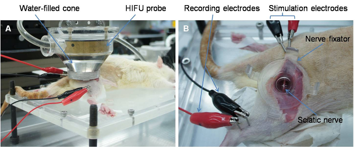
Figure 1: The Experimental Setup for In Vivo Nerve Conduction Block with High Intensity Focused Ultrasound (HIFU). (A) A custom-made acrylic spherical cone that is filled with reverse osmosis degassed water was combined with the nerve fixator to ensure the focal zone of HIFU transducer was at the focal plane of the nerves. (B) Stimulating and recording electrodes are shown in the figure. The nerve fixator positioned the sciatic nerve in the focal plane of the HIFU beam. Please click here to view a larger version of this figure.
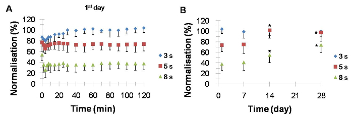
Figure 2: The CMAPs Responses in In Vivo Diabetic Neuropathic Rats after 3 s, 5 s, and 8 s of HIFU Sonication. (A) The temporal courses of the CMAPs during day 1 recording. After 3 s, 5 s, and 8 s of HIFU sonication, the CMAPs were decreased during the early 10 min and recovered after 120 min. (B) The temporal courses of the CMAPs during day 7, 14, and 28 recordings. The CMAPs were fully recovered by day 14 after 3 s and 5 s HIFU sonication on day 1 and partially increased by day 28 after 8 s HIFU sonication. Diamonds: 3 s of HIFU sonication, squares: 5 s of HIFU sonication, and triangles: 8 s of HIFU sonication. n= 6 for each HIFU parameter. *Significantly different from day 1. The data are expressed as the median (range), where the error bar is half of the range. This figure is modified from Lee, Y.F., 201513. Please click here to view a larger version of this figure.
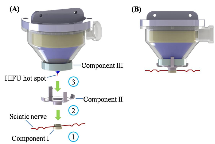
Figure 3: The Assembling Process to Ensure the Sciatic Nerve in the HIFU Hot Spot. (A) Three assembling steps are shown: (1) carefully settle the nerve in the slot of component I, (2) assemble component II and component I, (3) insert the front-end of component III with the HIFU transducer cone structure into component II. (B) A schematic drawing of the HIFU transducer integrated with the rat nerve. Please click here to view a larger version of this figure.
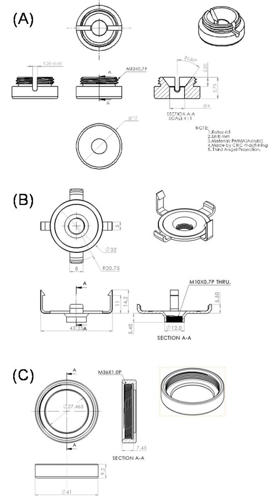
Figure 4: The Drawings of Components I (A), II (B) and III (C). Please click here to view a larger version of this figure.
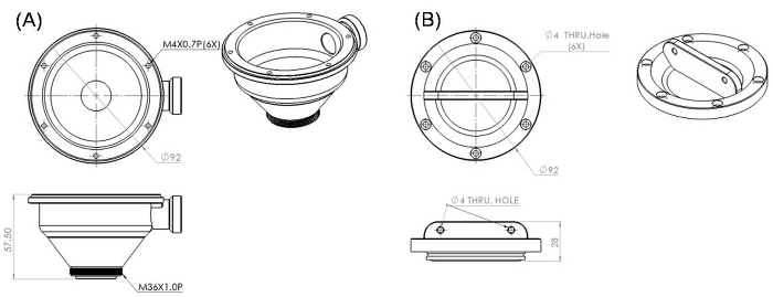
Figure 5: The Drawings of the Spherical Cone (A) and the Cone Cover (B). Please click here to view a larger version of this figure.
Discussion
Partial and temporary suppression of action potentials of the neuropathic nerves from diabetic rats in vivo and instant occurrence of the blocking effect after HIFU treatment both were observed. The 28-day follow-up study on CMAPs demonstrated that a safe blockade of the nerve conduction could be carried out at an appropriate HIFU exposure. As a result, the above protocol of the HIFU treatment can provide an alternative solution for the reversible conduction block of sciatic nerves in diabetic rats.
In this method, there is no neural degeneration, and the sensory nerves can recover completely over a period of hours to several days in mild nerve injuries14. For severe injuries, the temporal course of sensory recovery typically takes several months, if it occurs at all. Furthermore, the peripheral neural fibers regenerate more completely when the endoneurial tubes and Schwann cell basal lamina are intact following crushing injuries15. Therefore, we infer that the HIFU caused mild but reversible nerve injuries for the case because the suppressed CMAPs after the HIFU treatment returned to baseline over time. For severe nerve injuries, the CMAPs only recovered partially, even after 28 days.
The technique of this study provided an experimental platform for animal study of HIFU effects on peripheral nerves prior to clinical study. The HIFU focal zone can precisely aim at the target nerve because of the structural components and the protocol developed in this study, which solves the previous problem of positioning. Besides the normal nerve, the HIFU blocking technique can be also applied to the diseased nerve. However, the limitations of the current technique include the lack of temperature monitoring (leading to damage of the surrounding tissue), and the short blocking effect, although repeating the HIFU exposure may extend the analgesia. To translate the HIFU blocking technique to clinical trials, a noninvasive guidance of the HIFU focal zone is required, like ultrasound imaging or MR imaging, to identify the position of the target tissue and monitor the temperature in real time.
The existing oral medications result in only one in four patients with neuropathic pain experiences of over 50% pain relief and pose several significant side effects such as drowsiness, dizziness and somnolence16. Physical modalities are developed to hopefully improve analgesic efficacy like acupuncture, electrical and magnetic stimulations. However, the efficacy of acupuncture relies highly on the clinician experience, and the procedure is invasive. The efficacy of noninvasive electrical stimulation or magnetic stimulation is approximately 40% for pain freedom at 2 h. Both stimulations are not focused on the local site, which produces some adverse events17. Therefore, to satisfy clinical unmet needs for peripheral pain relief, the HIFU blocking technique is a promising tool because of instant effectiveness, reversible effect, physical therapy, non-invasive treatment, and potential home-use.
It is critical to aim the HIFU focal zone at the sciatic nerve accurately. Figure 3 illustrates the schematic procedure for positioning the nerve in the focal zone. The first step is to use a glass hook to slightly lift the sciatic nerve and put in the component I below the nerve, and then put down the nerve into the slot of component I (Figure 3A). The nerve passes the central site of component I through the first step. The second step is to assemble component II with component I via the screw cap structure. The assembly of components I and II is the nerve fixator shown in Figure 2B. Component II plays the role of linking components I and III. Before combining components II and III, they are filled with degassing Ringer's solution to transmit the ultrasound and preserve the nerve. The last step is to insert component III front-end structure of the HIFU transducer cone coupler into component II. Two pairs of flexible long and short pillars of component II provide sufficient fixing force. Assembly of components II and III is designed based on the mortise-tenon principle, which can ensure that the central point of the three components is in axis. The focal length of the HIFU transducer is equal to the distance between the central point of the transducer and the central point of component I. As a result, the nerve is certainly inside the focal zone, which is an ellipsoid with a width of 0.8 mm and a depth of 4 mm.
Disclosures
The authors have nothing to disclose.
Acknowledgements
The study was supported by the Ministry of Science and Technology (Project MOST 105-2221-E-400 -001) and the National Health Research Institutes (Project BN-105-PP-10), Taiwan.
Materials
| streptozotocin | Sigma | 85882 | |
| citric acid monohydrate | Sigma | C1909 | |
| trisodium citrate dihydrate | Sigma | W302600 | |
| glucose meters | Roche Accu-Check Active | GC | |
| electronic von Frey device | IITC Life Science | 2390 | |
| hot plate | IITC Life Science | ||
| Biopac MP36 acquisition system | Biopac Systems, Inc. | ||
| HIFU transducer | Sonic Concepts | H108 | |
| function generator | Agilent | 33250A | |
| power amplifier | Electronics & Innovation | 1040L | |
| Rats | Biolasco taiwan | Sprague-Dawley | |
| Puralube vet ointment | Dechra | ||
| isoflurane vaporizer | Parkland Scientific | V3000PS | |
| Isoflurance | Attane | ||
| Restraint bag (Decapicones) | Braintree Scientific | DC 200 |
References
- Abuaisha, B. B., Costanzi, J. B., Boulton, A. J. Acupuncture for the treatment of chronic painful peripheral diabetic neuropathy: A long-term study. Diabetes Res Clin Pract. 39 (2), 115-121 (1998).
- Hamza, M. A., et al. Percutaneous electrical nerve stimulation: A novel analgesic therapy for diabetic neuropathic pain. Diabetes Care. 23 (3), 365-370 (2000).
- Ballantine, H. T., Bell, E., Manlapaz, J. Progress and problems in the neurologic applications of focused ultrasound. J Neurosurg. 17, 858-876 (1960).
- Foley, J. L., Little, J. W., Vaezy, S. Image-guided high-intensity focused ultrasound for conduction block of peripheral nerves. Ann Biomed Eng. 35 (1), 109-119 (2007).
- Lee, Y. F., Lin, C. C., Cheng, J. S., Chen, G. S. High-intensity focused ultrasound attenuates neural responses of sciatic nerves isolated from normal or neuropathic rats. Ultrasound Med Biol. 41 (1), 132-142 (2015).
- Young, R. R., Henneman, E. Reversible block of nerve conduction by ultrasound. Arch Neurol. 4, 83-89 (1961).
- Lele, P. P. Effects of focused ultrasonic radiation on peripheral nerve, with observations on local heating. Exp Neurol. 8 (1), 47-83 (1963).
- Hong, C. Z. Reversible nerve conduction block in patients with poly- neuropathy after ultrasound thermotherapy at therapeutic dosage. Arch Phys Med Rehabil. 72 (2), 132-137 (1991).
- Okada, A., Morita, Y., Fukunishi, H., Takeichi, K., Murakami, T. Non-invasive magnetic resonance-guided focused ultrasound treatment of uterine fibroids in a large Japanese population: impact of the learning curve on patient outcome. Ultrasound Obstet Gynecol. 34 (5), 579-583 (2009).
- Huisman, M., et al. International consensus on use of focused ultrasound for painful bone metastases: current status and future directions. Int J Hyperthermia. 31 (3), 251-259 (2015).
- Dickinson, L., et al. Medium-term Outcomes after Whole-gland High-intensity Focused Ultrasound for the Treatment of Nonmetastatic Prostate Cancer from a Multicentre Registry Cohort. Eur Urol. 70 (4), 668-674 (2016).
- Foley, J. L., Little, J. W., Vaezy, S. Effects of high-intensity focused ultrasound on nerve conduction. Muscle Nerve. 37 (2), 241-250 (2008).
- Lee, Y. F., Lin, C. C., Cheng, J. S., Chen, G. S. Nerve conduction block in diabetic rats using high-intensity focused ultrasound for analgesic applications. Br J Anaesth. 114 (5), 840-846 (2015).
- Donoff, R. B. Nerve regeneration: basic and applied aspects. Crit Rev Oral Biol Med. 6 (1), 18-24 (1995).
- Fawcett, J. W., Keynes, R. J. Peripheral nerve regeneration. Annu Rev Neurosci. 13, 43-60 (1990).
- Nightingale, S. The neuropathic pain market. Nat Rev Drug Discov. 11 (2), 101-102 (2012).
- Lipton, R. B., et al. Single-pulse transcranial magnetic stimulation for acute treatment of migraine with aura: a randomised, double-blind, parallel-group, sham-controlled trial. Lancet Neurol. 9 (4), 373-380 (2010).
