Maintaining blood-brain barrier coverage is key for the homeostasis of the central nervous system. This protocol describes in vitro techniques to delineate the fundamental and pathological processes that modulate blood-brain barrier coverage.
Blood-brain barrier (BBB) coverage plays a central role in the homeostasis of the central nervous system (CNS). The BBB is dynamically maintained by astrocytes, pericytes and brain endothelial cells (BECs). Here, we detail methods to assess BBB coverage using single cultures of immortalized human BECs, single cultures of primary mouse BECs, and a humanized triple culture model (BECs, astrocytes and pericytes) of the BBB. To highlight the applicability of the assays to disease states, we describe the effect of oligomeric amyloid-β (oAβ), which is an important contributor to Alzheimer's disease (AD) progression, on BBB coverage. Further, we utilize the epidermal growth factor (EGF) to illuminate the drug screening potential of the techniques. Our results show that single and triple cultured BECs form meshwork-like structures under basal conditions, and that oAβ disrupts this cell meshwork formation and degenerates the preformed mesh structures, but EGF blocks this disruption. Thus, the techniques described are important for dissecting fundamental and disease-relevant processes that modulate BBB coverage.
The blood-brain barrier (BBB) of cerebral capillaries is the largest interface of blood-to-brain contact and plays a central role in the homeostasis of the central nervous system (CNS)1,2. Dynamic processes at the BBB prevent the uptake of unwanted molecules from the blood, remove waste products from the CNS, supply essential nutrients and signaling molecules to the CNS, and modulate neuroinflammation1,2,3,4,5. BBB damage is prevalent during aging and several neurodegenerative disorders including Alzheimer's disease (AD), multiple sclerosis and stroke1,2,3,4,5,6. Therefore, BBB dysfunction may play a key role in neurodegenerative disorders, including as a therapeutic target.
Maintaining vessel coverage is important for the homeostatic functions of the BBB. However, in vivo and in vitro data conflict on whether the processes involved in neurodegenerative disorders cause higher or lower BBB coverage6,7,8,9,10,11,12,13, particularly in AD. Therefore, there is a strong rationale for the development of in vitro models using relevant cell types to assess and more comprehensively understand the dynamics of BBB coverage. Cerebral capillaries are composed of astrocytes, pericytes and brain endothelial cells (BECs)3. All cell types contribute to the function of the BBB through structural support and via the secretion of effector molecules such as angiogenic growth factors, cytokines and chemokines that act in paracrine- and autocrine-like fashion. However, the major effector cells of the BBB are BECs3. In general, the cell culture techniques for assessing BBB function are permeability assays performed on cells grown on filter inserts, or assessing levels of key BEC proteins, both after the addition of stressors14,15,16. Although important, these assays do not focus on the cerebrovascular coverage.
Here, our previous methods17 are detailed to assess BEC coverage and meshwork-like structures using single cultures of immortalized human BECs, single cultures of primary mouse BECs, and a humanized triple culture model (BECs, astrocytes and pericytes) of the BBB. The goal was to demonstrate the detrimental effect of oAβ, which is considered an important contributor to AD progression, on BEC coverage. The protective effect of the epidermal growth factor (EGF) highlights the potential of the technique as a therapeutic screening tool. The technique has several broad applications for basic and applied research including: 1) delineating the role of specific pathways on angiogenesis and vessel coverage, 2) evaluating the effects of disease and aging-relevant factors on angiogenesis and vessel coverage, and 3) identifying pharmacological targets.
All experiments follow the University of Illinois, Chicago Institutional Animal Care and Use Committee protocols.
1. General Preparation
NOTE: The brain microvascular endothelial cell line (hCMEC/D3) is an extensively characterized immortalized human BEC line14,15,16,18,19. Culture the hCMEC/D3 cells on tissue culture flasks coated with collagen Type I (calf skin, 1:20 dilution of 0.1% solution in Hank's Balanced Salt Solution (HBSS) containing Ca2+ and Mg2+) in basal Endothelial Growth Basal Medium (EBM-2) containing 2-5% Fetal Bovine Serum (FBS), 10% ascorbic acid, 10% gentamicin sulphate, 25% hydrocortisone and 1/4 of the total volume of the supplied growth factor supplements per 500 mL of media [vascular endothelium growth factor (VEGF), epidermal growth factor (EGF), insulin-like growth factor 1 (IGF-1) and human basic fibroblastic growth factor (bFGF), see Table of Materials].
NOTE: EBM-2 medium with FBS and growth factors is referred to as "EBM-2 complete". EBM-2 without FBS and supplements is referred to as "EBM-2 basal". At full confluence, the hCMEC/D3 cells are ~1 x 10⁵ cells/cm2.
2. Meshwork-like Formation and Disruption Assays
3. Quantification
In single cultures, both the hCMEC/D3 cells (Figure 3A) and the primary mouse BECs (Figure 3B) form meshwork-like structures throughout the well. The structures are characterized by a meshwork of interlinked nodes (Figure 3). In all the paradigms described (Figure 1), the meshwork-like structures are similar after 24 h in the control groups, forming ~20 meshwork-like structures with a total cellular length of ~10,000 pixels.
To highlight the applicability of the methods to disease relevant stressors, oAβ was added to the hCMEC/D3 and the primary mouse BECs in two paradigms. In the disruption of cell meshwork formation (Paradigm 1), oAβ and cells are plated at the same time. In the disruption of preformed meshwork (Paradigm 2), oAβ is added 4 h after plating the cells (see Figure 1A). oAβ at 100 nM induces disruption of the meshwork-like formation and degeneration of preformed meshwork (Figure 3A, 3B). For example, using the hCMEC/D3 cells in both paradigms, quantification of the total cell coverage/length is 16-20% lower with 100 nM oAβ17. Further, the number of meshes is reduced by 40% with 100 nM oAβ in both paradigms17. Thus, a disease relevant stressor induces a similar detrimental effect on human and mice BEC coverage.
A key advantage of the cell culture system is the ability to identify factors or treatments that prevent disease-relevant damage, which can then advance to in vivo testing. As a proof of principle experiment, the effects of the main angiogenic growth factors on preventing oAβ-induced changes to vessel coverage were assessed17. The EGF prevented oAβ-induced damage to the hCMEC/D3 cells. Based on those data, EGF was tested in a prevention paradigm using a transgenic mouse model that recapitulates critical aspects of AD-like pathology. EGF treatment prevented the cognitive and BBB deficits, including vessel degeneration23. These data support the predictive potential of the in vitro system for in vivo activity. Currently, we utilize all three developed paradigms for screening: 1) mesh formation, 2) prevention of cell meshwork disruption, and 3) simultaneous treatment of meshwork disruption. As highlighted in Figure 3, EGF can protect against oAβ-induced damage in immortalized and primary mouse BEC cultures.
The BBB consists of BECs, pericytes and astrocytes that collectively contribute to overall cerebrovascular coverage. Therefore, one adaptation of the in vitro system is the incorporation of all three BBB cell types. Our triple culture assay paradigm incorporates the hCMEC/D3 cells, primary human pericytes and primary human astrocytes, which are added sequentially to the basement membrane matrix (Figure 1B). In the triple culture assay paradigm, the hCMEC/D3 cells form a similar meshwork pattern as in the single cultures, but with the pericytes and astrocytes attached to the nodes and connecting branches (Figure 4). The addition of 100 nM oAβ induces meshwork disruption (~10-15%) and a reduction in the number of pericytes contacting the BECs17. In correlation with the data derived from the single culture assay paradigms, oAβ-induced damage is prevented by EGF treatment. Thus, the triple culture BBB model is well suited for studies focused on the interactive effects of astrocytes, pericytes and BECs on vessel dynamics.
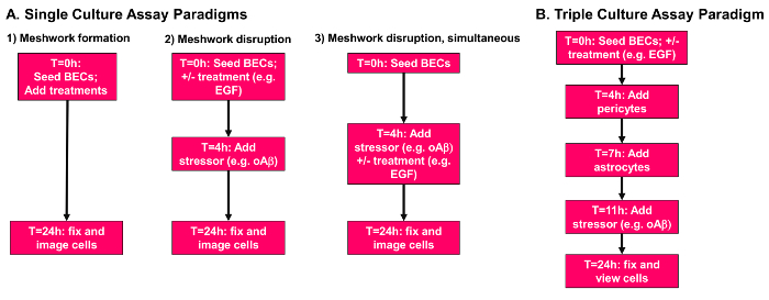
Figure 1: Overview of meshwork formation and disruption assays. (A) Single culture assay paradigms. Paradigm 1, meshwork formation. Cells, stressors and treatments are all added to the plate at the 0 h time point. Paradigm 2, prevention of meshwork disruption. Cells are plated in the presence of desired treatments, incubated for 4 h, and a stressor is added at the 4 h time point. Paradigm 3, simultaneous treatment of meshwork disruption. Cells are plated and allowed to form meshwork-like structures for 4 h before treatments and/or stressors are added simultaneously at the 4 h time point. All paradigms end at the 24 h time point, and cells are fixed with 4% paraformaldehyde. (B) Triple culture assay paradigm. BECs (10,000 cells/well) are plated in the presence of desired treatments at 0 h. At the 4 h time point pericytes (2,000 cells/well) are gently added to the plate. At 7 h astrocytes (10,000 cells/well) are added to the plate, followed by the addition of relevant stressors at 11 h. Cells are then incubated until the 24 h time point and fixed with 4% paraformaldehyde. Please click here to view a larger version of this figure.
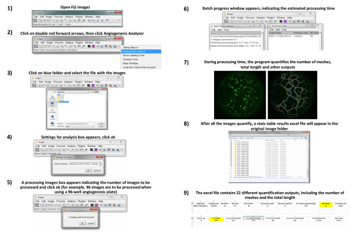
Figure 2: Quantitative analysis. All images are opened on Fiji ImageJ and batch-processed using the Angiogenesis Analyzer22. Quantification of total length and number of meshes are utilized. Please click here to view a larger version of this figure.
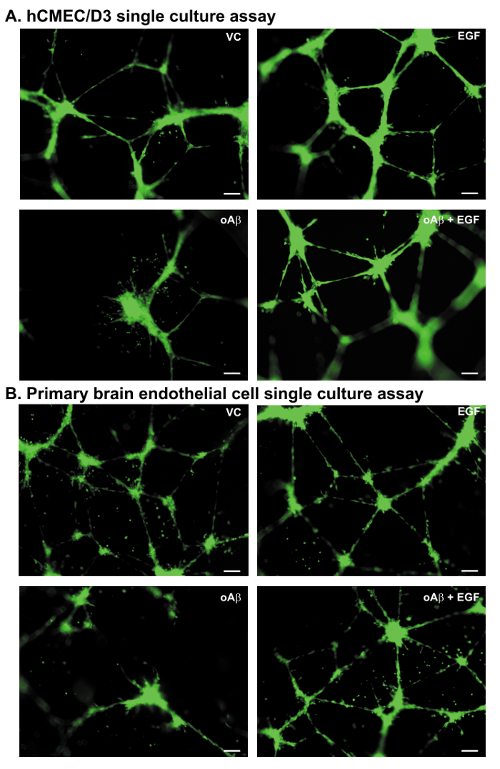
Figure 3: Meshwork disruption assays: representative images. All images are derived from experiments utilizing Paradigm 2, meshwork disruption. (A) Representative images of the hCMEC/D3 cells plated and treated with vehicle control (VC), EGF (100 nM), oAβ (100 nM), or oAβ (100 nM) + EGF (100 nM). Images at 10X magnification, Scale bar = 100 µm. (B) Representative images of the primary mouse BECs plated and treated with VC, EGF (100 nM), oAβ (100 nM) or oAβ (100 nM) + EGF (100 nM). Images at 10X magnification, green = BECs, Scale bar = 100 µm. Please click here to view a larger version of this figure.
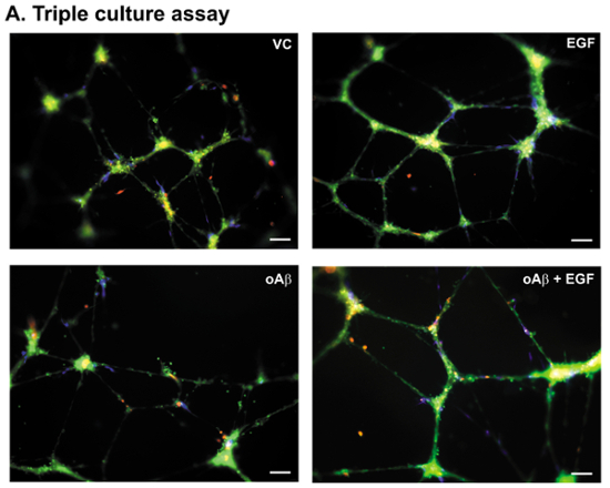
Figure 4. Triple culture assay: representative images. (A) Representative images of the hCMEC/D3 cells, primary human pericytes, and primary human astrocytes plated and treated with VC, EGF (100 nM), oAβ (100 nM), or oAβ (100 nM) + EGF (100 nM) according to the paradigm described in Figure 1B. Images at 10X magnification, green = BECs, blue = pericytes, and red (pseudocolor) = astrocytes. Scale bar = 100 µm. Please click here to view a larger version of this figure.
The methods described can be utilized to address several fundamental biological questions surrounding cerebrovascular coverage24. Specifically, they can identify which receptors and signaling pathways play a role in angiogenesis, vessel coverage in cancer tissue, and peripheral endothelial cells relevant to the brain. Examples include angiogenic growth factor receptors, nitric oxide, mitogen activated protein kinase signaling and calcium signaling25,26,27. The hCMEC/D3 and primary BECs are amendable to genetic knockdown approaches to facilitate this effort18. Mechanistic insight can be gained from culturing BECs isolated from mice with complete or endothelial cell-specific knockdown of key proteins with the methods described. Further, the triple culture assays enable in-depth analysis of the astrocyte and pericyte influence on the brain endothelial cell function. For example, pericytes and astrocytes can influence meshwork-like vessel formation through the production of soluble mediators (e.g. angiopoietin, cytokines) and also via structural support24.
The model system can also be applied to disease research. For example, the system can address whether disease- and aging-relevant stressors added exogenously promote angiogenesis and/or meshwork disruption. Stressors can be relevant to a specific disorder, e.g. oAβ, or more generalized, e.g. hydrogen peroxide, cytokines. The triple culture paradigm adds an additional layer of complexity that may improve the understanding of critical, disease-relevant mechanisms that influence BBB dynamics: delineating whether disease relevant stressors activate astrocytes and pericytes to disrupt BEC function. For both single and triple culture assays, the primary cells can be isolated from mouse models that mimic disorders of interest to further delineate functional effects.
There are a number of important considerations for adapting the meshwork-like formation and disruption assays to different cells types. The first is the seeding density. For each new cell line, a critical first step is optimizing the seeding densities in the basement membrane matrix for both the single and triple culture assays. The mesh formation is cell number dependent, as both too high and too low cell density result in a lack of meshwork formation. Further, even for the methods and cells described herein, it is prudent to conduct cell density comparisons to account for any laboratory variations in cell processing prior to conducting larger scale experiments. The second consideration is the temporal sequence of the meshwork formation. Time course experiments should be conducted to identify when meshwork-like structures form and degrade. Typically, a robust meshwork-like formation is observed at ~4 h. The third consideration is the choice of medium prior to seeding and during the experiment. BECs, pericytes and astrocytes are cultured in medium that contains FBS and a plethora of growth factors optimized for cell growth. Although it is preferred to serum and growth factor starve the BECs 24 h prior to the experiment, for certain cell types this may not be feasible. For example, primary cells with specific receptor knockdowns may require additional factors to facilitate growth. These considerations also apply during the experiment, and the choice of medium is dependent on the specific question under investigation. However, when investigating the role of exogenously added stressors and potential treatments, particularly growth factors or related compounds, the use of serum and growth factor free medium during the experiment is ideal. In addition, it may be possible to reduce the length of serum starvation of the BECs, but this is dependent on the signaling pathway under investigation. A fourth consideration is the timing of adding stressors or treatments. As for the choice of medium treatment, the timings are based on the scientific question. The meshwork formation assay is more analogous to angiogenesis, whereas the meshwork disruption assay may be more relevant for a mature BBB. In our experience, once established, the assays produce consistent data between experiments. Consistency issues are often due to seeding density discrepancies between personnel, and importantly, if components of the medium or relevant reagents are unknowingly altered.
There are limitations and areas of further development of the methods described. A strength of this procedure is that low concentrations of stressor (i.e. oAβ) can be utilized to induce meshwork disruption i.e. nM compared to µM, which are more physiologically relevant. However, this strength may also be a limitation. Indeed, higher, but still non-toxic, concentrations of oAβ may be required for drug screening purposes to increase sensitivity, which will apply to other disease relevant stressors. A limitation of the method is that the meshwork of cells is only stable over 24 h in the protocols described. Indeed, after 24 h, the cell meshworks begin to degenerate. Therefore, we add oAβ after a relatively short time (4 h) after the meshwork formation, to enable a total oAβ incubation time of 18 h. Perhaps lower cell seeding densities may enable longer treatment protocols. A further potential limitation is that the in vitro system may not mimic a stable meshwork of cells as would be observed in in vivo. Isolating the cells from aged mice may more closely mimic the in vivo scenario.
The authors have nothing to disclose.
Leon Tai is funded by University of Illinois Chicago start-up funds.
hCMEC/D3 cells | Milipore | SCC066 | |
EBM-2 basal media | Lonza | CC-3156 | |
Collagen Type 1 | ThermoFisher | A1064401 | |
HBSS, calcium, magnesium, no phenol red | ThermoFisher | 14025092 | |
HBSS, no calcium, no magnesium, no phenol red | ThermoFisher | 14175095 | |
Trypsin-EDTA (0.25%) | ThermoFisher | 25200056 | |
Final concentrations of the SingleQuot growth factor supplements for EBM2 media | Lonza | CC-4147 | |
5% FBS | Lonza | CC-4147 | |
10% Ascorbic acid | Lonza | CC-4147 | |
10% Gentamycin sulphate | Lonza | CC-4147 | |
25% Hydrocortisone | Lonza | CC-4147 | |
1/4 volume of the supplied growth factors: fibroblast growth factor, epidermal growth factor, insulin-like growth factor, vascular endothelial growth factor | Lonza | CC-4147 | |
Puromycin hydrochloride | VWR | 80503-312 | |
MEM-HEPES | Thermo Scientific | 12360-038 | |
Papain cell dissociation system (papain and DNase1) | Worthington Biochemical | LK003150 | |
Human pericytes | Sciencell | 1200 | |
Pericyte basal media | Sciencell | 1201 | |
Pericyte growth supplement | Sciencell | 1252 | |
Human Astrocytes | Sciencell | 1800 | |
Astrocyte media | Sciencell | 1801 | |
Astrocyte growth supplement | Sciencell | 1852 | |
Basement membrane (Matrigel Growth Factor Reduced) | Corning | 356231 | |
Angiogenesis m-plates (96-well) | ibidi | 89646 | |
Human Epidermal growth factor | Shenendoah Biotechnology | 100-26 | |
CellTracker green | ThermoFisher | C7025 | |
CellTracker orange | ThermoFisher | C34551 | |
CellTracker blue | ThermoFisher | C2110 | |
Poly-l-lysine | Sciencell | 0403 | |
10% Neutral Buffered Formalin | Sigma-Aldrich | HT5012-60ML | |
C57BL mice | Jackson Laboratory | na | |
PCR tube strips | GeneMate | T-3014-2 | |
Zeiss stereo discover v.8 dissecting microscope | Zeiss | na |