Here, we present a protocol for the isolation and cultivation of adult rat ventricular cardiomyocytes (ARVC). Isolated ARVC can be used for short and long-term cultivation. The isolation and cultivation of ARVC can play a key role in developing new treatment regimens for cardiac diseases.
In an intact heart, adjacent cells influence adult cardiomyocytes. With the method of isolation and cultivation of adult cardiomyocytes, a precise investigation of the behavior of these cells under specific treatments and environments is possible. This manuscript presents a protocol for successful isolation and cultivation of adult rat ventricular cardiomyocytes (ARVC).
The rat is sacrificed by cervical dislocation under deep anesthesia. Then, the heart is extracted and the aorta is uncovered. Subsequently, perfusion on the Langendorff perfusion system with calcium depletion and collagenase treatment is performed. Afterwards, ventricular tissue gets minced, re-circulated, and filtered, followed by three centrifugation steps with gradual addition of CaCl2 until physiological calcium concentration is reached. ARVC are plated on cell culture dishes. After refreshing the cell culture medium, ARVC can be cultivated for up to six days without changing the serum-containing culture medium. Isolation of ARVC is a calcium sensitive process. Small changes in the intracellular calcium concentration cause a decrease in the quality and viability of the isolated cells.
Freshly isolated ARVC are rod shaped. Within the first days of cultivation they lose the rod-shaped morphology and form pseudopodia-like structures (spreading). During this morphological formation ARVC initially degrade their contractile elements followed by a reformation through actin stress fibers and de novo sarcomerogenesis. After one week of cultivation, most ARVC show a widespread appearance with a clearly detectable cross striation. This process is sensitive to intracellular calcium concentration, as treatment with ionomycin attenuates spreading. Key markers in this process of de- and re-differentiation are β-myosin heavy chain (β-MHC), oncostatin M (OSM), and swiprosin-1 (EFHD2). Recent studies have suggested that cardiac re- and de-differentiation occurring under culture conditions mimics features seen in vivo during cardiac remodeling. Therefore, isolation and cultivation of ARVC play a key role in understanding the biology of cardiomyocytes.
Adult cardiomyocytes in vivo work as an electrical syncytium based on cell-cell contacts between myocytes. In addition, they are influenced by adjacent cells like cardiac fibroblasts, endothelial cells, neurons, and inflammatory cells1. In order to study the ability of cardiomyocytes to adapt their intracellular organization to altered load conditions, as seen during cardiac hypertrophy, which is an initial step leading to heart failure, the isolation and cultivation of adult ventricular rat cardiomyocytes (ARVC) is necessary2,3,4. Historically, cardiomyocytes were first isolated from embryonic chick hearts5,6. A few years later, the first isolation of terminally differentiated cardiomyocytes was described by using calcium depletion7. However, these adult cardiomyocytes were not calcium tolerant and could therefore not be used for functional assays. Finally, in 1976 a new protocol enabled Powell and Twist to investigate adult ventricular cardiomyocytes under physiological conditions8. As a first step, they isolated adult cardiomyocytes under low calcium concentrations and thereafter increased calcium to physiological concentrations in a stepwise procedure. Today, most protocols for the isolation and cultivation of adult cardiomyocytes work with this calcium protocol and use collagenase for the enzymatic digestion of the dense cell-cell contacts1.
For a successful cultivation, fetal calf serum (FCS) or oncostatin M (OSM) is required. ARVC perform a de- and re-differentiation with extensive structural changes including sarcomere disassembly and reformation9,10,11,12. This process is accompanied by a re-expression of fetal-type genes, like β-myosin heavy chain (β-MHC), as known from hypertrophy, and a formation of pseudopodia-like structures, also called spreading4,11,13. Furthermore, swiprosin-1 (EFHD2), a newly identified protein, plays a major role in the process of re-differentiation of cultivated ARVC11. As a result, ARVC in culture transform into widespread, polymorphic cells, which spontaneously show contractions after two to three weeks in culture2,4,14.
Recent discoveries have revealed that cardiac re- and de-differentiation as it occurs under culture conditions mimics features seen in vivo during cardiac remodeling10,15. Cardiac remodeling is a key process during cardiac diseases16. As cardiac diseases are still the main cause of death in industrialized societies, a better understanding of the biology of adult cardiomyocytes is important (WHO; 2015). Isolation and cultivation of ARVC can help to develop new strategies and medicines for the treatment of cardiac diseases. With this manuscript, a protocol for the isolation and cultivation of ARVC is provided. Furthermore, some critical parts of this method are highlighted in the discussion section.
The investigation is conducted according to the Guide for the Care and Use of Laboratory Animals published by the US National Institute of Health (NIH Publication No. 85-23, revised 1996). In general, male wistar rats aged 3 to 4 months and with an average weight of 250 – 350 g are used for this protocol. One rat heart is sufficient for 20 culture dishes (1 mL per dish; inner diameter: 35 mm) with an approximate cell density of 1.5 x 10⁴ cells/1000 mm2.
1. Preparation of Media and Reagents
2. Isolation of Adult Cardiomyocytes
3. Example Experiments
Adult cardiomyocytes in culture: Figure 1 shows an overview of freshly isolated adult cardiomyocytes 2 h after the last washing. Approximately 75% of all cardiomyocytes had a rod-shaped morphology. The remaining 25% showed an unusual appearance with a round morphology and no detectable intact cell membrane (Figure 1). At the end of cultivation (day 6), up to 15% of all cardiomyocytes showed spreading, about 10% remained in a round morphology without pseudopodia-like structures, and 75% of all cardiomyocytes presented an unusual appearance with an irregular surface and without a detectable intact cell membrane (data not shown).
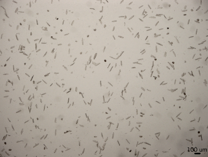
Figure 1: Overview of freshly isolated rat cardiomyocytes. The fraction of freshly isolated cardiomyocytes which showed a rod-shaped morphology amounted to 75% of cardiomyocytes, on average. The remaining 25% of cells presented an unusual appearance with an irregular surface and no detectable intact cell membrane. Recording was conducted by light microscopy 2 h after washing the freshly isolated cardiomyocytes. Light microscopy 2X magnification. Please click here to view a larger version of this figure.
With light microscopy, freshly isolated ARVC appeared rod shaped and around 100 µm in size (Figure 2A). Freshly isolated ARVC that contract spontaneously were not calcium tolerant. All cells that were round and without a detectable intact cell membrane were damaged and not viable (Figure 2A-B). In the following days, most of the rod shaped ARVC lost this morphology. Cells got rounded with a detectable intact cell membrane. These ARVC were viable. Starting at day three the latter cells formed pseudopodia-like structures. Some of these ARVC kept their rounded appearance during spreading (Figure 2B). Others converted into flat, polymorphic ARVC (Figure 2B).
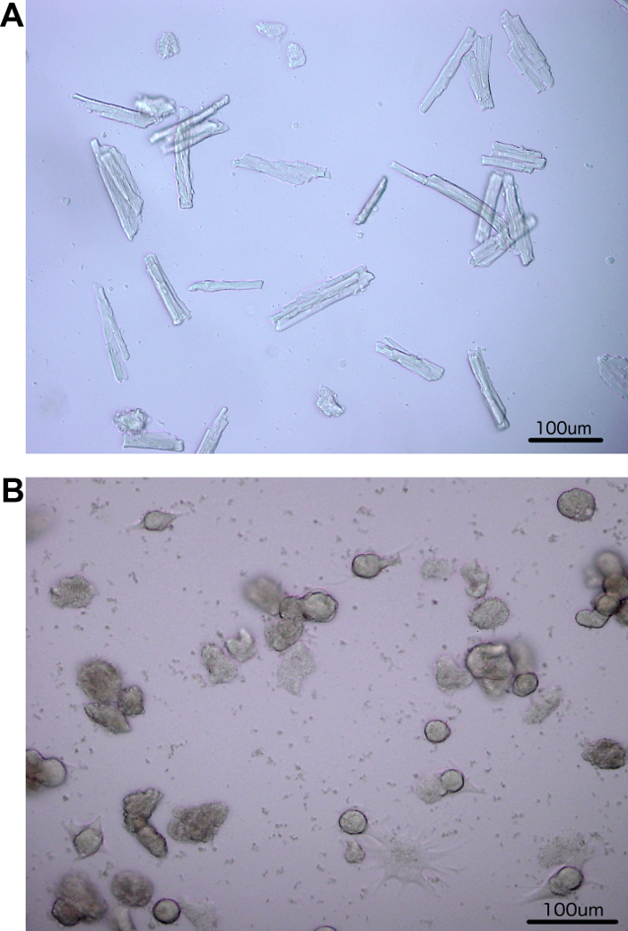
Figure 2: Isolated rat cardiomyocytes. (A) Freshly isolated ARVC were typically rod-shaped. (B) After six days in culture, pseudopodia-like structures (spreading) were clearly detectable in the now rounded ARVC. Some ARVC completely changed to a widespread morphology. ARVC with an unusual appearance displayed an irregular surface and no detectable intact cell membrane. Light microscopy 10X magnification. Please click here to view a larger version of this figure.
Freshly isolated ARVC were typically rod shaped with a clearly visible cross striation (Figure 3, Day 0). Changes in cell morphology were observed during the following days in culture. First, ARVC lost all their contractile elements (Figure 3, Days 1 and 2). This was followed by a reformation, implicating de novo sarcomerogenesis. The reformation was preceded by the formation of pseudopodia-like structures (spreading, Figure 3, Days 3 to 6). De novo sarcomerogenesis started with the appearance of actin stress fibers (Figure 3, Day 3). Additionally, actin bundles appeared in the perinuclear region and formed newly assembled sarcomeres (Figure 3, Days 4 and 5). The latter grew along the preformed actin stress fibers into the periphery (Figure 3, Day 6). At the end of the cultivation period (Day 6a), a typical cross striation from newly assembled sarcomeres in the spread ARVC was observed.
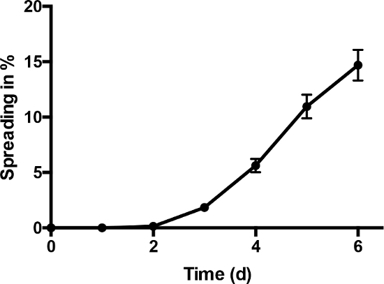
Figure 3: Fluorescence staining. The de- and re-differentiation of ARVC in culture with 20% FCS is shown. Freshly isolated ARVC with their typical rod shape (Day 0) became round by degrading sarcomeres during the first days of culture (Day 1). They lost all their contractile elements (Day 2) followed by formation of pseudopodia-like structures (spreading; Days 3 – 5) and subsequent reformation of their contractile elements indicating de novo sarcomerogenesis (Day 6). At day six in culture, cross striation was clearly detectable again (Day 6a). Staining with Phalloidin-TRITC according to the manufacturer's protocol; "arrows": pseudopodia-like structures (example shown); *: actin bundles in the perinuclear region (exemplary shown). Parts of this figure are published in11. Please click here to view a larger version of this figure.
Figure 4 displays the kinetic of the spreading process during cultivation. The fraction of ARVC showing pseudopodia-like structures at each time of examination is given as spreading in % (Figure 4). Spreading started around day three and increased constantly during the time of cultivation. 14.7% ± 1.39% of all counted ARVC showed pseudopodia-like structures after six days in cultivation.
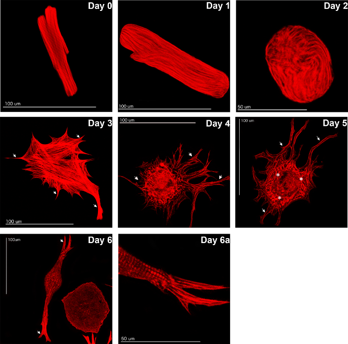
Figure 4: Spreading kinetic increase in cardiomyocytes with pseudopodia-like structures normalized to all counted cardiomyocytes (spreading in %) during six days of cultivation time (n = 33 cell preparations). Data are presented as means ± SEM. This figure is published in11. Please click here to view a larger version of this figure.
Effect of ionomycin on the spreading of ARVC: The isolation and cultivation of ARVC is a calcium sensitive process1,8. Treatment of ARVC with ionomycin (1 µM), which increases intracellular calcium concentration, caused a significant (p ≤0.01) decrease in the formation of pseudopodia-like structures compared to controls (Figure 5). When compared directly, 17.19% ± 2.45% of all counted ARVC showed spreading under control conditions but only 9.87% ± 2.77% of all counted ARVC formed pseudopodia-like structures in the presence of ionomycin (day 6 of cultivation). Thus, ionomycin reduced spreading by 42.58%.
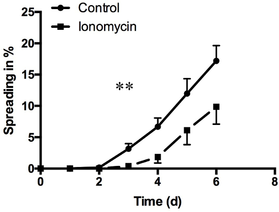
Figure 5: Spreading kinetics under treatment with ionomycin. Treatment with ionomycin (1 µM) at day 0 caused a highly significant reduction in cell spreading compared to control. Data are presented as means ± SEM; n = 4 cell preparations; Mann-Whitney-U test; * p ≤0.05; ** p ≤0.01 Please click here to view a larger version of this figure.
Additionally, ionomycin increased the percentage of ARVC with an unusual appearance compared to control conditions (Figure 6). At day six, 71.11% ± 4.65% of all counted ARVC treated with ionomycin showed an unusual appearance. However, under control conditions, only 51.35% ± 3.55% of the ARVC were categorized in this group.
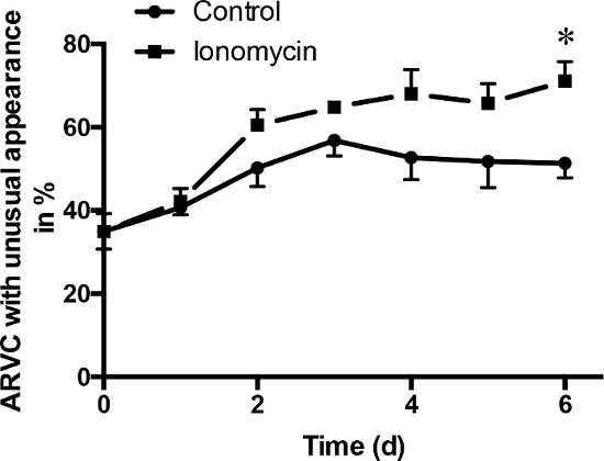
Figure 6: ARVC with an unhealthy appearance. Treatment with ionomycin (1 µM) at day 0 caused a significant increase in the number of ARVC, which showed an unusual appearance. At day 6, the difference between control and ARVC treated with ionomycin was significant. Data are presented as means ± SEM; n = 4 cell preparations; Mann-Whitney-U test; * p ≤0.05 Please click here to view a larger version of this figure.
At day 3 of cultivation, under treatment with ionomycin, qRT-PCR revealed a decrease in mRNA expression of β-MHC (p ≤0.01) and OSM, which both play a distinct role in the de-differentiation of ARVC (Figure 7A and C). Swiprosin-1, a marker for re-differentiation of ARVC was significantly downregulated, too (Figure 7B).
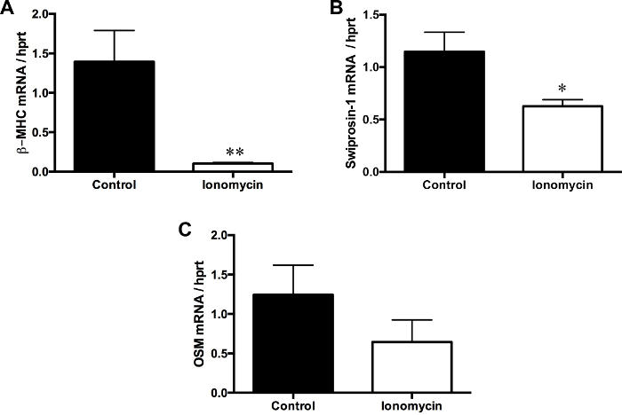
Figure 7: De- and re-differentiation of cultivated ARVC under treatment with ionomycin
(1 µM) at day 0 caused a decreased mRNA expression of oncostatin M (OSM) and β-MHC, which both play key roles in the de-differentiation of adult cardiomyocytes. Additionally, mRNA expression of Swiprosin-1, a key player in the re-differentiation of adult cardiomyocytes, was also decreased by ionomycin treatment. Day 3 of cultivation; Data are presented as means ± SEM; n = 30 cell culture plates per group; Mann-Whitney-U test; * p ≤0.05; ** p ≤0.01 Please click here to view a larger version of this figure.
Pre-plating medium |
20 mL CCT medium |
2 % Vol. Penicillin/Streptomycin (400 μL) |
4 % Vol. FCS (800 μL) |
Plating medium |
20 mL CCT medium |
2 % Vol. Penicillin/Streptomycin (400 μL) |
Washing medium |
20 mL CCT medium |
2 % Vol. Penicillin/Streptomycin (400 μL) |
Note: 4 %Vol. FCS in pre-plating medium can be replaced by 1 Vol.-% laminin (0.5 μg/cm2). Additionally, for cultivating cardiomyocytes for several days add 20 Vol.-% FCS to the washing medium. Store plating medium and washing medium by 4-8 °C until using. |
Table 1: Culture media used for cardiomyocyte isolation
The behavior of adult cardiomyocytes in vivo is influenced by many interactions with other cells (e.g., neurons, endothelial cells, fibroblasts, inflammatory cells) and the electrical syncytium which they form1. Therefore, studying stress adaptation of adult cardiomyocytes exclusively requires the isolation and cultivation of ARVC. The main effects of isolating and cultivating ARVC are: 1) disconnecting them from extracellular matrix and cell-cell contacts; 2) disconnecting them from contractile stimuli; 3) forcing them to adapt from a three-dimensional tissue to two-dimensional surroundings. Under these conditions, ARVC start de- and re-differentiation as described above and perform multiple adaptations, which are also seen during cardiac remodeling in vivo (β-adrenoceptor desensitization, reassembly of sarcomeres, etc.)4. Therefore, the isolation of adult cardiomyocytes represents a valid method to investigate these cells and their response to different treatments. These insights can be used afterwards for in vivo experiments, which would aid in avoiding unnecessary experiments and reducing the number of test animals. Certainly, some findings seen in vitro will not occur in vivo (e.g., the formation of pseudopodia-like structures). The existing cell-cell-contacts within the electrical syncytium will hamper excessive growth under physiological conditions17. Nevertheless, isolated and cultivated ARVC can be used to investigate the behavior of adult cardiomyocytes. Additionally, first trials of new treatment strategies against cardiac diseases in humans can be conducted with ARVC.
The described method for the isolation and cultivation of adult cardiomyocytes contains some critical points. To obtain successful results, the following items have to be considered.
1. Calcium tolerance: Historically, the calcium tolerance of adult cardiomyocytes was one of the most critical factors leading to a successful isolation and cultivation of adult cardiomyocytes1,7,8. Nowadays, protocols are established to ensure cultivation under physiological calcium conditions1,3. This manuscript shows the influence of a changed intracellular calcium concentration on the quality of isolated ARVC. Ionomycin, which increases intracellular calcium concentrations, caused a significant decrease in spreading and a significant increase in the number of cardiomyocytes with an unusual appearance. Furthermore, it caused a downregulation of the key markers for cardiac de- and re-differentiation: β-MHC, OSM, and Swiprosin-1. Therefore, changing the intracellular calcium concentration during cultivation hampers the capability of ARVC to adapt to new environments. Although some ARVC were able to spread and adapt (9.87% ± 2.77% of all counted ARVC; Figure 5), an accurate investigation of ARVC under these conditions is not possible. Consequently, for a precise isolation and cultivation of ARVC an established calcium protocol should be used. Additionally, it should be ensured that none of the investigated treatments interfere with the calcium hemostasis of ARVC.
2. Collagenase: There are different batches of collagenase available. Each batch shows differences in quality and effectiveness1. Therefore, the authors recommend ordering and testing samples of different batches. Additionally, the time of digestion and the amount of collagenase of each new batch used needs to be evaluated separately. Accordingly, the concentration and time of digestion in the protocol described can differ slightly to other protocols.
3. Time until heart perfusion: To ensure a high quality of ARVC, the time between extracting the heart from the body and the start of perfusion with the Langendorff system should be as short as possible. A prolonged time causes damage to the heart and results in a higher number of non-viable ARVC.
Additionally, warming the perfusion solution during the perfusion and digestion for 5 minutes after chopping the tissue is essential to yielding a good result. To avoid unnecessary damage of the biological tissue, it should be handled carefully at all-time points. Furthermore, it should be pointed out that treatment with OSM or lower concentrations of FCS also enable ARVC to de- and re-differentiate4,10,18,19. However, without these nutritive treatments, ARVC degenerate within a few days2.
In conclusion, the isolation and cultivation of ARVC is a sensitive method that offers a variety of possibilities to investigate the behavior of adult cardiomyocytes exclusively.
The authors have nothing to disclose.
The authors thank Nadine Woitasky and Peter Volk for technical assistance. Additionally, the authors thank Mrs. Claudia Lorenz (medical writer, ACCEDIS) for her help in preparing the manuscript. This manuscript was financially supported by DFG (Schlu 324/7-1).
Langendorff perfusion system | inhouse construction | double-walled with a water based heating system |
|
Tissue chopper Mc Ilwain | Cavey Laboratory Engeneering Co. Ltd. | ||
Aortic Cannula, OD 1,8 mm | inhouse construction | ||
Abdominal shears | Aeskulap | BC772R | |
Capsule forceps | Eickemeyer | 171307 | |
Dissecting scissor large | Aeskulap | BC562R | |
Dissecting scissor small | Aeskulap | BC163R | |
Mash with Polyamid | Neolab | 4-1413 | mash size 200 μm |
plastic disc | Cavey Laboratory Engeneering Co. Ltd. | ||
Collagenase Typ II | Worthington | LS004177 |