Summary

体外延时活细胞成像探索细胞向科尔蒂器官迁移

Published: December 04, 2020
doi:

Summary

在这项研究中,我们提出了一种实时成像方法,使用共聚焦显微镜观察细胞通过与含有Corti器官的耳蜗上皮的外活体孵化向受损组织移动。

Abstract

为了研究间质干细胞(MSCs)对细胞再生和治疗的影响,该方法跟踪MSC迁移和与声蜗上皮共同培养后的形态变化。Corti 的器官通过按压解剖过程中产生的里斯纳膜的一部分,在塑料盖上固定。当气缸被移除时,被玻璃缸限制的 MSC 迁移到高音表皮。它们的主要定位是在科尔蒂器官的修饰中观察到的,其方向与神经纤维的方向相似。然而,一些MSC在limbus区域被本地化,并显示出水平拉长的形状。此外,进入毛细胞区的迁移增加,在卡那霉素治疗后,MSC的形态转变为各种形式。最后,这项研究的结果表明,MSC与人工增生上皮的共生将有助于通过细胞移植和细胞再生研究,可以检查各种条件和因素的治疗发展。

Introduction

听力损失可能是先天发生的,也可以是由几个因素逐渐引起的,包括衰老、药物和噪音。听力损失往往很难治疗,因为一旦负责听力的毛细胞受损,恢复受损的功能是非常具有挑战性。据世界卫生组织统计,全世界估计有4.61亿人听力损失,占世界人口的6.1%。在听力损失者中,93%是成年人,7%是儿童。

已尝试采取若干方法治疗听力损失:值得注意的是,使用MSC的再生方法已成为一种很有前途的治疗方法。当组织受损时,MSC自然释放到循环系统中,并迁移到损伤部位,在那里它们分泌各种分子,形成促进再生的微环境2。因此,重要的是要开发一种方法,通过外部植入的MSC迁移来治疗受损组织的目标器官及其随后的分子分泌,导致强大的免疫调节,血管生成和抗凋亡,以加强恢复受损细胞功能3,4,5。

MSC 迁移到受损组织的回程过程可能是最重要的障碍。MSC 具有系统性定位机制,具有连续的系绳/滚动、激活、逮捕、迁移/迁移和迁移6、7、8等步骤。目前,正在努力确定改进这些步骤的方法。各种策略,包括基因改造,细胞表面工程,体外启动,磁导,已经测试了6,7。此外,还多次试图通过将MSC吊到受损的科克菌部位来促进听觉毛细胞的保护和再生。然而,在体内跟踪MSC是耗时和劳动密集型的,需要高度专业化的技能9。

为了解决这个问题,开发了一种方法,通过延时共焦显微镜观察耳蜗中MSC的生长情况,该显微镜可拍摄细胞在几个小时内的迁移(图1)。它开发于20世纪初 ,最近已成为研究特定细胞迁移的有力工具。
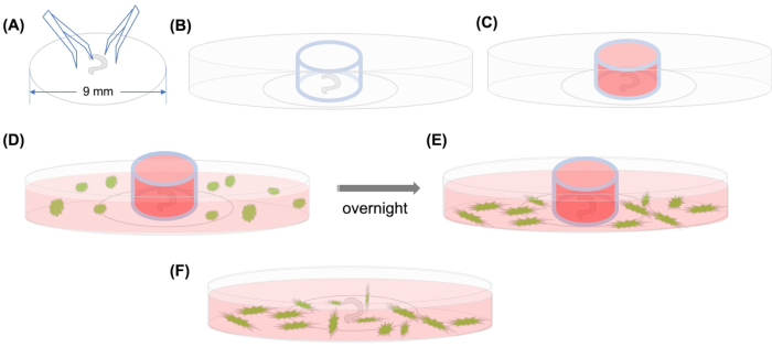
Figure 1
图1:图形摘要。A) Corti 的解剖器官使用钳子粘附在塑料盖片上后,盖片被放置在 35 毫米玻璃底的圆角微碟上,并且(B) 玻璃缸被定位。(C) 在玻璃缸内填充中等物后,在气缸外小心添加带有中型的 GFP 标签的 MSC。(E) 在一夜之间孵化后,(F) 玻璃缸被移除,图像用共聚焦显微镜拍摄。缩写:GFP = 绿色荧光蛋白;MSC=中性干细胞。请单击此处查看此图的较大版本。

Protocol

所有涉及ICR小鼠的研究协议都得到了元州医学院延世大学机构动物护理与使用委员会(IACUC)的批准。实验是根据世界医学协会的道德规范进行的。在本协议中,怀孕的ICR小鼠被保存在12/12小时的光/暗循环中,可以自由获得食物和水。 1. 科克利解剖 通过打开紫外线约30分钟,对层层流动组织培养罩进行消毒,并在使用前用70%乙醇喷洒所有表面。让表面干燥。 将…

Representative Results

在三维模式下,MSC的体外迁移已通过Transwell系统或传统的伤口愈合方法进行评估,以观察二维(2D)模式11中的迁移。科尔蒂的器官是一个复杂的结构,由各种细胞组成,如博彻细胞,克劳迪斯细胞,神细胞,支柱细胞,汉森的细胞,外毛细胞,内毛细胞,神经纤维,罗勒膜,和网状拉米纳12。当MSC被移植到由这种复杂细胞组成的组织中?…

Discussion

对MSC移植到受损部位,促进受损细胞再生进行了广泛的研究,治疗效果明显。据报道,MSC的移植和随后的分化,以恢复大鼠的听力损失引起的3硝基丙酸13。虽然李等人将MSC应用于人类,但他们在听力14方面并没有取得任何显著改善。直到最近,通过MSC移植,还进行了近12项实验,以恢复啮齿动物模型的听力。虽然由于异质性,结果有些不清楚,但有迹象表明,MSC…

Disclosures

The authors have nothing to disclose.

Acknowledgements

这项工作得到了韩国国家研究基金会(NRF-2018-R1D1A1B07050175,HURF-2017-66)和韩国国家研究基金会(NRF)和哈莱姆大学研究基金的研究赠款的支持。

Materials

10X PBS Buffer GenDEPOT P2100-104
4% Formalin T&I BPP-9004
Ampicillin sigma  A5354-10ml
BSA sigma  A4503-100G
confocal dish SPL 200350
confocal microscope  ZEISS LSM800
coverslip SPL 20009
DMEM/F12 Gibco 10565-018
Fetal Bovine Serum Thermo Fisher scientific 16140071
Fluorsheild with DAPI sigma  F6057
Forcep Dumont 0508-L5-P0
HBSS Thermo Fisher scientific 14065056
HEPES Thermo Fisher scientific 15630080
N2 supplement Gibco 17502-048
Phalloidin-iFluor 647 Reagent abcam ab176759
Stage Top Incubator TOKAI HIT WELSX
Strain C57BL/6 mouse messenchymal stem cells with GFP cyagen MUBMX-01101
Triton X-100 sigma  T8787

References

  1. Brown, C. S., Emmett, S. D., Robler, S. K., Tucci, D. L. Global hearing loss prevention. Otolaryngologic Clinics of North America. 51 (3), 575-592 (2018).
  2. Chamberlain, G., Fox, J., Ashton, B., Middleton, J. Concise review: mesenchymal stem cells: their phenotype, differentiation capacity, immunological features, and potential for homing. Stem Cells. 25 (11), 2739-2749 (2007).
  3. Fu, X., et al. Mesenchymal stem cell migration and tissue repair. Cells. 8 (8), (2019).
  4. Uder, C., Brückner, S., Winkler, S., Tautenhahn, H. M., Christ, B. Mammalian MSC from selected species: Features and applications. Cytometry A. 93 (1), 32-49 (2018).
  5. Rojewski, M. T., et al. Translation of a standardized manufacturing protocol for mesenchymal stromal cells: A systematic comparison of validation and manufacturing data. Cytotherapy. 21 (4), 468-482 (2019).
  6. Ullah, M., Liu, D. D., Thakor, A. S. Mesenchymal stromal cell homing: Mechanisms and strategies for improvement. iScience. 15, 421-438 (2019).
  7. Ahn, Y. J., et al. Strategies to enhance efficacy of SPION-labeled stem cell homing by magnetic attraction: a systemic review with meta-analysis. International Journal of Nanomedicine. 14, 4849-4866 (2019).
  8. Alon, R., Ley, K. Cells on the run: shear-regulated integrin activation in leukocyte rolling and arrest on endothelial cells. Current Opinion in Cell Biology. 20 (5), 525-532 (2008).
  9. Sykova, E., Jendelova, P. In vivo tracking of stem cells in brain and spinal cord injury. Progress in Brain Research. 161, 367-383 (2007).
  10. Landegger, L. D., Dilwali, S., Stankovic, K. M. Neonatal murine cochlear explant technique as an in vitro screening tool in hearing research. Journal of Visualized Experiments. (124), e55704 (2017).
  11. Pijuan, J., et al. In vitro cell migration, invasion, and adhesion assays: From cell imaging to data analysis. Frontiers in Cell and Developmental Biology. 7, 107 (2019).
  12. Rask-Andersen, H., et al. Human cochlea: anatomical characteristics and their relevance for cochlear implantation. The Anatomical Record. 295 (11), 1791-1811 (2012).
  13. Kamiya, K., et al. Mesenchymal stem cell transplantation accelerates hearing recovery through the repair of injured cochlear fibrocytes. The American Journal of Pathology. 171 (1), 214-226 (2007).
  14. Lee, H. S., Kim, W. J., Gong, J. S., Park, K. H. Clinical safety and efficacy of autologous bone marrow-derived mesenchymal stem cell transplantation in sensorineural hearing loss patients. Journal of Audiology and Otology. 22 (2), 105-109 (2018).
  15. Vanden Berg-Foels, W. S. In situ tissue regeneration: chemoattractants for endogenous stem cell recruitment. Tissue Engineering Part B: Reviews. 20 (1), 28-39 (2014).
  16. Parker, M., Brugeaud, A., Edge, A. S. Primary culture and plasmid electroporation of the murine organ of Corti. Journal of Visualized Experiments. (36), e1685 (2010).
  17. Ogier, J. M., Burt, R. A., Drury, H. R., Lim, R., Nayagam, B. A. Organotypic culture of neonatal murine inner ear explants. Frontiers in Cellular Neuroscience. 13, 170 (2019).
  18. Oshima, K., et al. Mechanosensitive hair cell-like cells from embryonic and induced pluripotent stem cells. Cell. 141 (4), 704-716 (2010).
check_url/61947?article_type=t

Play Video

Cite This Article
Park, J., Lee, S. H., Park, D. J., Seo, Y. J., Kim, S. K. In vitro Time-lapse Live-Cell Imaging to Explore Cell Migration toward the Organ of Corti. J. Vis. Exp. (166), e61947, doi:10.3791/61947 (2020).

View Video