Summary

通过表面等离子体共振 设计 抗病毒药物

Published: June 14, 2022
doi:

Summary

本协议描述了用于SPR结合测定的新工具,以检查CV-N与HA,S糖蛋白,相关杂交型聚糖和高甘露糖寡糖的结合。SPR用于测定将二聚体或单体CV-N与这些聚糖结合的KD

Abstract

表面等离子体共振(SPR)用于测量血凝素(HA)与结构域交换的氰基蛋白-N(CV-N)二聚体的结合,并监测甘露糖基化肽与CV-N高亲和力结合位点之间的相互作用。据报道,病毒包膜加标 gp120、HA 和埃博拉糖蛋白 (GP) 1,2 可结合二聚体 CVN2 上的高亲和力和低亲和力结合位点。二甘露糖基化的HA肽也在两个低亲和力结合位点与CVN2的工程分子结合,CVN2具有相应配体的高亲和力位点并发生突变以取代碳水化合物结合口袋中的稳定二硫键,从而确认多价结合。HA与假抗体CVN2的一个高亲和力结合位点显示,解离常数(KD)为275 nM,通过寡聚化进一步中和人类免疫缺陷病毒1型(HIV-1)。将结构域交换的CVN2中的二硫键数量从4个减少到2个,通过将胱氨酸取代为谷氨酸和精氨酸的极性残基对,导致与HA的结合亲和力降低。在最强的相互作用中,埃博拉GP1,2与CVN2结合,使用没有跨膜结构域的包膜聚糖在较低的纳摩尔范围内具有两个高亲和力结合位点。在本研究中,多特异性单体CV-N与严重急性呼吸综合征冠状病毒2(SARS-CoV-2)刺突(S)糖蛋白的结合在K D = 18.6μM处测量,与纳摩尔KD与其他病毒尖峰相比,并通过其在中摩尔范围内的受体结合域μ中摩尔范围内。

Introduction

Tetherin相关的抗病毒活性由干扰素-α诱导,它包含基于蛋白质的Tether,导致完全形成的病毒粒子保留在感染的细胞表面1。Tetherin糖基化在抑制病毒释放方面的必要性仍然不确定,这意味着糖基化模式对重组表达聚糖的重要性对于体外研究12,这取决于(在流感病毒的情况下)表面表达的流感血凝素HA34的构象.已经注意到,与N-连接糖基化相关的寡糖的修饰足以限制HIV-1型释放2,而二聚化在防止病毒释放中起着至关重要的作用,因此涉及跨膜结构域或糖基磷脂酰肌醇(GPI)锚,用于拴系出芽病毒粒子5.描述了人和鼠系留蛋白的独特功能,以阻断多种包膜病毒、逆转录病毒和丝状病毒。BST-2/tetherin是一种先天免疫1,6的干扰素诱导抗病毒蛋白具有广谱抗病毒活性,并被包膜糖蛋白5拮抗以易位系留蛋白或破坏Tetherin6的结构。例如,表面表达的包膜糖蛋白HA和神经氨酸酶对甲型流感病毒的拮抗作用是众所周知的,以菌株特异性方式7,促进宿主受体结合位点8的识别。在与HA上快速定制的聚糖屏蔽相互作用的化学计量学中研究聚糖靶向抗体,从而与甲型H3N2和H1N1亚型4结合亲和力。

为了阐明抗病毒药物与病毒包膜尖峰(即碳水化合物配体)之间的结合机制,以及互补的免疫学和光谱学方法,对单甘露糖、二甘露糖和三甘露糖部分进行了化学合成。甘露糖基化肽是通过糖基{β}-过乙酸酯的叠氮基化为1,2-反式糖基叠氮化物转化9创建的,模仿了在危及生命的病毒表面上通常发现的N-乙酰氨基葡萄糖和高甘露糖低聚糖。三唑生物等排体用于模拟形成 HA 肽10 甘露糖基化残基的键,并促进与 HA 头结构域上第二个 N 连接糖基化点周围的抗病毒 CV-N 衍生物的位点特异性相互作用(HA 顶部有 4 个 N-连接聚糖 N54、N97、N181、N301)81112.谷氨酸(Glu)和精氨酸(Arg)之间的相互作用以及由此产生的螺旋偶极子表现出模型肽和蛋白质的良好稳定性,但使用SPR可视化。如果与通过直接抑制聚糖部分上的受体结合来识别HA10上的单个化学合成糖基化位点相比,则显示四位点突变Fc结构与其受体的更高亲和力可在体内引发效应功能,揭示连接到Fc突变体的N-连接聚糖的不相关组成有待机械确定13

CV-N 显示出针对 HIV 14,15、流感 16 和埃博拉病毒的抗病毒活性,这是通过纳摩尔结合包膜刺突蛋白12,17,1819 上的高甘露糖寡糖修饰介导的。确定流感HA与CV-N中的一个高亲和力碳水化合物结合位点(H)或共价连接的二聚体CVN2中的两个Hs结合分别具有平衡解离常数(K D)= 5.7 nM(图1A和KD = 2.7 nM。CV-N和CVN2都含有另外一个或两个低亲和力碳水化合物结合位点(L)s12172021埃博拉GP1,2与CVN2的2H结合,亲和力在较低的纳摩尔范围(KD = 26 nM)。与埃博拉GP1,2和HA结合的CV-N WT表现出从K D = 34 nM到KD = 5.7 nM的亲和力(A/New York/55/04)12。凝集素,如CV-N,专门针对病毒包膜上的高甘露糖聚糖,进一步抑制丙型肝炎病毒、SARS-CoV、疱疹病毒、马尔堡病毒和麻疹病毒22的复制。

CV-N小分子已经彻底研究了20多年,因为它的功能可以结合多种病毒以抑制病毒进入1618。结构分析和结合亲和力测定表明,通过微摩尔范围内的二价结合,在结构域交换的CVN2二聚体中交联两个L,以增强对病毒包膜糖蛋白1019的亲和力。Manα1-2Manα在Man(8)D1D3臂上的选择性结合,Man(9)包括位于相反蛋白质原型体20上的两个具有不同亲和力的结合位点,从而达到纳摩尔结合亲和力(图1B)。因此,CVN2被认为是一种假抗体,因为它在HIV gp120上结合表位的应用,类似于病毒中和抗体17。在本文中,作者有兴趣研究CVN2通过其受体结合域(RBD) SARS-CoV-2尖峰的潜在结合。固定化人血管紧张素转换酶 (ACE)-2 与 SARS-CoV-2 RBD 的结合曲线导致这种生物学相关的结合相互作用的 KD = 4.7 nM23

相比之下,选定的免疫球蛋白类别可识别特定且一致的结构蛋白模式,这些模式为膜锚定的HA区域赋予亲和力成熟的底物24。CV-N在几乎所有甲型和乙型流感病毒中都显示出高效的活性16,它是一种广泛的中和抗病毒剂。我们对HA1和HA2茎上靶向表位位置的了解不完整,这些表位可能涉及通过高度中和抗体靶向聚糖的表位结构,并与凝集素结合25相比。

Figure 1
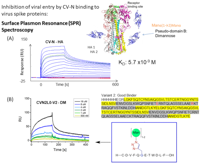
图 1:CV-N 与病毒包膜尖峰的 SPR 结合测定示意图。 (A)用于CV-N与配体结合的SPR测定:HA全长蛋白(90kDa)。动力学数据集(5120、2560、1280、640、320、160、80、40、20、10、5、2.5、0 nM)显示与甲型流感HA/纽约/55/04(H3N2)的实时双重参照结合。(B)CVN2L0变体V2与固定配体DM结合,浓度范围为500nM至16μM。 序:L残基以黄色突出显示。H 残基以灰色突出显示。E58和R73是野生型蛋白中半胱氨酸的替代品,使V2成为稳定的蛋白质折叠,具有三个而不是四个二硫键 请单击此处查看此图的大图。

虽然膜-远端 HA 顶部的聚糖屏蔽诱导与 CV-N 12 的高亲和力结合,但在其低亲和力位点1012 进一步观察到CVN2 与 HA 顶部二硫键相邻的 HA 结合。通过CV-N在碳水化合物结合中鉴定各种极性相互作用和相互作用位点。通过在结合位点生成敲除变体来验证这些相互作用,以将结合亲和力与计算机预测的糖基化相关联12。因此,该项目旨在比较先前测试的化学甘露糖基化HA肽与SARS相关2019-nCoV尖峰和SARS-CoV-2的短肽序列的结合亲和力和特异性,这些短肽序列由少量不同的N-连接糖基化位点和O-连接糖基化修饰。使用冷冻电子显微镜和结合测定,Pinto及其同事报告了一种单克隆抗体S309,该抗体可能识别SARS-CoV-2刺突蛋白上的表位,该表位含有沙贝病毒亚属内的保守聚糖,而不会与受体附着竞争26。本研究的方案描述了设计、表达和表征 CV-N 变体对于使用 SPR 技术研究 CV-N 和 CVN2 如何与糖基化蛋白质和合成甘露糖基化肽结合的重要性1012.

串联连接的二聚体CVN2L027和结合位点变体(V2-V5)重组表达,变体具有二硫键替代物(C58E和C73R)(图2A)。此外,制备具有单点突变E41A的突变体,因为该位置已被视为分子间交叉接触残基。这种突变体是另一种有趣的分子,用于凝集素和高甘露糖寡糖之间的SPR结合测量,破译结合结构域,并允许与二聚体形式进行比较。CVN2的结构域交换晶体结构显示出一个灵活的接头,其延伸在49到54个残基之间。这两个结构域可以作为刚体继续围绕铰链移动,通过分子内结构域相互作用(结构域A-残基1-39;90-101-与结构域B-残基40-89)或通过分子间结构域交换产生二聚体[域A(第一个单体)与结构域B(第二个),以及域B(第一个单体)与结构域A(第二个拷贝)]。除了Glu4128之外,两个原型的A和B结构域之间没有密切的相互作用。CV-N的基因可以使用重复PCR方法与40-mer合成的寡核苷酸29进行开发,然后亚克隆到pET11a的NdeI和BamHI位点中,以转化为(电穿孔)为电感受态细胞,如Keeffe,J.R.27所述。该蛋白质用于实现各自的晶体结构(PDB ID 3S3Y),包括一个N-末端6-组氨酸纯化标签,后跟一个因子Xa蛋白酶切割位点。定点诱变用于进行点突变、切换密码子以及插入或删除单个或多个碱基或密码子以进行氨基酸交换。这些转化为蛋白质功能和结构提供了宝贵的见解。重组表达和纯化的CV-N、CVN2和CVN3已经过生物物理研究20,2127,生产成本低廉,因此用于表征固定在SPR传感器芯片上的聚糖的结合测定。传统的酶联免疫吸附测定(ELISA)在聚糖配体的固定化技术方面提供了较低的重现性,并将各种结合位点变体的实时结合(如SPR所示)转化为终点测定。

结合亲和变体CVN2L0-V2(具有二硫键取代10的同源二聚体CV-N的完整折叠)在大肠杆菌(大肠杆菌)中用His标签表达,在Ni-NTA 柱上应用亲和色谱纯化并使用SPR测试与HA(H3N2),单甘露糖基化HA肽和二甘露糖基化HA肽的结合。 化学甘露糖基化肽或HA和S蛋白都是配体和胺偶联到亲水芯片表面通过反应性酯或生物素-链霉亲和素蛋白工程。将相同的顺序运行程序应用于这些配体,注入CV-N的各种稀释液和CV-N(和CVN2)的变体,以获得分子相互作用分析的动力学信息,如下所述30。RBD固定化SPR传感器芯片用于CV-N与S肽的结合研究,并将亲和力与SARS-CoV-2与人ACE2的结合进行比较。

Protocol

在本研究中,CVN小泛素样修饰剂(SUMO)融合蛋白已用于酶联免疫吸附测定而不是CV-N,适用于基于细胞的测定。根据标准方案12,商业获得重组全长甲型流感病毒HA H3蛋白(见材料表)或在哺乳动物HEK293细胞系和杆状病毒感染的昆虫细胞中表达。武汉-1刺突蛋白在哺乳动物HEK293细胞中表达。单甘露糖基化肽(MM)和二甘露糖基化肽(DM)的合成允许检测CVN2和单甘露糖基?…

Representative Results

在三个独立的SPR实验中测试二聚结构域交换的CVN2L0分子是否与HA顶部区域结合,并以KD值表示结合亲和力。假设结构域B包含H结合位点,其通过取代二硫键进入离子残基而受到影响,并且结构域A形成L10,18。首先测试CVN2L0和变体V2(三个二硫键)和V5(两个二硫键)的单次进样是否与HA耦合传感器芯片结合,以估计结合能力,然后再使用自动进样器…

Discussion

CV-N的结合亲和力与功能结合位点的数量相关[当工程为结构域交换二聚体时,结构域B上为2H,结构域A为2L]。在大肠杆菌中表达具有改变的结合亲和力的变体(CVN2L0-V2,CV-N的同源二聚体稳定折叠,包括二硫键敲除),纯化,并使用SPR10对HA蛋白(H3N2)结合进行阳性测试,并且在HA与H或L碳水化合物结合位点和KD1 = 49 nM和KD2 = 8μM38时显示出构象…

Disclosures

The authors have nothing to disclose.

Acknowledgements

作者感谢维也纳工业大学生物技术和微生物学系和维也纳医科大学肾脏病学和透析学系第三医学系的Christian Derntl博士,特别是Markus Wahrmann博士的技术和科学支持。哺乳动物细胞中的蛋白质表达得到了维也纳自然资源与生命科学大学(BOKU)生物技术系的支持。作者希望对来自德国杜塞尔多夫XanTec生物分析的Nico Dankbar博士表示深深的感谢,感谢他对进行SPR结合测定的有益科学讨论。

Materials

Äkta primeplus Cytiva
Amicon tubes Merck C7715
Ampillicin Sigma-Aldrich A5354
Beckmann Coulter Cooler Allegra X-30R centrifuge Beckman Coulter B06320
Cell spreader Sigma-Aldrich HS86655 silver stainless steel, bar L 33 mm
Custom DNA Oligos Sigma-Aldrich OLIGO
Custom Gensynthesis GenScript #1390661  cloning vector: pET27b(+) 
Cytiva HBS-EP+ Buffer 10, 4x50mL Thermo Scientific 50-105-5354
Dionex UlitMate 3000 Thermo Scientific IQLAAAGABHFAPBMBFB
Dpn I restriction enzyme (10 U/μL)  Fisher Scientific ER1701
DTT Merck DTT-RO
EDC Merck 39391
EDTA Merck E9884
Eppendorf Safe-Lock Tubes Eppendorf 30120086
Eppendorf Safe-Lock Tubes Eppendorf 30120094
Eppendorf Minispin and MiniSpin Plus personal microcentrifuge Sigma-Aldrich Z606235
Ethanol Merck 51976
Ethanolamine HCl Merck E6133
Falcon 50mL Conical Centrifuge Tubes Fisher Scientific 14-432-22
Falcon 14 mL Round Bottom Polystyrene Test Tube, with Snap Cap, Sterile, 25/Pack Corning 352057
Glucose Merck G8270
Glycine HCl Merck 55097
HA H3 protein Abcam ab69751
HEPES Merck H3375
His-select Ni2+ Merck H0537
Imidazole Merck I2399
IPTG Merck I6758
Kanamycin A Sigma-Aldrich K1377
Kromasil 300-5-C4 Nouryon
LB agar Merck 52062
LB agar Merck 19344
LB Lennox Merck L3022
Lysozyme Merck 10837059001
Magnesium chloride Merck M8266
Magnesium sulfate Merck M7506
NaH2P04 Merck S0751
NanoDrop UV-Vis2000c spectrophotometer Thermo Scientific ND2000CLAPTOP
NaOH Merck S5881
NHS Merck 130672
NZ amine (casein hydrolysate) Merck C0626
PBS Merck 806552
PD MidiTrap G-10 Sigma-Aldrich GE28-9180-11
Peptone Merck 70171
pET11a Merck Millipore (Novagen) 69436 
PMSF Merck PMSF-RO
QIAprep Spin Miniprep Kit (1000) Qiagen 27106X4
Reichert Software Package Autolink1-1-9 Reichert
Reichert SPR SR7500DC Dual Channel System Reichert
Scrubber2-2012-09-04 for data analysis Reichert
SDS Merck 11667289001
Site-directed mutagenesis kit incl pUC18 control plasmid Stratagene #200518
Sodim chloride Merck S9888
Sodium acetate.Trihydrate Merck 236500
SPR sensor chip C19RBDHC30M XanTec bioanalytics SCR C19RBDHC30M
SPR sensor chip CMD500D XanTec bioanalytics SCR CMD500D
Sterilin Standard 90mm Petri Dishes Thermo Scientific 101R20
TBS Merck T5912 10x, solution
Triton-X100 Merck T8787
Tryptone Merck 93657
Tween20 Merck P1379
Vortex-Genie 2 Mixer Merck Z258423
X-gal Merck XGAL-RO
XL1-Blue Supercompetent Cells Stratagene #200236
Yeast extract Merck Y1625

References

  1. Perez-Caballero, D., et al. Tetherin inhibits HIV-1 release by directly tethering virions to cells. Cell. 139 (3), 499-511 (2009).
  2. Waheed, A. A., Gitzen, A., Swiderski, M., Freed, E. O. High-mannose but not complex-type glycosylation of tetherin is required for restriction of HIV-1 release. Viruses. 10 (1), 26 (2018).
  3. Wilson, I. A., et al. Structure of the haemagglutinin membrane glycoprotein of influenza virus at 3 A resolution. Nature. 289 (5796), 366-373 (1981).
  4. Otterstrom, J. J., et al. Relating influenza virus membrane fusion kinetics to stoichiometry of neutralizing antibodies at the single-particle level. Proceedings of the National Academy of Sciences of the United States of America. 111 (48), 5143-5148 (2014).
  5. Kaletsky, R. L., Francica, J. R., Agrawal-Gamse, C., Bates, P. Tetherin-mediated restriction of filovirus budding is antagonized by the Ebola glycoprotein. Proceedings of the National Academy of Sciences of the United States of America. 106 (8), 2886-2891 (2009).
  6. Tokarev, A., Skasko, M., Fitzpatrick, K., Guatelli, J. Antiviral activity of the interferon-induced cellular protein BST-2/tetherin. AIDS Research and Human Retroviruses. 25 (12), 1197-1210 (2009).
  7. Gnirss, K., et al. Tetherin sensitivity of influenza A viruses is strain specific: Role of hemagglutinin and neuraminidase. Journal of Virology. 89 (18), 9178-9188 (2015).
  8. Fleury, D., et al. A complex of influenza hemagglutinin with a neutralizing antibody that binds outside the virus receptor binding site. Nature Structural Biology. 6 (6), 530-534 (1999).
  9. Salunke, S. B., et al. Iron(III) chloride as an efficient catalyst for stereoselective synthesis of glycosyl azides and a cocatalyst with Cu(0) for the subsequent click chemistry. Chemical Communication. (Camb). 47 (37), 10440-10442 (2011).
  10. Schilling, P. E., et al. Mannosylated hemagglutinin peptides bind cyanovirin-N independent of disulfide-bonds in complementary binding sites. RSC Advances. 10 (19), 11079-11087 (2020).
  11. Fleury, D., Wharton, S. A., Skehel, J. J., Knossow, M., Bizebard, T. Antigen distortion allows influenza virus to escape neutralization. Nature Structural Biology. 5 (2), 119-123 (1998).
  12. Maier, I., Schiestl, R. H., Kontaxis, G. Cyanovirin-N binds viral envelope proteins at the low-affinity carbohydrate binding site without direct virus neutralization ability. Molecules. 26 (12), 3621 (2021).
  13. Ahmed, A. J., Keremane, S. R., Vielmetter, J., Bjorkman, P. J. Structural characterization of GASDALIE Fc bound to the activating Fc receptor FcγRIIIa. Journal of Structural Biology. 194 (1), 78-89 (2016).
  14. Boyd, R. Discovery of cyanovirin-N, a novel human immunodeficiency virus-inactivating protein that binds viral surface envelope glycoprotein gp120: potential applications to microbicide development. Antimicrobial Agents and Chemotherapy. 41 (7), 1521-1530 (1997).
  15. Bolmstedt, A. J., O’Keefe, B. R., Shenoy, S. R., McMahon, J. B., Boyd, M. R. Cyanovirin-N defines a new class of antiviral agent targeting N-linked, high-mannose glycans in an oligosaccharide-specific manner. Molecular Pharmacology. 59 (5), 949-954 (2001).
  16. O’Keefe, B. R., et al. Potent anti-influenza activity of cyanovirin-N and interactions with viral hemagglutinin. Antimicrobial Agents and Chemotherapy. 47 (8), 2518-2525 (2003).
  17. Shenoy, S. R., et al. Multisite and multivalent binding between cyanovirin-N and branched oligomannosides: calorimetric and NMR characterization. Chemical Biology. 9 (10), 1109-1118 (2002).
  18. Bewley, C. A., Kiyonaka, S., Hamachi, I. Site-specific discrimination by cyanovirin-N for alpha-linked trisaccharides comprising the three arms of Man(8) and Man(9). Journal of Molecular Biology. 322 (4), 881-889 (2002).
  19. Barrientos, L. G., Matei, E., Lasala, F., Delgado, R., Gronenborn, A. M. Dissecting carbohydrate-Cyanovirin-N binding by structure-guided mutagenesis: functional implications for viral entry inhibition. Protein Engineering Design & Selection. 19 (12), 525-535 (2006).
  20. Bewley, C. A., Otero-Quintero, S. The potent anti-HIV protein cyanovirin-N contains two novel carbohydrate binding sites that selectively bind to Man(8) D1D3 and Man(9) with nanomolar affinity: implications for binding to the HIV envelope protein gp120. Journal of the American Chemical Society. 123 (17), 3892-3902 (2001).
  21. Bewley, C. A. Solution structure of a cyanovirin-N:Man alpha 1-2Man alpha complex: structural basis for high-affinity carbohydrate-mediated binding to gp120. Structure. 9 (10), 931-940 (2001).
  22. Jensen, S. M. R., et al. Differential inhibitory effects of Cyanovirin-N, Griffithsin, and Scytovirin on entry mediated by envelopes of gammaretroviruses and deltaretroviruses. Journal of Virology. 88 (4), 2327-2332 (2014).
  23. Lan, J., et al. Structure of the SARS-CoV-2 spike receptor-binding domain bound to the ACE2 receptor. Nature. 581 (7807), 215-220 (2020).
  24. Lingwood, D., et al. Structural and genetic basis for development of broadly neutralizing influenza antibodies. Nature. 489 (7417), 566-570 (2012).
  25. Ekiert, D. C., et al. Antibody recognition of a highly conserved influenza virus epitope. Science. 324 (5924), 246-251 (2009).
  26. Pinto, D., et al. Cross-neutralization of SARS-CoV-2 by a human monoclonal SARS-CoV antibody. Nature. 583 (7815), 290-295 (2020).
  27. Keeffe, J. R., et al. Designed oligomers of cyanovirin-N show enhanced HIV neutralization. Proceedings of the National Academy of Sciences of the United States of America. 108 (34), 14079-14084 (2011).
  28. Yang, F., et al. Crystal structure of cyanovirin-N, a potent HIV-inactivating protein, shows unexpected domain swapping. Journal of Molecular Biology. 288 (3), 403-412 (1999).
  29. Stemmer, W. P., Crameri, A., Ha, K. D., Brennan, T. M., Heyneker, H. L. Single-step assembly of a gene and entire plasmid from large numbers of oligodeoxyribonucleotides. Gene. 164 (1), 49-53 (1995).
  30. Fischer, M. J. E., Mol, N., Fischer, M. Amine coupling through EDC/NHS: A practical approach. Surface Plasmon Resonance. Methods in Molecular Biology (Methods and Protocols). 627, (2010).
  31. Novoradovsky, A., et al. Computational principles of primer design for site directed mutagenesis. Technical Proceedings of 2005 NSTI Nanotechnology Conference and Trade Show. , 532-535 (2005).
  32. . QuikChange Site-Directed MutagenesisKit, User Manual Available from: https://users.drew.edu/jliu3/Docs/Stratagene%20Quikchange%20mutagenesis.pdf#:~:text=The%20QuikChange%20sitedirected%20mutagenesis%20kit%20is%20used%20to (2005)
  33. Laemmli, U. K. Cleavage of structural proteins during the assembly of the head bacteriophage T4. Nature. 227 (5259), 680-685 (1970).
  34. . Expasy.org Available from: https://web.expasy.org/cgi-bin/protparam/protparam (2022)
  35. Karlsson, R. Real-time competitive kinetic analysis of interactions between low-molecular-weight ligands in solution and surface-immobilized receptors. Analytical Biochemistry. 221 (1), 142-151 (1994).
  36. Schuck, P., Zhao, H. The role of mass transport limitation and surface heterogeneity in the biophysical characterization of macromolecular binding processes by SPR biosensing. Methods Molecular Biology. 627, 15-54 (2020).
  37. Barnes, O., et al. SARS-CoV-2 neutralizing antibody structures inform therapeutic strategies. Nature. 588 (7839), 682-687 (2020).
  38. . Using reichert Surface Plasmon Resonance (SPR) for Antiviral Development, Application Note 28 Available from: https://www.reichertspr.com/clientuploads/directory/application_notes/Application_Note_28__Using_Reichert_Surface_Plasmon_Resonance_for_Antiviral_Testing.pdf (2022)
  39. Sundberg, E. J., Andersen, P. S., Gorshkova, I. I., Schuck, P., Schuck, P. Surface plasmon resonance biosensing in the study of ternary systems of interacting proteins. Protein Interactions: Biophysical Approaches for the Study of Complex Reversible Systems. 5, 97-141 (2007).
  40. . Method Development Notes Available from: https://www.reichertspr.com/applications/method-development-notes/ (2022)
  41. Angulo, J., Enríquez-Navas, P. M., Nieto, P. M. Ligand-receptor binding affinities from saturation transfer difference (STD)-NMR spectroscopy: the binding Isotherm of STD initial growth rates. Chemistry. 16 (26), 7803-7812 (2010).
  42. Goldflam, M., Tarragó, T., Gairí, M., Giralt, E. NMR studies of protein-ligand interactions. Methods in Molecular Biology. 831, 233-259 (2012).
  43. Kumar, S., Maurya, V. K., Prasad, A. K., Bhatt, M. L. B., Saxena, S. K. Structural, glycosylation and antigenic variation between 2019 novel coronavirus (2019-nCoV) and SARS coronavirus (SARS-CoV). Virusdisease. 31 (1), 13-21 (2020).
check_url/63541?article_type=t&slug=engineering-antiviral-agents-via-surface-plasmon-resonance

Play Video

Cite This Article
Maier, I. Engineering Antiviral Agents via Surface Plasmon Resonance. J. Vis. Exp. (184), e63541, doi:10.3791/63541 (2022).

View Video