Although challenging, the isolation of pulmonary endothelial cells is essential for studies on lung inflammation. The present protocol describes a procedure for the high-yield, high-purity isolation of macrovascular and microvascular endothelial cells.
The availability of cells isolated from healthy and diseased tissues and organs represents a key element for personalized medicine approaches. Although biobanks can provide a wide collection of primary and immortalized cells for biomedical research, these do not cover all experimental needs, particularly those related to specific diseases or genotypes. Vascular endothelial cells (ECs) are key components of the immune inflammatory reaction and, thus, play a central role in the pathogenesis of a variety of disorders. Notably, ECs from different sites display different biochemical and functional properties, making the availability of specific EC types (i.e., macrovascular, microvascular, arterial, and venous) essential for designing reliable experiments. Here, simple procedures to obtain high-yield, virtually pure human macrovascular and microvascular endothelial cells from the pulmonary artery and lung parenchyma are illustrated in detail. This methodology can be easily reproduced at a relatively low cost by any laboratory to achieve independence from commercial sources and obtain EC phenotypes/genotypes that are not yet available.
The vascular endothelium lines the inner surface of the blood vessels. It plays key roles in regulating blood coagulation, vascular tone, and immune-inflammatory responses1,2,3,4. Although the culture of endothelial cells (ECs) isolated from human specimens is essential for research purposes, it must be remarked that the ECs from different blood vessels (arteries, veins, capillaries) have specific functions. These cannot be fully recapitulated by human umbilical vein endothelial cells (HUVEC), which are easily available and widely used in studies on vascular endothelium pathophysiology5,6. For instance, human lung microvascular endothelial cells (HLMVECs) play key roles in lung inflammation by controlling leukocyte recruitment and accumulation4,7. Thus, an experimental setting aimed at reproducing lung inflammation with high fidelity should include HLMVECs. On the other hand, EC dysfunction can be observed in several pathologies; therefore, ECs from the patient are fundamental to building a reliable in vitro model of the disease. For instance, the isolation of fragments of ECs from the pulmonary artery (HPAECs), dissected from the explanted lungs of people affected by cystic fibrosis (CF), have enabled us to uncover mechanisms of endothelial dysfunction in this disease8,9.
Thus, protocols aimed at optimizing the isolation of ECs from different sources/organs also in disease states are essential to provide investigators with valuable research tools, particularly when these tools are not commercially available. HLMVEC and HPAEC isolation protocols have been previously reported10,11,12,13,14,15,16,17,18,19. In all cases, the enzymatic digestion of the lung specimens resulted in mixed cell populations, which were purified using ad hoc selective media and magnetic beads- or cytometric-based cell sorting. Further optimizations of these protocols must address two main issues in EC isolation: (1) cell and tissue contamination, which should be resolved at the earliest possible culture passages to minimize EC replicative senescence20; and (2) the low yield of primary EC isolates.
This study describes a new protocol for the high-yield, high-purity isolation of HLMVECs and HPAECs. This procedure can be easily applicable and give virtually pure macrovascular and microvascular ECs in a few steps.
This study was approved, and the protocol followed the guidelines of the human research ethics committee of the University of Chieti-Pescara (#237_2018bis). Figure 1 illustrates the isolation of endothelial cells from segments (1-3 cm long) of pulmonary parenchyma or pulmonary artery from deidentified human subjects (with written consent) undergoing thoracic surgery for various reasons, such as pneumothorax or lobectomy. In this latter case, the surgeons also collected a pulmonary artery segment. Notably, the surgeons were accurately instructed to collect cancer-free samples. The presented protocol was optimized to obtain the greatest possible yield and purity.
1. Experimental preparation
2. Sample preparation
3. Enzymatic digestion
4. Recovery of the digested cells
5. Non-EC removal by filtration
6. Sedimentation of cell clusters and cell seeding
7. Cell expansion
8. Cell sorting
9. Post-sorting expansion
HLMEC isolation
The main problem during the isolation of HLMVECs is the presence of contaminating cells since the microscopic capillaries cannot be easily separated from the stromal tissue. Therefore, achieving the highest possible purity at the earliest stages of the isolation process is crucial in order to reduce the culture passages and, thus, the cell aging. Likewise, an optimal isolation protocol should give the highest possible yield of pure HLMVECs. To achieve these goals, a new procedure was set up based on previously described protocols10,11,12,14,15.
Although the lung parenchyma is highly vascularized, thus representing an excellent source of HLMVECs, it is largely populated by smooth muscle cells (SMCs), collagen fibers, and fibroblasts. These cells, especially SMCs, can adapt to a variety of culture media and, in almost all cases, replicate in vitro much faster than other cell populations, thus meaning they take over the mixed cell culture. Therefore, our first concern was to calibrate the enzymatic digestion of the parenchyma in order to reduce the carryover of unwanted cells. Since type 2 collagenase rapidly detaches groups of endothelial cells from the rest of the capillary, we reasoned that the digestion time of the lung sample could be crucial for minimizing cell contamination. Therefore, as a first step, the time of exposure to collagenase of the excised lung fragment was reduced to a maximum of 10 min instead of the suggested 20 min15 (Figure 2A). After this treatment, it was observed that part of the cell aggregates, including the SMCs and fibroblasts, were still bound to long fibers (Figure 2B), while SMCs represented the majority of non-blood cell singlets. Small, compact clusters of cells, with diameters generally not exceeding 10-20 µm, were also observed without elements attributable to fragments of stromal tissue (i.e., large fibers) (Figure 2B). It was hypothesized that these small cell aggregates could be composed mainly of HLMVECs.
Following this hypothesis, serial filtration steps were carried out to maximize the HLMVEC recovery. For the first filtration, a metal strainer was used, and the sample was massaged with tweezers to facilitate the escape of HLMVECs from the capillaries (Figure 2C, right). The aim of this step was to remove the large clusters of smooth muscle cells and fibroblasts still bound to the extracellular matrix. The outflow was then filtered using a sequence of 70 µm and 40 µm mesh size strainers to remove the residual large, although not eye-visible, aggregates (Figure 2C, middle, left). To remove the cell singlets, the samples were centrifuged at 30 x g for 5 min, a speed that sediments cells and aggregates with a diameter >7-10 µm, thus leaving the smaller cells in suspension. These centrifugation parameters were chosen based on those reported by Miron et al. for pelleting circulating blood cells22. Notably, after this centrifugation, the supernatant was red-colored (Figure 2D), indicating the presence of many red blood cells (~5 µm in diameter). At this point, the supernatant was carefully removed and placed in a second tube. Both tubes containing the supernatant and the pellet were filled with PBS−− and centrifuged at 300 x g for 5 min. After centrifugation, the tube containing the supernatant showed a red pellet enriched in blood cells, suggesting that it also contained most of the explanted cell singlets (Figure 2E, right).
The obtained pellets were suspended with the endothelial cell growth medium and separately seeded in two wells of a 6-well plate, which were pre-coated with 1.5% gelatin from porcine skin (Figure 2E, middle). After 3 days, the cells were washed with PBS++, fed with fresh medium, and imaged. As shown on the left of Figure 2E, EC islands were more abundant in the well containing the cells from "tube 1" (top), while the well containing the cells from "tube 2" (bottom) was mostly populated by single and non-endothelial-like cells.
The same procedure was applied to the less challenging HPAEC isolation protocol that was reported in our previous work, in which the HPAEC separation involved exploiting their shorter adhesion time compared with that of contaminant cells8. Due to their large size, arteries can be easily cleaned from residual connective and fat tissues, thus eliminating a large proportion of non-ECs. However, a large proportion of fibroblasts and extracellular matrix fibers was still present in the samples at this stage, in addition to vascular SMCs8. The new procedure further increased the initial EC purity during the HPAEC explants (results not shown).
Purification and phenotypic and functional characterization of freshly isolated HLMVECs
At the end of the isolation protocol, the purity of the HLMVECs was analyzed by the flow cytometric detection of cell membrane CD31. To correctly identify the CD31+ cell compartment, a gate was set on a forward-scatter (FSC)/side-scatter (SSC) dot plot, and a morphologically homogeneous population of cells was identified. Such a population was represented on an FSC-Area/FSC-Height dot plot, and single cells were gated and analyzed for the surface expression of CD31 (hierarchical gating strategy, not shown). In the best-case scenario, ~70% pure HLMVEC single cells (Figure 3A,B) were obtained. Of note, the HLMVEC yield was significantly reduced when the sample was not processed immediately after surgery or when the donor was elderly or a smoker.
To obtain purer HLMVEC preparations, the CD31+ cells were sorted and seeded in 1.5% gelatin-coated culture plates. The sorted cells assumed the characteristic endothelial-like shape (Figure 3C), and as reported in Figure 3D, confocal microscopy showed that almost 100% of the FACS-purified cells stained positive for EC antigens, namely CD31 and the more specific von Willebrand factor (vWF)23. Moreover, when these cells were seeded in Matrigel, they generated tubular structures, as well as HUVECs (Figure 3E), thus confirming their endothelial phenotype.
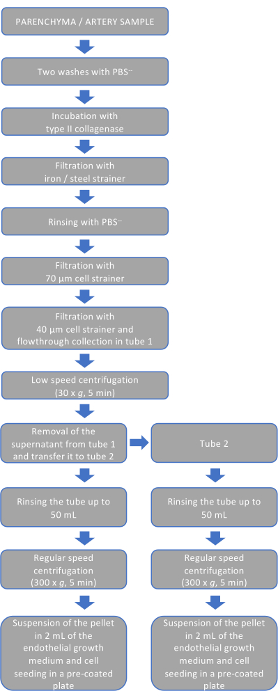
Figure 1: Schematic representation of the procedure. The flowchart shows the procedure for endothelial cell isolation and non-endothelial cell removal. Each step recapitulates the events described in steps 2-6 of the experimental protocol. Please click here to view a larger version of this figure.
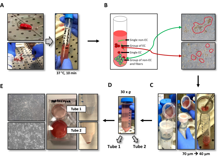
Figure 2: Illustration of the optimized EC isolation procedure. (A) Representative image of a lung parenchyma specimen diced in small fragments before collagenase digestion (left) and incubated in a 15 mL tube containing 5 mL of type 2 collagenase (right). (B) Schematic (left) and actual (right; magnification: 100x) representation of the cell populations obtained after collagenase digestion. These populations include singlets of non-ECs and ECs, groups of ECs, and groups of non-ECs bound to fibers, which are removed using strainers. (C) Serial filtrations using metal 70 µm and 40 µm mesh strainers. (D) Image of the 50 mL tube after low-speed centrifugation and before separating the supernatants from the pellets in "tube 1" and "tube 2". Cell clusters are contained in the pellet, while the single cells are mostly contained in the supernatant. (E) Image showing the different colors and compositions of the pellet and the isolated cells from "tube 1" (top) and "tube 2" (bottom); magnification: 100x. Abbreviation: EC = endothelial cells. Please click here to view a larger version of this figure.
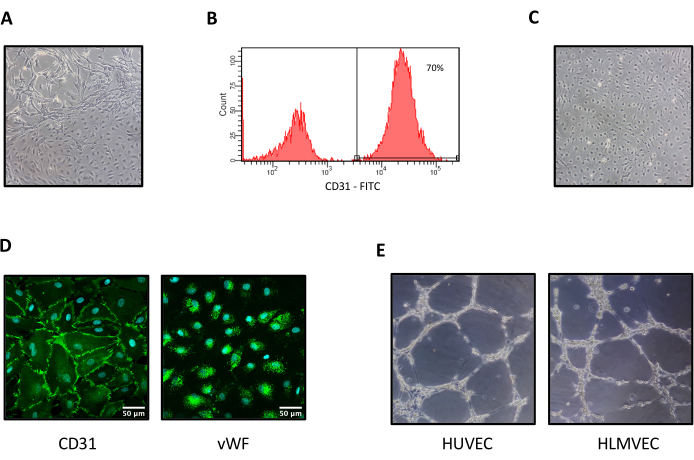
Figure 3: HLMVEC phenotypic and functional characterization. (A) A mixed cell population obtained from a lung parenchyma explant. (B) CD31 detection by flow cytometry on the surface of cells at the end of the first passage. The percentage purity (70%) refers to the used hierarchical gating strategy. (C) Pure HLMVECs after being live-sorted for the CD31 antigen. (D) Representative immunofluorescence confocal microscopic images of CD31 (left) and vWF (right) positive staining (scale bar: 50 µm). (E) Brightfield images were captured 6 h after seeding HUVECs or HLMVECs on Matrigel.Magnification (A, C, E): 100x. Abbreviations: HLMVECs = human lung microvascular endothelial cells. Please click here to view a larger version of this figure.
The multiple roles played by vascular endothelial cells in human pathophysiology make these cells an indispensable tool for in vitro pathogenetic and pharmacological studies. Since ECs from different vascular sites/organs display peculiar features and functions, the availability of healthy and diseased ECs from the organ of interest would be ideal for research purposes. For instance, HLMVECs are essential for studies on lung inflammation; therefore, a methodology for the high-yield, high-purity isolation of these cells will benefit varying research fields.
Methods to isolate HLMVECs have been reported by various groups10,11,12,14,15,17,18,19. Each method has introduced innovative approaches, such as cell sorting with magnetic beads11,12,15,17,18 or mechanical dissociation using a blunt cannula17,18. The present protocol was designed to collect the small islets of endothelial cells obtained after collagenase digestion and used sedimentation time-based serial centrifugations to increase the purity of the ECs in the initial mixed cell population. This point is crucial since higher starting cell numbers mean lower numbers of in vitro expansions are needed to reach the number of cells required for the experiments, which, in turn, means more investigations can be completed before the ECs senesce. This aspect is strictly related to the degree of purity of the HLMVEC isolates, which is also relevant for obtaining reliable results. Moreover, if the lung sample is very small in size and there is no possibility of performing EC sorting immediately after the explant, which was, in our experience, the most frequent scenario, the contaminating SMCs will grow faster than the ECs, thus taking over the culture and reducing the yield of ECs after cell sorting.
In this study, we tried to address these points. For this, what happened to the lung specimens after collagenase digestion was initially monitored, and significant contamination were identified by varying cell types, particularly SMCs (Figure 2), which grow fast and can overtake HLMVECs in mixed cultures. Therefore, a strategy was set to minimize this contamination immedaitely after digestion (Figure 1 and Figure 2). Remarkably, HLMVECs with ~70% purity (Figure 3A,B) were consistently obtained at the first isolation phase. This excellent outcome facilitated the subsequent purification step by FACS, leading to the final result of obtaining virtually pure, functional HLMVECs (Figure 3C-E). Similar results were obtained when pulmonary artery segments were used as the starting material and HPAECs were the isolated cells, thus meaning this methodology represents an improvement of the methodology reported earlier8.
Although simple and reproducible, this procedure has key requirements. For instance, we succeeded at HLMVEC isolation by only processing lung fragments obtained within 3-6 h from surgical excision, whereas longer intervals were associated with poor outcomes (data not shown). The collagenase digestion time could also introduce variability in the EC yield due to the lot-to-lot variations in enzymatic activity24. Another critical point relates to the sample condition. Lung parenchyma derived from smokers or elderly donors gave low HLMVEC yields. Moreover, the supernatant of low-speed centrifugation may contain some aggregates that are transferred into "tube 2". For this reason, it is recommended to keep the contents of "tube 1" and "tube 2" in culture, at least during the early stage of cell isolation. The direct sorting of CD31+ cells, as previously described in EC isolation protocols11,12,15,17,18, can also represent a good solution for increasing the purity from the start. We did not apply this early sorting stage due to the small size of the specimens analyzed. However, this possibility will be tested in the future in the implementation of the protocol.
A simple and reliable isolation protocol for isolating HLMVECs and HPAECs will encourage investigators to utilize this route to obtain cells for their studies, particularly when these cells are not commercially available. For instance, isolating HLMECs from CF lungs will allow the construction of a full CF airway on a chip25. Moreover, making their own EC collection will enable researchers to plan and run more experiments at a lower cost.
In conclusion, this method, which resolves some critical points that may occur during EC isolation from surgical specimens, improves the existing methods, thus facilitating research on EC pathobiology and inflammation.
The authors have nothing to disclose.
This work was supported by funds from the Italian Ministry of the University and Research (ex 60% 2021 and 2022) to R. P. and by grants from the Italian Cystic Fibrosis Foundation (FFC#23/2014) and from the Italian Ministry of Health (L548/93) to M. R.
0.05% trypsin-EDTA 1X | GIBCO | 25300-054 | Used to detach cells from the culture plates |
Anti CD31 Antibody, clone WM59 | Dako | M0823 | Used for CD-31 staining in immunocytochemistry. Dilution used: 1:50 |
Anti vWF Antibody | Thermo Fisher Scientific | MA5-14029 | Used for von Willebrand factor staining in immunocytochemistry. Working dilution: 1:100 |
Autoclavable surgical scissors | Any | Used for chopping specimens | |
Cell strainers 40 µm | Corning | 431750 | Used during the second filtration |
Cell strainers 70 µm | Corning | 431751 | Using during the first filtration |
Collagenase, Type 2 | Worthington | LS004177 | Type 2 Collagenase used for enzymatic digestion. Working concentration: 2 mg/mL |
Conjugated anti CD31 Antibody | BD Biosciences | 555445 | Used for cell sorting (1:20 dilution) |
Dulbecco′s Phosphate Buffered Saline (PBS) with CaCl2 and MgCl2 | Sigma-Aldrich | D8662 | Used for cell washing before medium change |
Dulbecco′s Phosphate Buffered Saline (PBS) without CaCl2 and MgCl2 | Sigma-Aldrich | D8537 | Used for washing surgical specimens and cells before trypsinization |
Endothelial Cell Growth Medium MV | PromoCell | C-22020 | HLMVEC growth medium |
Fibronectin | Sigma-Aldrich | F0895 | Fibronectin from human plasma used for plate coating. Working concentration: 50 µg/mL |
Gelatin from porcine skin, type A | Sigma-Aldrich | G2500 | Used for plate coating |
Type A gelatin | Sigma-Aldrich | g-2500 | Gelatin from porcine skin used for plate coating. Working concentration: 1.5% |