Here, we present a protocol that can be applied in the neonatal intensive care unit and the delivery room in relation to three scenarios: cardiac arrest, hemodynamic deterioration, or respiratory decompensation. This protocol can be performed with a state-of-the-art ultrasound machine or an affordable handheld device; an image acquisition protocol is carefully detailed.
The use of routine point-of-care ultrasound (POCUS) is increasing in neonatal intensive care units (NICUs), with several centers advocating for 24 h equipment availability. In 2018, the sonographic algorithm for life-threatening emergencies (SAFE) protocol was published, which allows the assessment of neonates with sudden decompensation to identify abnormal contractility, tamponade, pneumothorax, and pleural effusion. In the study unit (with a consulting neonatal hemodynamics and POCUS service), the algorithm was adapted by including consolidated core steps to support at-risk newborns, aiding clinicians in managing cardiac arrest, and adding views to verify correct intubation. This paper presents a protocol that can be applied in the NICU and the delivery room (DR) in relation to three scenarios: cardiac arrest, hemodynamic deterioration, or respiratory decompensation.
This protocol can be performed with a state-of-the-art ultrasound machine or an affordable handheld device; the image acquisition protocol is carefully detailed. This method was designed to be learned as a general competence to obtain the timely diagnosis of life-threatening scenarios; the method aims to save time but does not represent a substitute for comprehensive and standardized hemodynamic and radiological analyses by a multidisciplinary team, which might not universally be on call but needs to be involved in the process. From January 2019 to July 2022, in our center, 1,045 hemodynamic consultation/POCUS consults were performed with 25 patients requiring the modified SAFE protocol (2.3%), and a total of 19 procedures were performed. In five cases, trained fellows on call resolved life-threatening situations. Clinical examples are provided that show the importance of including this technique in the care of critical newborns.
Ultrasound is a tool that allows an immediate evaluation at the patient's bedside without having to transfer them to another room or floor in the hospital. It can be repeated, it is simple, economical, and precise, and it does not emit ionizing radiation. Ultrasound has been increasingly used by emergency physicians1, anesthesiologists2, and intensivists3 to obtain anatomical and functional images at the patient's bedside. It is a practical tool that is considered by some authors as the fifth pillar of physical examination, as an extension of the human senses4 (inspection, palpation, percussion, auscultation, and insonation)5.
In 2018, the SAFE protocol (for the acronym sonographic algorithm for life threatening emergencies) was published, which allows the assessment of neonates with sudden decompensation (respiratory and/or hemodynamic) to identify alterations in contractility, pericardial effusion with cardiac tamponade (PCE/CT), pneumothorax (PTX), and pleural effusion (PE)6. Our unit is a tertiary-level referral hospital, with most babies needing mechanical ventilation and central catheters; in this context, the SAFE protocol was modified by evaluating the consolidated core steps for a critically ill newborn8, adapting the assistance for cardiac arrest7, taking calcium and glucose, and adding ultrasonographic views to verify intubation. Since 2017, a hemodynamic consultation (HC) and POCUS team has been available in the NICU with dedicated equipment.
Compared to adults, most cases of cardiac arrest in newborns are due to respiratory causes, resulting in pulseless electrical activity (PEA) or asystole. Ultrasound might be a valuable tool adjuvant to traditional resuscitation skills to assess intubation, ventilation, and heart rate (HR)9 and rule out hypovolemia, PCE/CT, and tension PTX. Electrocardiograms have been found to be misleading during neonatal resuscitation, as some newborns may have PEA10,11,12.
The overall goal of this method was to adapt the cited literature to create a sonographic algorithm that can be applied in the NICU and the DR in relation to three scenarios: cardiac arrest, hemodynamic deterioration, or respiratory decompensation. This allows for the expansion of the physical examination by the critical care team to provide a timely diagnosis with correct intubation, including diagnoses of PEA or asystole, abnormal contractility, PCE/CT, PTX, or PE, either using high-end ultrasound equipment (HEUE) or an affordable handheld device (HHD). This algorithm was adapted from the SAFE protocol to be applied both in tertiary level care centers with a NICU-dedicated machine and in the DR and secondary level care centers with reasonably priced portable equipment. This method was designed as a general competence to obtain opportune diagnoses of life-threatening scenarios; the method aims to save time but does not represent a substitute for comprehensive, standardized hemodynamic and radiological analyses performed by a multidisciplinary team, which is essential but not always universally available.
Figure 1 depicts the protocol: a modified sonographic algorithm for life-threatening emergencies in the critically ill newborn. This procedure can be performed with an HEUE or an HHD depending on the healthcare center's resources. In this method, the POCUS team is considered an adjuvant to the attending team; patient management, especially during newborn resuscitation, should be performed according to the latest International Liaison Committee on Resuscitation (ILCOR) recommendations13 and local guidelines, while the sonographer helps as an extra member.
This protocol was approved by the institution's human research ethics committee; written consent was obtained for acquiring and publishing anonymized images. Never substitute a traditional maneuver, such as auscultating, for an ultrasound image (they can be done simultaneously or alternately by different operators). The consolidated core steps for a critically Ill newborn are a rapid series of supportive actions that need to be remembered as the POCUS team assesses the patient. Always have a second member of the POCUS team securing the endotracheal tube (ETT). Adapt the scanning to the patient's needs without interfering with resuscitation maneuvers.
1. Ultrasound preparation, specification, and settings14
2. Newborn handling
3. Verify intubation using the HEUE/HHD in cricothyroid membrane view
4. Verifying the ETT depth (HEUE) with the aortic arch suprasternal view
5. Cardiac arrest assessment based on HEUE with subcostal views, an HHD in parasternal long axis view, and an HEUE/HHD LUS
NOTE: While the attending team is performing neonatal resuscitation according to the ILCOR recommendations, the POCUS team prepares the ultrasound equipment. Intubation may be verified by documenting the endotracheal tube in situ and assessing the depth with the weight + 6 formula. Ultrasound may be used to identify the HR21, qualitatively assess the contractility, and rule out PCE/CT.
6. Hemodynamic instability (hypoperfusion, hypotension, with or without respiratory deterioration)24
7. Exclusive respiratory symptoms (normal blood pressure and perfusion)
8. Drainage (HEUE/HHD)
NOTE: In all cases, use sterile technique.
The inspection of cardiac function by "eyeballing" can be applied to qualitatively assess the global cardiac systolic function. Any suspicion of impaired cardiac function should lead to an urgent HC with pediatric cardiology for the assessment of congenital heart disease (CHD). Treatment must be started according to the pathophysiology, and the treatment should be integrated and modified according to a comprehensive anatomical and functional echocardiography study27. If ductal-dependent CDH is suspected, prostaglandins must be started, and a pediatric cardiology consult must be scheduled. In the study center, pediatric cardiology and neonatal hemodynamics consultation services are available.
From January 2019 to July 2022, a total of 1,045 HC/POCUS studies were carried out in our hospital, of which 25 corresponded to the protocol (2.3%). The type of decompensation was classified as respiratory in 14 newborns, hemodynamic in 8 newborns, and cardiac arrest-related (one PEA and one tamponade) in 3 newborns. The ultrasound protocol diagnoses were PTX (12), PE (4), PCE/CT (3), altered contractility (2), cardiac arrest-related (2), mobilization of the endotracheal tube (1), and hypoglycemia (1).
The protocol and interventions were performed by an expert neonatologist with advanced ultrasound training in 8 patients, by neonatology fellows supervised by an expert in 12 patients, and by fellows exclusively in 5 patients (including the resolution of three tension PTX cases and two tamponade drains). Most (96%) of the patients survived the event, and 68% survived to discharge. Overall, 19 procedures were performed (five chest tubes, three chest tube corrections, four pneumothorax needle drainages, four pleural effusion needle drainages, and three tamponade needle drainages), an endotracheal tube adjustment was performed, and one glucose bolus was administered. The chest X-ray (CXR) corresponding to each event was found in the electronic system at a median (interquartile range) of 58 (27-97) min. Table 3 details the institution's experience with this protocol.
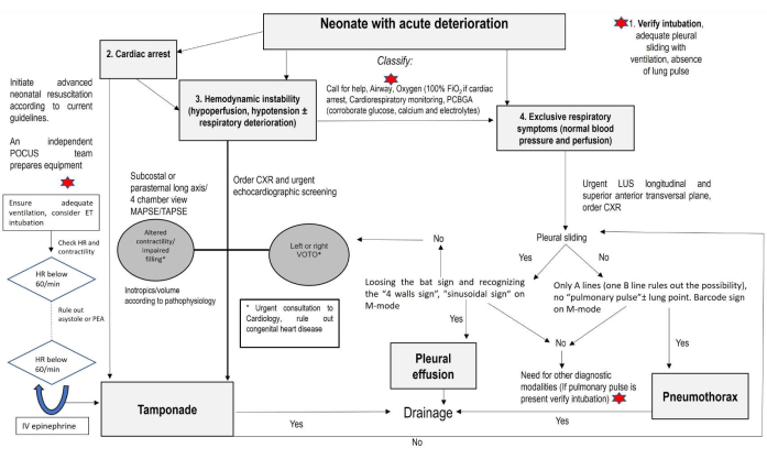
Figure 1: Algorithm: A modified sonographic algorithm for life-threatening emergencies in the critically ill newborn. Start by assessing the airway if the newborn is intubated, perform the consolidated core steps to ensure the newborn is monitored, and obtain the PCBGA. If the infant is in cardiac arrest, assistance (image acquisition) can be provided in two steps: a) performing corrective steps to detect the HR and effective cardiac output and ensure a real asystole; b) performing advanced CPR to rule out PCE/CT and hypovolemia and performing LUS to detect PTX. If hemodynamic instability (hypoperfusion, hypotension, with or without respiratory deterioration) is present, assess contractility, assess the left or right VOTO, and rule out PCE/CT. If negative or exclusive respiratory symptoms (normal blood pressure and perfusion) are present, rule out PTX and PE. Abbreviations: PCBGA = point of care blood gas analysis; POCUS = point of care ultrasound; ET = endotracheal; HR = heart rate; PEA = pulseless electrical activity; MAPSE = mitral annular systolic excursion; TAPSE = tricuspid annular systolic excursion; CXR = chest X-ray; VOTO = ventricular outflow tract obstruction; PCE/CT = pericardial effusion/cardiac tamponade; PTX = pneumothorax; PE = pleural effusion. Please click here to view a larger version of this figure.
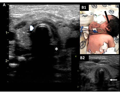
Figure 2: Verifying intubation. (A) Observe the outline of the ETT (double rail image, arrowhead), which generates a posterior shadow. The esophagus on the left of the screen is collapsed (asterisk). (B1) Difficult airway in a newborn with lymphangioma. (B2) The ETT is observed in situ; a small orogastric tube is observed (arrow). Abbreviation: ETT = endotracheal tube. Please click here to view a larger version of this figure.
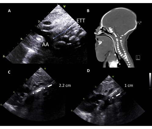
Figure 3: ETT depth. (A) The aortic arch is considered an orientation point to locate the carina, and the ETT is located at 1 cm from the AA. (B) Difficult airway in a newborn with lymphangioma; a high ETT is detected. (C) A high ETT (2.2 cm from the AA) is seen on the ultrasound and corrected. (D) Correctly placed ETT (1 cm from the AA). Abbreviations: AA = aortic arch; ETT = endotracheal tube. Please click here to view a larger version of this figure.
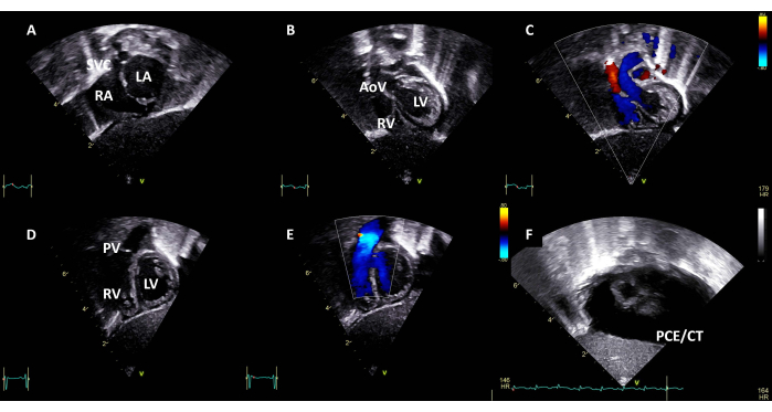
Figure 4: Subcostal long axis view. Sweeping from posterior to anterior, identify (A) the superior vena cava, the right and left atriums; (B) the right and left ventricles and the aortic valve; (C) color Doppler, indicating left ventricular outflow tract without obstruction; (D) and the crossing right ventricle and pulmonary valve. (E) Color doppler, indicating right ventricular outflow tract without obstruction. (F) Subcostal view with PCE/CT. Abbreviations: SVC = superior vena cava; RA = right atrium; LA = left atrium; RV = right ventricle; LV = left ventricle; AoV = aortic valve; PV = pulmonary valve; PCE/CT = pericardial effusion with cardiac tamponade. Please click here to view a larger version of this figure.
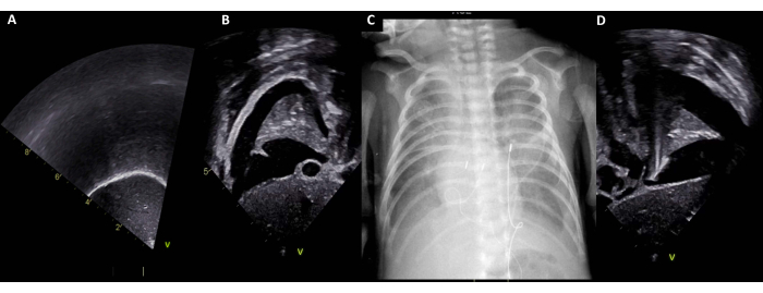
Figure 5: Transdiaphragmatic window. (A) Normal right transdiaphragmatic window. (B) Right PE. (C) Corresponding CXR with bilateral PE. (D) Left PE. Abbreviations: PE = pleural effusion; CXR = chest X-ray. Please click here to view a larger version of this figure.
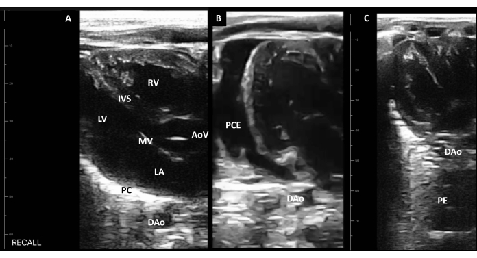
Figure 6: Handheld device long axis view. (A) Identify the right ventricle, the interventricular septum, the aortic valve, the left ventricle, the mitral valve, the left atrium, the pericardium, and the descending aorta. (B) The PCE identified as fluid anterior to the DAo. (C) The PE posterior to the DAo. Abbreviations: LA = left atrium; RV = right ventricle; LV = left ventricle; AoV = aortic valve; IVS = interventricular septum; MV = mitral valve; PC = pericardium; DAo = descending aorta; PCE = pericardial effusion; PE = pleural effusion. Please click here to view a larger version of this figure.
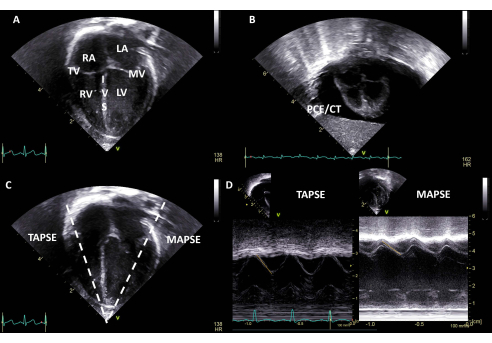
Figure 7: Four chamber view. (A) Identify the right atrium, the tricuspid valve, the right ventricle, the interventricular septum, the left atrium, the mitral valve, and the left ventricle. (B) Four chamber view with PCE/CT. (C) An M-mode image can be obtained on the tricuspid and mitral annulus to calculate the TAPSE/MAPSE. (D) TAPSE and MAPSE are depicted; the measurement in millimeters (mm) can be compared to gestational age nomograms. Abbreviations: SVC = superior vena cava; RA = right atrium; LA = left atrium; RV = right ventricle; LV = left ventricle; PCE/CT = pericardial effusion with cardiac tamponade; TV = tricuspid valve; MV = mitral valve; IVS = interventricular septum; TAPSE = tricuspid annular systolic excursion; MAPSE = mitral annular systolic excursion. Please click here to view a larger version of this figure.
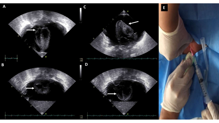
Figure 8: Pericardial effusion with cardiac tamponade. Large circumferential pericardial effusion. (A,B) A systolic right atrial collapse and (C,D) diastolic right ventricular collapse are observed qualitatively. (E) Pericardiocentesis. Please click here to view a larger version of this figure.
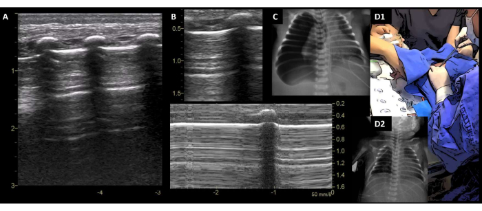
Figure 9: Pneumothorax. (A) PTX is diagnosed with absent pleural sliding, only A-lines, and no "lung pulse". (B) M-mode image shows the "Bar code sign". (C) Corresponding X-rays. (D1) Chest tube insertion. (D2) PTX resolved on a control CXR. Abbreviations: PTX = pneumothorax; CXR = chest X-ray. Please click here to view a larger version of this figure.
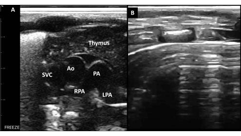
Figure 10: Anterior-superior transverse plane. (A) In a healthy newborn, the sternum and mediastinal structures, including the thymus, the superior vena cava, the aorta, and the pulmonary artery with its right and left branch, can be observed. (B) A-lines in the anterior transverse plane without sliding is a sensitive sign of anterior PTX. Abbreviations: SVC = superior vena cava; Ao = aorta; PA = pulmonary artery; RPA = right PA branch; LPA = left PA branch. Please click here to view a larger version of this figure.
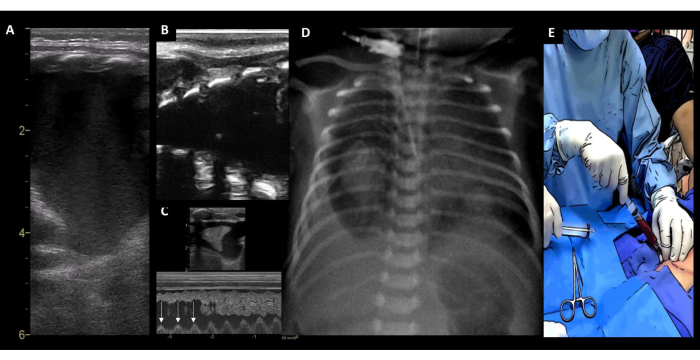
Figure 11: Pleural effusion. (A) PE identified by the absence of the bat sign and the "four walls sign" (high-end ultrasound equipment). (B) Same PE identified with a hand-held device. (C) M-mode image showing the "sinusoidal sign" (with each respiratory cycle, the lung surface line moves toward the pleural line, arrow). (D) Corresponding CXR. (E) Drainage of the hemothorax. Abbreviations: PE = pleural effusion; CXR = chest X-ray. Please click here to view a larger version of this figure.
Video 1: Lung pulse, deep ETT, and pneumothorax. A preterm newborn with respiratory decompensation and a suspected PTX, but a lung pulse was encountered; in verifying the ETT depth, a deep tube was recognized and retracted. The lung pulse disappeared, and a PTX was diagnosed. Parenchymal signs appeared after chest tube placement. The corresponding X-rays are shown. Please click here to download this Video.
Table 1: Ultrasound settings. Please click here to download this Table.
Table 2: Lung ultrasound semiology29,30,31,32,33,34,35,36,37,38,39,40,41,42,43,44,45. Abbreviations: PTX = pneumothorax; SVC = superior vena cava; PE = pleural effusion; ETT = endotracheal tube. Please click here to download this Table.
Table 3: Center experience. Abbreviations: DT = deterioration type; GA = gestational age; PDL = postnatal day of life; SF = supervised fellow; A = attending neonatologist; NF = neonatology fellow; SE = survived event; SD = survived discharge; Y = yes; N = no; RDS = respiratory distress syndrome; PDA = patent ductus arteriosus; VSD = ventricular septal defect; PO = post operated; ROP = retinopathy of prematurity; IVH = intraventricular hemorrhage; ETT = endotracheal tube; NEC = necrotizing enterocolitis. Please click here to download this Table.
Compared to children and adults, most cases of acute deterioration/cardiac arrest are due to respiratory causes in newborns. The original SAFE protocol was modified in our unit, a tertiary referral care neonatal center, due to this unit expecting several ventilated patients with indwelling catheters. The protocol has been adapted to different scenarios and equipment for use in low- and medium-income countries. As an institution with a neonatal hemodynamics and POCUS program, and after giving LUS workshops in different states of the Republic, we noted the need to integrate ultrasound to enhance neonatal care.
The critical steps in the protocol include the categorization of the patient into three starting scenarios (cardiac arrest, hemodynamic deterioration, or respiratory decompensation) and the addition of some steps where ultrasound might help the critical care/resuscitation team.
One of the steps included is verifying intubation, which can be performed at several points of the algorithm according to the patient's needs. Transtracheal ultrasound has a sensitivity of 98.7% (95% confidence interval [CI]: 97.8%-99.2%) and a specificity of 97.1% (95% CI: 92.4%-99.0%)47. Once the ETT is detected in situ, the depth can be checked with the Tochen formula18. Additionally, correct intubation is confirmed by documenting adequate pleural sliding on both sides, as well as the presence of parenchymal signs (B-lines, consolidation) and the absence of a lung pulse. Ultrasound can be used to verify the depth of the ETT only if a skilled sonographer is present, the condition of the patient allows it, and deterioration is considered dependent on the airway (e.g., the presence of a lung pulse). In a study with neonates weighing 1,282 g ± 866 g, considering a tube "deep" (<1 cm) compared to CXR showed a sensitivity of 86% and a specificity of 96%48. In this work, the tube was demonstrated in situ in all the cases with an intubated patient. Only in one case was a displaced ETT the cause of respiratory decompensation.
We consider the POCUS team as a valuable adjuvant to the attending team performing neonatal resuscitation. As mentioned earlier, the POCUS team might help by detecting the HR and effective cardiac output and ensuring a real asystole or PEA in the first step10,11,12,21,22. After advanced CPR, the POCUS team may help rule out PCE/CT and hypovolemia (empty right and left ventricles) and perform LUS to detect PTX21,22. In one of our cases, the POCUS team was called to a preterm infant who was being ventilated. The cardiac monitor indicated an HR of 80 bpm, but the ultrasound image detected asystole (PEA). Immediate chest compressions were started as the attending team was ventilating only because the monitor indicated an HR ˃60 bpm.
Ultrasound provides useful, additional information to the conventional treatment of a crashing infant. Modern PCBGA provides the levels of glucose, calcium, and electrolytes, so reversible causes can be immediately addressed considering the 7Hs, including hypovolemia (POCUS), hypoxia (PCBGA), hydrogenation/acidosis (PCBGA), hypothermia (clinical), hypoglycemia (PCBGA), hypo/hyperkalemia (PCBGA), hypocalcemia (PCBGA), and the 2Ts, including tamponade and tension pneumothorax. In one of our cases, in a newborn classified with hemodynamic decompensation (pale, hypotensive, lethargic), the etiology was hypoglycemia detected with PCBGA.
PCE/CT is infrequent but linked to high mortality. PCE/CT is closely related to the presence of a central line and the tip position (as the pericardial fluid found is normally consistent with the infusate) and commonly affects very-low-birthweight (VLBW) infants49. Survival improves when PCE/CT is detected early and treated promptly50,51. In units caring for VLBW infants and surgical patients, a dedicated ultrasound machine is recommended for immediate access. When a significant PCE causing CT is found, normally a blind procedure can be performed safely. Nevertheless, the fact that the same probe used for diagnosing helps in guiding the procedure improves patient safety and decreases the complication rate to a minimum52. In our series, three PCE/CT cases were diagnosed, with two survivors (draining with parenteral nutrition in one case and normal saline with antibiotics in the other) and one death (hemopericardium). A large PE causing hemodynamic instability or cardiac arrest is infrequent, but in case it presents, ultrasound diagnostic performance for fluid is high, and drainage can be performed safely. In some scenarios of neonatal resuscitation, such as hydrops, ultrasound guidance is essential.
The subjective evaluation of cardiac contractility, ventricular filling, and outflow assessment can guide the neonatologist to begin with a pathophysiology-suitable treatment and to perform an appropriate pediatric cardiology and hemodynamic consultation. It is of great value to identify an underfilled heart and differentiate it from volume overload and altered contractility, as the treatment is different24. In our unit, we advocate for the practice of advanced neonatal hemodynamics with highly trained members of the team; however, all our neonatology fellows must acquire basic POCUS skills as they are the primary care providers. In this series, one newborn was observed to have altered contractility and ventricular dilatation, which led to a prompt diagnosis of an aortic coarctation.
The LUS diagnostic accuracy for PTX is very high and can even reach 100% in terms of sensitivity, specificity, and positive and negative predictive values. As its superiority is staggering compared to CXR and transillumination with respect to time, there is enough evidence to consider LUS as the first-line diagnostic test53. Either with HEUE or an HHD, procedures can be safely performed while avoiding sliding portions where aerated lung is present. Using this algorithm, 12 PTX cases were successfully diagnosed and treated.
There is mostly moderate evidence regarding the use of cardiac, lung, vascular, cerebral, and abdominal POCUS54. POCUS protocols need to be individualized according to different centers' needs in close collaboration with cardiology and radiology to ensure quality care. It is fundamental to include POCUS skills on the curriculum for neonatology fellows as many complications occur on call. Immediate equipment availability is essential to ensure a successful program.
This protocol warrants further external validation to prove its generalizability. This modified protocol has limitations as it is focused on cardiopulmonary deterioration in the NICU and relies on prompt expert consultation (HC, pediatric cardiology). Recently, a protocol has been published on hemodynamic precision in the neonatal intensive care unit using targeted neonatal echocardiography (TnECHO)55. This expert consultative model in which a neonatologist performs an HC (a comprehensive and standardized echocardiographic assessment with a recommendation based on advanced hemodynamics knowledge) needs advanced training. The objective of this protocol is to present it as a general competence to ensure that the neonatologist on call (in a unit with an ultrasound in the NICU) has the ability to diagnose and treat life threatening emergencies. Additionally, the recently published sonographic assessment of life-threatening emergencies-revised (SAFE-R)56 has added the recognition of acute critical aortic occlusion, acute abdominal complications, and severe intraventricular hemorrhage.
The authors have nothing to disclose.
We thank Dr. Nadya Yousef, Dr. Daniele De Luca, Dr. Francesco Raimondi, Dr. Javier Rodriguez Fanjul, Dr. Almudena Alonso-Ojembarrena, Dr. Shazia Bhombal, Dr. Patrick McNamara, Dr. Amish Jain, Dr. Ashraf Kharrat, the Neonatal Hemodynamics Research Center, Dr. Yasser Elsayed, Dr. Muzafar Gani, and the POCUSNEO group for their support and feedback.
Conductivity gel | Ultra/Phonic, Pharmaceutical innovations, New Jersey, United States | 36-1001-25 | |
Handheld linear probe, 10.0 MHz | Konted, Beijing, China | C10L | handheld device |
Hockey stick probe 8–18 MHz, L8-18I-SC Probe | GE Medical Systems, Milwaukee, WI, United States | H40452LZ | high-end ultrasound equipment |
iPad Air 2 | Apple Inc | MGWM2CL/A | electronic tablet |
Phased array probe 6-12 MHz, 12S-D Phased Array Probe | GE Medical Systems, Milwaukee, WI, United States | H45021RT | high-end ultrasound equipment |
Vivid E90 v203 Console Package | GE Medical Systems, Milwaukee, WI, United States | H8018EB | Vivid E90 w/OLED monitor v203 Console |