Summary

The TD Drive: A Parametric, Open-Source Implant for Multi-Area Electrophysiological Recordings in Behaving and Sleeping Rats

Published: April 26, 2024
doi:

Summary

Here, we present a unique, 3D-printable implant for rats, named TD Drive, capable of symmetric, bilateral wire electrode recordings, currently in up to ten distributed brain areas simultaneously.

Abstract

Intricate interactions between multiple brain areas underlie most functions attributed to the brain. The process of learning, as well as the formation and consolidation of memories, are two examples that rely heavily on functional connectivity across the brain. In addition, investigating hemispheric similarities and/or differences goes hand in hand with these multi-area interactions. Electrophysiological studies trying to further elucidate these complex processes thus depend on recording brain activity at multiple locations simultaneously and often in a bilateral fashion. Presented here is a 3D-printable implant for rats, named TD Drive, capable of symmetric, bilateral wire electrode recordings, currently in up to ten distributed brain areas simultaneously. The open-source design was created employing parametric design principles, allowing prospective users to easily adapt the drive design to their needs by simply adjusting high-level parameters, such as anterior-posterior and mediolateral coordinates of the recording electrode locations. The implant design was validated in n = 20 Lister Hooded rats that performed different tasks. The implant was compatible with tethered sleep recordings and open field recordings (Object Exploration) as well as wireless recording in a large maze using two different commercial recording systems and headstages. Thus, presented here is the adaptable design and assembly of a new electrophysiological implant, facilitating fast preparation and implantation.

Introduction

The multi-area nature of brain interactions during wake and sleep makes it difficult to exhaustively study the ongoing physiological processes. While approaches such as functional MRI (fMRI) and functional ultrasound (fUS) allow sampling of brain activity from whole brains1,2, they exploit neurovascular coupling to infer brain activity from hemodynamic activity, limiting their temporal resolution2. In addition, fMRI requires the placement of the research subject in an MRI scanner, prohibiting experiments with freely moving animals. Optical imaging of calcium dynamics with single or multiphoton imaging enables cell type-specific recordings of hundreds of neurons simultaneously3. However, head-mounted microscopes such as the Miniscope3, which do allow freely moving behavior, are usually limited to imaging superficial cortical areas in intact brains4. While the diameter of their field of view on the cortex can be in the order of 1 mm, the space requirements of these head-mounted microscopes can make it difficult to target several, especially adjacent, areas. Therefore, to capture multi-area brain dynamics in wake and sleep accurately, extracellular electrophysiology, recorded with electrodes implanted in the brain areas of interest, is one of the methods of choice due to its high temporal resolution and spatial precision5. In addition, it allows the characterization of sleep dynamics in animals compatible with analyses obtained from human EEG, increasing the translational value of this method6.

Classically, studies recording brain activity with extracellular electrodes have employed individual wire electrodes or electrode bundles, such as tetrodes7. State-of-the-art probes such as the Neuropixels probe8 allow targeting several areas simultaneously, given that they are aligned on an axis that allows implanting the probe along that axis without impairing the animal. However, accurate simultaneous recordings of multiple, spatially separated areas still remain challenging, with existing methods being either costly or time-intensive.

In recent years, additive manufacturing methods such as stereolithography have become broadly available. This allowed researchers to develop novel electrode implants that were adaptable to their experimental requirements9, for example, simplified repeatable targeting of multiple brain areas. Frequently, these implant designs are also shared with the academic community as open-source hardware, allowing other researchers to adapt them to their own purposes. The degree of adaptability of specific implants varies both as a result of how the implant is designed and how it is shared. Parametric modeling10 is a popular approach in computer-aided design, in which different components of the design are linked by interdependent parameters and a defined design history. Implementing a parametric approach for designing implants increases their reusability and adaptability10, as changing individual parameters automatically updates the complete designs without the need for complex re-modeling of the design. A consequential necessity is that the design itself is shared in an editable format that preserves the parametric relationships and design history. File formats that only represent geometric primitives, such as STL or STEP, make subsequent parametric modifications of published models unfeasible.

While tetrode hyperdrives11,12,13 enable recordings from dozens of tetrodes, their assembly and implantation are time-intensive, and their quality is largely dependent on the skill and experience of the individual researcher. In addition, they usually combine the guide tubes that direct the recording electrodes to their target location in one or two larger bundles, therefore limiting the number and spread of areas that can be targeted efficiently.

Other implants14,15 expose the complete skull and allow for the free placement of multiple individual microdrives that carry the recording electrodes. While the placement of independent microdrives16 during surgery time maximizes flexibility, it increases surgery time and can make it difficult to target multiple adjacent areas due to the space requirements of the individual microdrives. In addition, while the implants are open source, they are only published as STL files, making modification difficult.

An example of a drive with a more inherent parametric philosophy is the RatHat17. By providing a surgical stencil that covers the whole dorsal surface of the skull, it allows precise targeting of multiple brain targets without the use of a stereotactic frame during surgery. Multiple implant variations for cannulas, optrodes, or tetrodes are available. However, while the drive is free to use for academic purposes, it is not published open-source, creating a hurdle for researchers to evaluate and use the implant.

Presented in this article is the TD Drive (see Figure 1), a novel 3D-printable implant for extracellular electrode recordings in rats. The TD Drive aims to overcome some of the drawbacks of existing solutions: it allows to target multiple brain areas, mirrored across both hemispheres, with independent wire electrodes simultaneously. Due to its simple design, it can be assembled in a few hours at a relatively low cost by less experienced researchers. The TD Drive is published open-source, in easily modifiable file formats to allow researchers to adjust it to their specific needs. Incorporating a parametric 3D modeling approach from the beginning of the TD Drive's design process allows the parameters necessary to be changed to be abstracted: to change target locations, researchers can simply edit the parameters representing their dorsoventral and anteroposterior coordinates, without the need for re-designing the drive themselves. The files to modify and manufacture the TD Drive can be found at https://github.com/3Dneuro/TD_Drive.

Figure 1
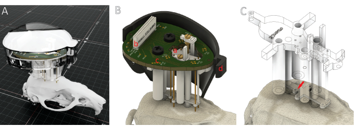
Figure 1: Overview of the TD Drive. (A) Rendering of a TD Drive with a protective cap. (B) Rendering with inner parts shown. The TD Drive features (a) multiple, parametrically adjustable recording locations for fixed and moveable electrode wires, an EIB with (b) a high-density Omnetics connector compatible with common tethered and wireless data acquisition systems, and (c) an intuitive channel mapping optimized for recordings with Intan/Open Ephys systems (see Supplementary Figure 1) and (d) a cap to protect the implant during tethered recordings and when no headstage is connected. (C) A guide stencil on the bottom of the TD Drive facilitates the placement of guide cannulas and serves as a redundant verification of implant locations during surgery. Please click here to view a larger version of this figure.

The implant design was piloted in n = 4, validated in n = 8, and confirmed in n= 8 Lister Hooded rats that performed different tasks. The first 4 animals were used to develop the drive and adjust parameters. Then, a full pilot was run with 8 animals (shown in results). A second cohort of 8 animals was run and included in the implant survival analysis. The implant was compatible with tethered sleep recordings and open field recordings (Object Exploration) as well as wireless recording in a large maze (HexMaze 9 m x 5 m) using two different commercial recording systems and headstages. The two cohorts of 8 were recorded with two different acquisition systems – tethered for longer sleep recordings and wireless for large maze exploration recordings. We can conclude that this simple wire drive allows for long-running experiments with larger cohorts by less experienced researchers to enable sleep stage analysis as well as oscillation analysis in multiple brain areas. This is in contrast to most electrophysiology implants to date, which, due to difficulty and time intensity, allow for smaller animal cohorts and usually need very experienced experimenters. However, with this drive, no individual neuron activity can be recorded; thus, the use is limited to investigations of local field potential (LFP) and summation activity.

Protocol

The present study was approved by the Dutch Central Commissie Dierproeven (CCD) and conducted according to the Experiments on Animals Act (protocol codes: 2020-0020-006 & 2020-0020-010). Male Lister Hooded rats of 9-12 weeks on arrival were used. The reagents and the equipment used in the protocol are listed in the Table of Materials. See Supplementary Figure 1 and Supplementary Figure 2 for the steps of the drive-building process. 1…

Representative Results

Using the instructions provided in the protocol, the TD Drive could be built easily by multiple experimenters. After drive development (n = 4), a full pilot was run with eight animals. An additional batch of eight animals was implanted, and experimental data collection was performed. As data analysis has not been completed on these animals, they have been included in the survival analysis, but not in other analyses (e.g., targeting or histology). Implant surgery was performed 2 weeks after arrival (see <strong class="xfi…

Discussion

Presented in this article is an adaptable implant for bilateral, symmetric multi-area wire electrode recordings for freely-moving rats.

The ability to easily adjust the implant by changing predefined parameters was one of the motivations for the creation of the TD Drive. While aiming to maximize the flexibility for changing parameters, inherent constraints in the relations between them necessarily impose limits to this adaptability. No limits are set by default for the anteroposterior paramete…

Disclosures

The authors have nothing to disclose.

Acknowledgements

The authors would like to thank Angela Gomez Fonseca for the inspiration to develop the drive and all the students who ran pilot experiments with the animals, Milan Bogers, Floor van Ravenswoud, and Eva Severijnen. This work was supported by the Dutch Research Council (NWO; Crossover Program 17619 "INTENSE").

Materials

0.5 mm drill bit  McMaster 2951A38
1.27 mm pitch interconnected SIP/DIP socket (Mill-Max) Mouser Electronic 575-003101 For essembling and connection of EEG & GND screws
5 minute epoxy  Bison Commercially available regular off-the-shelf epoxy
cyanoacrylate glue Loctite Super Glue-3 
EEG wire Science Products GmbH 7SS-2T
Electrode wire Science Products GmbH NC7620F
Ethanol LC For standard pre-operative sterilization procedure of drive
Fine forceps (5) FST 91150-20 For wire bundle preperation and handling
Form 3B Formlabs 3D printer used to 3D print the self-printed parts of the TD drive
Gold pins (small) Neuralynx, Inc. 9885 Attachment of electorde wires to EIB board
Ground wire Science Products GmbH SS-3T/A
High-density connector  LabMaker GmbH/Omnetics A79026-001
Lister Hodded rats Charles River Laboratories Crl:LIS we used male rats, 9-12 weeks of age at arrival
M1 brass insert AliExpress Commercially available https://aliexpress.com/item/33047616164.html
M1 tap McMaster 2504A33
M1x16 screw Bossard 1096613
M1x3 stainless steel screws  Screws and More 84213_14985
M2.5×5 polyimide screws Screws and more 7985PA25S_50
mineral oil McMaster 1244K14
Nail polish Etos Commercially available For color coding EEG and GND wires
painter's tape Gamma Commercially available For wire bundle preperation
Pin vise McMaster 8455A16
plotting paper Canson Commercially available For wire bundle preperation
polyimide tubes Amazon / Small Parts TWPT-0159-30-50 AWG, 0.0159" ID, 0.0219" OD, 0.0030" Wall, 30" Length
RHD 32-channel headstage with accelerometer Intan Technologies, LLC C3324 For tethered recordings in the sleepbox
RHD 3-ft (0.9 m) standard SPI cables Intan Technologies, LLC C3203 From commutator to headstage
RHD 6-ft (1.8 m) standard SPI cables Intan Technologies, LLC C3206 From OpenEphys box to commutator
Slip Ring with Flange Adafruit 1196 Commutator: 22 mm diameter, 12 wires
Solder flux  Griffon S-39 50 ml Commercially available For soldering EEG & GND screws
soldering paste Amazon B08CBZ5HC5
stainless steel M2 nut  McMaster 93935A305
Tethered recording setup  OpenEphys Acquasition Board
Wireless recording logger SpikeGadgets miniLogger 32 For wireless recordings in the task
Wireless recording setup SpikeGadgets Main Control Unit (MCU) incl. breakout board and RF transceiver For wireless recordings in the task

References

  1. Deffieux, T., Demené, C., Tanter, M. Functional Ultrasound Imaging: A New Imaging Modality for Neuroscience. Neuroscience. 474, 110-121 (2021).
  2. Finn, E. S., Poldrack, R. A., Shine, J. M. Functional neuroimaging as a catalyst for integrated neuroscience. Nature. 623 (7986), 263-273 (2023).
  3. Aharoni, D., Federico Guo, C. Aharoni-Lab/Miniscope-v4: Release for generating. GitHub. , (2023).
  4. Takasaki, K., Abbasi-Asl, R., Waters, J. Superficial bound of the depth limit of two-photon imaging in mouse brain. eNeuro. 7 (1), (2020).
  5. Buzsáki, G., et al. Tools for probing local circuits: High-density silicon probes combined with optogenetics. Neuron. 86 (1), 92-105 (2015).
  6. Lacroix, M. M., et al. Improved sleep scoring in mice reveals human-like stages. bioRxi.v. , (2018).
  7. Wilson, M. A., McNaughton, B. L. Dynamics of the hippocampal ensemble code for space. Sci New Ser. 261 (5124), 1055-1058 (1993).
  8. Jun, J. J., et al. Fully integrated silicon probes for high-density recording of neural activity. Nature. 551 (7679), 232-236 (2017).
  9. Headley, D. B., DeLucca, M. V., Haufler, D., Paré, D. Incorporating 3D-printing technology in the design of head-caps and electrode drives for recording neurons in multiple brain regions. J Neurophysiol. 113 (7), 2721-2732 (2015).
  10. Camba, J. D., Contero, M., Company, P. Parametric CAD modeling: An analysis of strategies for design reusability. Comput Aided Des. 74, 18-31 (2016).
  11. Kloosterman, F., et al. Micro-drive array for chronic in vivo recording: Drive fabrication. J Vis Exp. (26), e1094 (2009).
  12. Voigts, J., Siegle, J. H., Pritchett, D. L., Moore, C. I. The flexDrive: An ultra-light implant for optical control and highly parallel chronic recording of neuronal ensembles in freely moving mice. Front Syst Neurosci. 7, (2013).
  13. Voigts, J., Newman, J. P., Wilson, M. A., Harnett, M. T. An easy-to-assemble, robust, and lightweight drive implant for chronic tetrode recordings in freely moving animals. J Neural Eng. 17 (2), 026044 (2020).
  14. Sheng, T., et al. A novel 3D-printed multi-driven system for large-scale neurophysiological recordings in multiple brain regions. J Neurosci Methods. 361, 109286 (2021).
  15. Vöröslakos, M., Petersen, P. C., Vöröslakos, B., Buzsáki, G. Metal microdrive and head cap system for silicon probe recovery in freely moving rodent. eLife. 10, e65859 (2021).
  16. Mishra, A., Marzban, N., Cohen, M. X., Englitz, B. Dynamics of neural microstates in the VTA-striatal-prefrontal loop during novelty exploration in the rat. bioRxiv. , (2020).
  17. Allen, L. M., et al. RatHat: A self-targeting printable brain implant system. eNeuro. 7 (2), (2020).
  18. . Soldering Safety Available from: https://safety.eng.cam.ac.uk/safe-working/copy_of_soldering-safety (2018)
  19. . Harvard Soldering Safety Guidelines Available from: https://www.ehs.harvard.edu/sites/default/files/soldering_safety_guidelines.pdf (2019)
  20. Samanta, A., et al. CBD lengthens sleep but shortens ripples and leads to intact simple but worse cumulative memory. iScience. 26 (11), 108327 (2023).
  21. Machado, F., Malpica, N., Borromeo, S. Parametric CAD modeling for open source scientific hardware: Comparing OpenSCAD and FreeCAD Python scripts. PLOS One. 14 (12), e0225795 (2019).
  22. Schwarz, C., et al. The head-fixed behaving rat: Procedures and pitfalls. Somatosens Mot Res. 27 (4), 131-148 (2010).
  23. Gardiner, T. W., Toth, L. A. Stereotactic surgery and long-term maintenance of cranial implants in research animals. Contemp Top Lab Anim Sci. 38 (1), 56-63 (1999).
  24. França, A. S. C., van Hulten, J. A., Cohen, M. X. Low-cost and versatile electrodes for extracellular chronic recordings in rodents. Heliyon. 6 (9), e04867 (2020).
This article has been published
Video Coming Soon
Keep me updated:

.

Cite This Article
Schröder, T., van der Meij, J., van Heumen, P., Samanta, A., Genzel, L. The TD Drive: A Parametric, Open-Source Implant for Multi-Area Electrophysiological Recordings in Behaving and Sleeping Rats. J. Vis. Exp. (206), e66457, doi:10.3791/66457 (2024).

View Video