TMS를 사용한 동작 관찰 중 운동 흥분성 측정
English
Share
Overview
출처: 조나스 T. 카플란과 사라 I. 짐벨의 연구소 – 서던 캘리포니아 대학
경두개 자기 자극 (TMS)은 두피에 대해 배치 된 절연 코일을 통해 전류를 통과하는 비 침습적 뇌 자극 기술입니다. 간단한 자기장은 코일의 전류에 의해 생성되며, 유도의 물리적 과정 으로 인해, 이것은 가까운 신경 조직의 전류로 이어집니다. 이러한 자기 펄스의 지속 시간, 주파수 및 크기에 따라 기본 신경 회로는 여러 가지 방법으로 영향을 받을 수 있습니다. 여기서, 우리는 하나의 간단한 자기 펄스가 신피질을 자극하기 위하여 이용되는 단 하나 펄스 TMS의 기술을 보여줍니다.
TMS의 한 가지 관찰 가능한 효과는 모터 피질 위에 적용될 때 근육 경련을 일으킬 수 있다는 것입니다. 모터 피질의 소토토피 조직으로 인해 코일의 정확한 배치에 따라 다른 근육을 표적으로 삼을 수 있습니다. 이러한 근육 경련을 일으키는 전기 신호, 모터 는 잠재력을 불러 일으켰다, 또는 MEP, 기록 및 대상 된 근육을 통해 피부에 배치 전극에 의해 정량화 될 수있다. MEP의 진폭은 모터 피질의 근본적인 흥분성을 반영하도록 해석될 수 있다; 예를 들어, 모터 피질이 활성화되면 관찰된 MEP가 더 큽습니다.
이 실험에서는, Fadiga와동료에 의해 원래 수행된 연구 결과에 근거하여 1 그리고 많은 다른 사람에 의해 복제된 이후,2 우리는 행동 관측 도중 모터 피질의 흥분성을 시험하기 위하여 단 하나 펄스 TMS를 이용합니다. 우리가 움직일 때뿐만 아니라 다른 사람들이 움직임을 수행하는 것을 볼 때 모터 피질을 활성화 할 수 있다고 알려져 있습니다. 이 현상의 일반적인 해석은 다른 사람의 행동을 이해하는 데 역할을 할 수있는 시뮬레이션 프로세스를 반영한다는 것입니다. 여기서 우리는 주 모터 피질에 TMS에 의해 연상 된 MEP를 기록할 것이고 피험자는 제어 자극에 비해 다른 사람의 움직임을 관찰합니다.
Procedure
Results
A comparison of MEP amplitudes reveals a facilitation effect (Figure 1). MEP amplitude recorded from the FDI muscle is significantly greater during the hand action videos compared with control videos. This result suggests that the motor cortex increases in excitability during action observation.
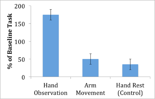
Figure 1: MEP amplitude during action observation. Motor evoked potentials from the first-dorsal interosseous muscle are largest when observing a hand movement, compared with an arm movement or a control video that displays no action.
Notably, the facilitation effect is relatively selective for the videos that involve a grasping action, as MEPs recorded during observation of the arm movement video show smaller MEPs compared with the hand action video. This suggests that the motor facilitation that occurs during action observation does not affect the entire motor cortex, but instead is specific to the muscle movements that are observed. In fact, the motor facilitation effect appears to be specific not only for which muscle is observed, but also to when the muscle is observed. For instance, Gangitano et al. have demonstrated temporal correlation between motor excitability and observed action dynamics.3
Applications and Summary
The single-pulse TMS technique lends itself well to the study of the motor cortex, both because of the accessible location of this cortex on the frontal surface of the brain, and also because of the directly observable reaction produced in the muscle in the form of MEPs. The measurement of cortico-spinal motor excitability has provided support further for the phenomenon of motor simulation during action observation in humans. This resonant motor activity may have implications for social behavior, for example in contributing to the process of understanding what others are doing. Furthermore, this same technique has provided evidence for motor activation during the imagination of action,4 a process that may be important for improving performance through mental rehearsal.
The robustness and specificity of the motor facilitation effect may reflect the sophistication of an individual’s motor representations. For example, the temporal dynamics of motor facilitation are directly related to motor expertise.5 This effect is also disrupted with disorders of movement, opening up the possibility that measurement of TMS-induced motor potentials can be used as way to assess the health of the motor cortex, as in recovery from stroke or other brain disease.6
References
- Fadiga, L., Fogassi, L., Pavesi, G. & Rizzolatti, G. Motor facilitation during action observation: a magnetic stimulation study. J Neurophysiol 73, 2608-2611 (1995).
- Fadiga, L., Craighero, L. & Olivier, E. Human motor cortex excitability during the perception of others' action. Curr Opin Neurobiol 15, 213-218 (2005).
- Gangitano, M., Mottaghy, F.M. & Pascual-Leone, A. Phase-specific modulation of cortical motor output during movement observation. Neuroreport 12, 1489-1492 (2001).
- Wright, D.J., Williams, J. & Holmes, P.S. Combined action observation and imagery facilitates corticospinal excitability. Front Hum Neurosci 8, 951 (2014).
- Aglioti, S.M., Cesari, P., Romani, M. & Urgesi, C. Action anticipation and motor resonance in elite basketball players. Nat Neurosci 11, 1109-1116 (2008).
- Koski, L., Lin, J.C., Wu, A.D. & Winstein, C.J. Reliability of intracortical and corticomotor excitability estimates obtained from the upper extremities in chronic stroke. Neurosci Res 58, 19-31 (2007).
Transcript
Transcranial Magnetic Stimulation, abbreviated as TMS, is a technique that can be used to investigate not only the connections between the brain and different muscles, but also how brain activity changes when a person observes motions in others.
Everyday, a person consciously moves their muscles to perform certain actions, like waving their right arm to hit a tennis ball with a racket.
All such voluntary muscle motions are the result of excitation in the motor cortex, which is located at the surface of the brain, just beneath the scalp.
Importantly, the movement of different body parts—whether it’s the right arm or left leg—is controlled by sets of neurons in distinct motor cortex regions.
For example, when neurons near the top right of the head are excited, they can produce electrical signals that travel through the brain to the spinal column, and then to muscles in the left arm.
In response, muscle cells produce their own electrical signals, which leads to contraction and movement.
Interestingly, research has shown that regions of the motor cortex are activated not only when a person performs some action themselves, but also when they watch someone else move—like a mechanic hitting a machine with her left arm.
TMS provides researchers with a way to probe the facilitation that action observation causes in motor cortex neurons.
Through the TMS techniques of Luciano Fadiga and colleagues, this video demonstrates how to investigate the relationship between action observation, motor cortex excitation, and muscle activity.
In this procedure, participants are subjected to two phases—TMS localization and calibration, and an experimental task—to identify whether areas of their motor cortex are excited when they observe an action performed by someone else.
The aim of the first phase is to identify the brain region responsible for moving a specific muscle in the participant’s right hand—the first-dorsal interosseous, abbreviated as FDI—located between the thumb and forefinger.
A figure-eight TMS coil is then placed against their scalp above the motor cortex. It is positioned on the left of their head, as this hemisphere of the brain directs movement on the right side of the body.
At the same time, electrodes are positioned over the FDI and nearby bone on the subject’s right hand, so that any electrical activity in this muscle can be detected and recorded.
The coil is then used to deliver a single pulse to the scalp, which creates a brief magnetic field that—through induction—leads to an electrical current in the underlying neural tissue.
Neurons, primarily those beneath the center of the coil, are activated by this and as a result, generate signals that—similar to those for conscious motions—travel to target muscles.
In response, additional electrical signals called motor-evoked potentials, MEPs, are created and can be recorded and visualized as peaks on a graph, where millivolts are plotted on the Y-axis and time on the X-axis.
These MEPs cause the muscles to physically twitch—movement that can be observed. If such a spasm occurs in a muscle other than the FDI—like in the upper arm—the correct region of the motor cortex has not been localized.
In this instance, the coil is moved slightly and used to deliver another single pulse, with any resulting motion being noted. This is repeated until twitches are seen in the right FDI—an indicator that its representation in the motor cortex has been found.
Once this region has been localized, the strength of the TMS stimulus is modified, a new pulse is administered, and the resulting MEP is recorded. Careful attention is paid to the amplitude—measured from the highest positive peak to the lowest negative point—of this signal.
These adjustments continue until a setting is found that produces MEPs with amplitudes greater than 50 µV in half of the phases—a stimulus intensity referred to as the participant’s motor threshold.
Several individual TMS pulses at 120% of motor threshold are then administered above the right-FDI region of the motor cortex, and baseline MEPs are recorded for each.
In the second phase, the experimental task, participants are asked to watch three types of movies involving arm or hand movements, while keeping their own body parts still.
The first type of footage, called hand-action videos, show a right hand reaching towards and grasping a cup—an action that requires the FDI. These clips will assess how observation of an FDI-based motion affects activity in the FDI-associated region of the motor cortex.
In contrast, arm-action videos involve a right arm lifting and moving around an area with a cup—motions that are independent of the FDI. This footage will evaluate specificity—whether the motor cortex area responsible for right FDI contraction can be excited by the observation of movements not involving this muscle.
The third type are control videos, which show a still right hand resting next to a cup, without any type of muscle movement.
When the actual task is performed, a TMS coil is again placed above the region of the brain responsible for right FDI movement. Then, participants watch the videos in a random order, with each type being repeated 10 times.
When muscle movement occurs in the footage—approximately 2 s after a hand or arm action video starts—the TMS coil is used to deliver a single electromagnetic pulse.
Similarly, although no movement is depicted in the control videos, a pulse is administered 2 s after the start of such clips. In all instances—whether control, arm, or hand action videos—MEPs generated by the right FDI are recorded.
Here, the dependent variable is the MEP amplitudes. Based on previous work, it is expected that MEPs recorded from the FDI upon observation of hand action movies will be greater than those recorded while watching arm action or control movies, reflecting higher FDI activity and thus greater motor cortex excitability.
Before the experiment begins, recruit 20 participants who are right-handed, have normal vision, and do not have any history of neurological disorders and obtain written consent from them.
Explain that they will be watching a series of videos, during which time regions of their brain will be stimulated via TMS. Emphasize that they may feel a slight tap on the head from the TMS coil, but should not experience great discomfort.
To start, have the participant sit in a chair positioned in front of a computer screen. Guide their right elbow to rest at a 90° angle, and make sure that their right arm and hand are comfortably prone.
Then, place their chin in a chinrest, so that their eyes will be at least 50 cm from the screen.
For MEP recording, clean the skin of the participant’s right hand with alcohol, and have them flex their right FDI muscle by pressing their forefinger and thumb together. Identify the peak location of muscular tension, and place a recording electrode on it. Afterwards, position a reference electrode on a nearby bone in the hand. Also securely fasten the ground electrode to their right elbow.
Once the electrodes have been positioned, instruct the participant to relax their hand so that there is no muscle tension.
Then, place a figure-8 TMS coil against the left side of the participant’s scalp, and use it to locate the right FDI representation in the motor cortex. When this occurs, expect to observe twitching of the right FDI, and stable MEPs recorded from this muscle.
For calibration, proceed to adjust the settings of the TMS coil until the minimum output strength—one that produces an MEP of greater than 50 µV on 5 out of 10 stimulations—is determined.
Record this value, which represents the participant’s motor threshold. Once it has been determined, use these settings to deliver a series of 10 TMS pulses—each separated by 15 s—to the participant, in order to determine the baseline MEP amplitude.
Afterwards, have the participant perform the experimental task by showing them the three types of movies—hand action, arm action, and control—in a random order. Between each, include a 15 s rest period.
Ensure that each video is played 10 times, for a total of 30 movies. As the participant views the action in each clip—roughly 2 s after the start of movie—administer a TMS pulse at 120% of the motor threshold.
To analyze the data, for each MEP recorded—whether from baseline, action observations, or control video conditions—calculate the peak-to-peak amplitude. To eliminate spurious spikes, discard MEPs that occur either before TMS stimulation or more than 100 ms after stimulation.
Calculate the average MEP amplitude for the hand- and arm-movements, as well as the hand-rest videos, and express these as the percentage above the average baseline.
Notice that, for hand-action videos, the MEP amplitude recorded from the FDI muscle was significantly greater compared to the control amplitude, which suggests a facilitation effect whereby the motor cortex increases in excitability during action observation.
However, MEPs recorded during arm-action observation were much smaller than those generated by watching hand-movement videos. This indicates that the facilitation effect is relatively selective, and is specific to the region of the motor cortex responsible for the observed muscle movements.
Now that you know how researchers are using TMS to investigate excitability of the motor cortex in response to action observation, let’s look at how this technique is applied in other applications.
Up until now, we’ve focused on the relationship between watching a movement and motor cortex function. However, some researchers are looking at whether imagining an action can also have effects on recorded MEPs.
Such work requires a participant to picture themselves physically moving a body part—such as bending their right arm—when single TMS pulses are administered.
When the resulting MEPs from muscles are assessed, they are found to be greater than those recorded during control scenarios—when the participant imagines no such movement.
This effect is further facilitated when the participant observes the motion they are imagining. Collectively, these results provide evidence for a relationship between imagination and motor activation.
Other neuropsychologists are exploring whether modifications of TMS can be used for therapeutic purposes.
For example, there is interest in whether TMS can help treat aphasia—a condition where patients have difficultly conveying verbal information, like the name of an object—resulting from stroke.
Here, a region of the brain called the right inferior frontal gyrus was stimulated in stroke patients using repetitive TMS—a method where pulses are repeatedly and rapidly administered.
When patients were asked to verbally identify objects—like lobsters or fireplaces—months after therapy, this revealed that repetitive TMS had long-term, positive effects on individuals’ naming capacities, providing insight into how this method can be used to treat cognitive deficits.
You’ve just watched JoVE’s video on TMS and action observation. By now, you should understand how TMS can be used to evaluate activity in the motor cortex after viewing a muscle movement. You should also know how to stimulate regions of the brain with a TMS coil, present observation stimuli, and collect and interpret MEP data. Finally, you should realize how TMS is being used in other applications, such as in therapies for stroke victims.
Thanks for watching!