הדמיה של ניוון מפרק הברך לאחר פציעת ACL לא פולשנית בחולדות
English
Share
Overview
מקור: לינדסי ק. לפלי1,2,סטיבן מ. דאבי1,טימותי א. באטרפילד3,4 וסינה שהבזמוהמדי5,
1 המחלקה לקינסיולוגיה, אוניברסיטת קונטיקט, סטורים, CT; 2 המחלקה לכירורגיה אורתופדית, מרכז הבריאות של אוניברסיטת קונטיקט, פרמינגטון, CT; 3 המחלקה למדעי השיקום, אוניברסיטת קנטאקי, לקסינגטון, KY; 4 המרכז לביולוגיה של שרירים, המחלקה לפיזיולוגיה, אוניברסיטת קנטאקי, לקסינגטון, KY; 5 המחלקה להנדסה ביו-רפואית, אוניברסיטת קונטיקט, סטורים, CT
פגיעה ברצועה הצולבת (ACL) של הברך מגבירה באופן דרמטי את הסיכון לדלקת מפרקים ניוונית פוסט טראומטית (PTOA), שכן כשליש מהאנשים יפגינו PTOA רדיוגרפי בעשור הראשון לאחר פציעת ACL. למרות ששחזור ACL (ACLR) משחזר בהצלחה את יציבות מפרק הברך, ACLR וטכניקות השיקום הנוכחיות אינן מונעות את תחילת PTOA. לכן, פגיעת ACL מייצגת את המודל האידיאלי ללמוד את התפתחות PTOA לאחר פגיעה משותפת טראומטית.
מודלים חולדה שימשו בהרחבה כדי לחקור את ההשפעה וההשפעה של פגיעת ACL על PTOA. המודל הנפוץ ביותר של פגיעת ACL הוא טרנספרקציה ACL, שהוא מודל אקוטי המערער בניתוח את המפרק. למרות מעשי, מודל זה אינו מחקה בנאמנות פגיעה אנושית ACL עקב הליכי פגיעה פולשניים ולא פיזיולוגיים להסוות את התגובה הביולוגית המקומית לפציעה. כדי לשפר את התרגום הקליני של התוצאות שלנו, פיתחנו לאחרונה מודל חדשני לא פולשני של פגיעת ACL שבו ACL נקרע באמצעות עומס אחד של דחיסת טיבי. פציעה זו משכפלת מקרוב את תנאי הפציעה הרלוונטיים לבני אדם והיא ניתנת לשחזור רב.
הדמיה של ניוון מפרקים באמצעות טומוגרפיה מיקרו-ממוחשבת (μCT) מספקת מספר התפתחויות משמעותיות על פני טכניקות הכתמת OA מסורתיות, כולל הדמיה תלת-ממדית מהירה, ברזולוציה גבוהה ולא הרסנית של ניוון מפרקים שלם. מטרת הדגמה זו היא להציג את פציעת ACL הלא פולשנית במודל מכרסמים ולהשתמש ב- μCT כדי לכמת ניוון מפרק הברך.
Principles
Procedure
Results
Smaller trabecular number, reduced trabecular thickness and greater trabecular spacing, all hallmark characteristics of PTOA onset, were evident 4 weeks after the non-invasive ACL tear (Table 1 and Figure 3). An image of a dissected ACL of healthy limb versus an acute injured limb is shown in Figure 5. The novel non-invasive model of ACL injury, where the ACL is ruptured through a single load of tibial compression, was able to produce an isolated proximal tear of the ACL.
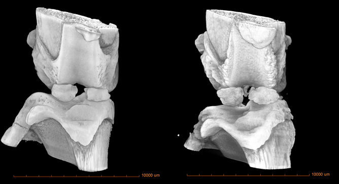
Figure 3: 3-D reconstructed μCT image of an acute ACL-injury (left) and 4 weeks post-ACL injury (right) in a rat.
Table 1: Characteristic measurements of PTOA onset.
| Animal | Tb.N (1/mm) |
Tb.Th (µm) |
TB.Sp (µm) |
| Acute ACL injured | 3.11 | 168.5 | 217 |
| 4 wks post-ACL injury | 2.63 | 166.7 | 213 |
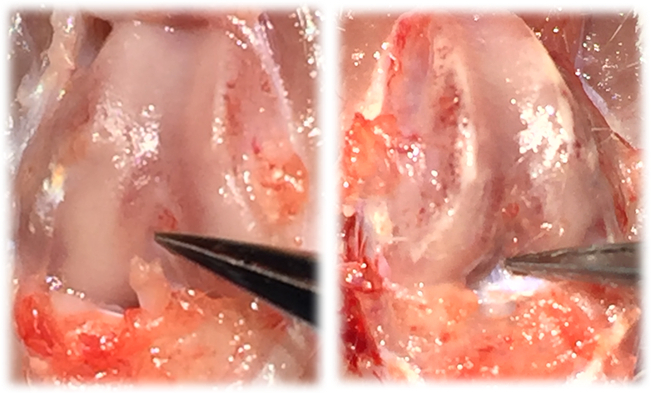
Figure 4: Image of an acute injured ACL-limb (left) and image of intact, healthy ACL (right).
Applications and Summary
This video demonstrates how a linear actuator can be used to produce an isolated non-invasive ACL rupture in rats. This injury closely replicates injury conditions relevant to humans and is highly reproducible. To overcome several of the major limitations of traditional OA staining techniques, this method uses µCT to quantify whole joint degeneration and trabecular structure.
Evidence-based interventions to improve musculoskeletal rehabilitative outcomes is a highly significant area that has changed little in the past two decades, even though significant advances in basic biology have suggested that alterations to rehabilitation protocols are long overdue. The issue is that classically rehabilitation specialists have used anecdotal reports to shape clinical practice rather than basic science to provide informed hypotheses that are tested in model organisms before translation to the clinic. The procedures described here provide scientists with a method to closely replicate a traumatic joint injury that is relevant to humans and use µCT to track the progression of joint health.
Materials List:
| Equipment | Company | Catalog Number | Comments |
| Linear actuator | Phidgets | L16-63-12-P | |
| Load Cell | HDM Inc. | PW6D | |
| μCT | Zeiss | XRM Xradia 520 |
References
- Maerz T, Kurdziel MD, Davidson AA, Baker KC, Anderson K, Matthew HW. Biomechanical Characterization of a Model of Noninvasive, Traumatic Anterior Cruciate Ligament Injury in the Rat. Ann Biomed Eng. 2015;43(10):2467-2476.
- Christiansen BA, Anderson MJ, Lee CA, Williams JC, Yik JH, Haudenschild DR. Musculoskeletal changes following non-invasive knee injury using a novel mouse model of post-traumatic osteoarthritis. Osteoarthritis Cartilage. 2012;20(7):773-782.
- Lockwood KA, Chu BT, Anderson MJ, Haudenschild DR, Christiansen BA. Comparison of loading rate-dependent injury modes in a murine model of post-traumatic osteoarthritis. J Orthop Res. 2014;32(1):79-88.
- Blair-Levy JM, Watts CE, Fiorentino NM, Dimitriadis EK, Marini JC, Lipsky PE. A type I collagen defect leads to rapidly progressive osteoarthritis in a mouse model. Arthritis Rheum. 2008;58(4):1096-1106.
- Mohan G, Perilli E, Kuliwaba JS, Humphries JM, Parkinson IH, Fazzalari NL. Application of in vivo micro-computed tomography in the temporal characterisation of subchondral bone architecture in a rat model of low-dose monosodium iodoacetate-induced osteoarthritis. Arthritis Res Ther. 2011;13(6):R210.
- Jones MD, Tran CW, Li G, Maksymowych WP, Zernicke RF, Doschak MR. In vivo microfocal computed tomography and micro-magnetic resonance imaging evaluation of antiresorptive and antiinflammatory drugs as preventive treatments of osteoarthritis in the rat. Arthritis Rheum. 2010;62(9):2726-2735.
Transcript
One of the most common knee injuries is the rupture or tear of the anterior cruciate ligament, also called the ACL, with almost one third of ACL injuries resulting in post-traumatic osteoarthritis, or PTOA, within a decade.
Rat models have been extensively used to study the effect of ACL injury on PTOA, as the rat knee joint is a close model to the human knee joint. The most widely used model of ACL injury is ACL transection, where the joint is surgically destabilized. However, this model does not accurately replicate ACL injury conditions in humans.
In this video, we will discuss a novel, non-invasive rat ACL injury model, demonstrate the injury, and imaging of injured joint, and finally review research in the biomedical engineering field on ligament repair.
The knee consists of three bones, the femur, patella, and tibia. The anterior cruciate ligament, or ACL, is a band-like structure of dense connective tissue that ascends from the anterior intercondylar space of the tibia and extends superiorly and laterally to the posterior aspect of the lateral condyle of the femur.
The other ligaments in the knee include the posterior cruciate ligament, the lateral collateral ligament, and the medial collateral ligament. Structurally, all of the ligaments, especially the ACL, serve as passive stabilizers of the knee along with the thigh musculature to help control the joint during dynamic movement.
The greatest stress on the ACL occurs when the knee is near extension, and it is during this time that the ACL is at the highest risk of injury. Animal models provide both a practical and clinically relevant way to study joint injury and treatment. The rat knee model in particular is widely used to study knee injury, as the rat knee closely resembles the human knee. To model a clinically-relevant ACL injury in humans, a single load of tibial compression is applied. When done correctly, this causes full rupture of the ACL.
ACL-injured hind limbs can then be imaged using micro-computed tomography, or Micro CT, to visualize joint injury and degeneration. Micro CT is an imaging technique that uses x-rays to create images of an object, like a joint. These cross-sections are measured across the object, and combined to create a three-dimensional reconstruction. For more information on micro CT, please watch the video in this collection.
Now that we’ve discussed the novel, non-invasive rat ACL injury model, let’s take a look at how the injury is done, followed by micro CT visualization of the joint.
The ACL injury will be performed using a custom device, which will induce a single load of compression on the tibia of an anesthetized rat. First, place a rat in an induction chamber with five percent isoflurane and one liter per minute of oxygen. Once anesthetized, move the rat to the device using a nose cone to maintain a flow of one to three percent isoflurane. Position the right hind limb at 30 degrees of dorsaflexion and 100 degrees of knee flexion.
Move the top knee stage, which is mounted to a linear actuator, at one millimeter per second. Make sure to provide room for anterior subluxation of the tibia, relative to the femur. Then, position the flexed knee on the bottom stage, which is mounted directed above a load cell. Once the rat is properly positioned, turn the custom device on, open lab view, and input a compression speed of eight millimeters per second. Then, run the test to induce ACL rupture using a single load of tibial compression. As you run the test, monitor the procedure. The ACL injury is noted by the release of compressive force.
After injury, remove the rat from the device and place it on a flat surface. Then perform Lachman’s test to assess the integrity of the ACL. While stabilizing the femur, pull the tibia forward. An intact ACL produces a firm endpoint, whereas an injured ACL produces a soft end feel. Once Lachman’s test has been performed, return the rat to its housing to allow it to wake up from anesthesia.
Now let’s image the damaged joint. To prepare for micro CT imaging, euthanize the rat in a humane way according to AVMA guidelines. Then extend and secure the ACL-injured hind limbs using several plastic zip ties, and carefully maneuver them into the custom device. The hind limb should be fully extended within the conical tube.
Secure the rest of the rat body in an appropriate container that is compatible with the micro CT stage. Then place the secured joint in the micro CT instrument and acquire two-dimensional images of the bones in the joint using scanner settings of 70 kilovolts at a current of 85.5 microangstroms and resolution of 11.5 microns for 180 degrees. Use an exposure time of five seconds at 0.6 degree rotation. Collect two-dimensional images, rotating every 0.6 degrees through the entire 180 degrees. Then reconstruct the images using an algorithm to create a three-dimensional image of the joint. To determine trabecular bone characteristics, first use a software plugin to acquire a volume rendering of the joint.
Then view orthogonal projections and move through slices to select the desired location between the epiphyseal plate of the medial and lateral tibial plateaus, and the medial and lateral condyles of the femur. Next, crop the knee at the desired location and mask it with a 1.53 millimeter sphere. Use interactive thresholding to label the bone and binarize the image. Now, compute the trabecular bone thickness, which is a measurement of the onset of osteoarthritis.
Repeat for different locations and to quantify other trabecular bone characteristics. After imaging, you may want to confirm ACL rupture by visual inspection and by opening up the knee. To do this, first remove the skin. You should see a hemarthrosis, which means there is blood in the capsule and is characteristic of an ACL injury.
Now, continue to open up the joint to expose the anterior distal femur, the patella, and the ACL. Perform a Lochman’s test to open up the joint even further and observe blood in the joint and the isolated proximal tear of the ACL.
Now, let’s compare the joint degeneration and trabecular bone structure in a rat knee with an acute ACL injury and a rat knee four weeks post-ACL injury. Here, we see reconstructed 3-D images of a rat knee with an acute ACL injury and at four weeks post ACL-injury. The trabecular bone thickness, number, and spacing is calculated at four different locations in the center of the epiphyseal plate and compared.
A smaller trabecular number, reduced trabecular thickness, and greater trabecular spacing, was evident four weeks after the noninvasive ACL tear, compared to the rat knee with an acute ACL injury. All of these are hallmark characteristics of the onset of post-traumatic osteoarthritis.
Various animal models are important not only for the study of the ACL injuries, but also to evaluate new treatments. One of the current treatments for ACL injury is the ligament reconstruction using a tissue graft. In this study, researchers created a fibrous tissue graft using polycaprolactone. The acellular graft was then implanted into rats, replacing the natural ligament.
The graft was secured to the knee joint by drilling holes in the femur and tibial plateau, and then passing the graft through the holes and securing with sutures. After 16 weeks, the histological analysis demonstrates that the scaffold matrix became infiltrated by fibroblasts and that the polymer was largely resorbed with little evidence of it remaining. Engineered ligaments can also be studied in vitro.
In this study, human cells were isolated from ACL remnants and expanded in culture. The cells were then cultured on coated plates with anchors to form engineered ligament constructs. After adding fibrinogen to encourage fibrin formation, the plates were cultured in an incubator.
After 28 days, the fibrin formed linear tissue between the two anchors. This type of study enables researchers to understand the role of different types of growth factors and hormones, to synthesize ACL replacement tissue, and determine ways to encourage ACL repair in vivo.
You’ve just watched Jove’s introduction to the use of a rat model to induce and visualize ACL injury. You should now understand how the rat model is used to study and image ligament injury and several applications of this field of study.
Thanks for watching!