This protocol provides a step-by-step guide for the procurement of a porcine pancreas for islet isolation and purification.
Pancreatic islet transplantation is an emerging treatment for type I diabetes; however, it is limited by donor matching and availability. Porcine islet xenotransplantation offers a promising alternative to allotransplantation, with the potential for large-scale production of on-demand, functional islets. The yield and viability of isolated islets is highly susceptible to the quality of the donor pancreas and the method of procurement, particularly the duration of warm-ischemia time. To improve organ preservation and subsequent islet yield and viability, we have developed a protocol for surgical perfusion and resection of the porcine pancreas. This protocol employs direct infrarenal aortic cannulation and organ perfusion to both minimize warm-ischemia time and simplify the procedure for operators who do not have extensive surgical expertise. Subsequent arterial perfusion of the pancreas via the aorta flushes stagnant blood from the microvasculature, thereby reducing thrombosis and oxidative damage to the tissue. This manuscript provides a detailed protocol for surgical perfusion and resection of the porcine pancreas, followed by islet isolation and purification.
Type 1 diabetes is caused by the autoimmune destruction of pancreatic beta cells1. Consequently, patients are dependent on exogenous insulin, placing them at high-risk glycemic fluctuations with episodes of hyper- and hypoglycemia2. Islet allotransplantation is a potential cure for type I diabetes; however, the limited availability of pancreatic organ donors remains a major barrier to widespread adoption of the procedure2,3. Islet xenotransplantation from porcine donors is a promising alternative as these animals are readily available. Optimization and scaling of porcine pancreatic islet isolation will be critical for progressing islet xenotransplantation.
Although several porcine pancreas procurement techniques have been previously published, many of these procedures describe pancreatectomy after induction of cardiac death or exsanguination4,5,6,7,8,9,10. One major disadvantage of these techniques is the variable warm ischemic time (WIT) occurring between the time of cardiac death and the initiation of intraarterial and/or intraductal infusion of preservation solution. Even 10 min of WIT will negatively impact islet yield and viability5. Minimizing WIT requires prompt perfusion of the pancreas with a preservation solution. During abdominal organ procurement, aortic cannulation followed by intracorporeal perfusion with University of Wisconsin (UW) solution is crucial for removing blood, preventing intravascular thrombosis, protecting against ischemic damage, and minimizing cellular injury11,12.
Previous studies have demonstrated that during porcine pancreas procurement, low-pressure flushing of the celiac trunk and superior mesenteric artery with UW solution improves islet yield and purity4. However, previously published methods for porcine pancreas procurement include open aortic cannulation, which can be technically challenging, particularly for smaller pigs13,14,15. In this manuscript, we present a detailed, step-by-step protocol with accompanying visual aids for the surgical perfusion and procurement of the porcine pancreas, followed by islet isolation and purification. This new technique of aortic cannulation during pancreatic isolation was developed specifically to minimize WIT and allow the pancreatic procurement to be performed in a bloodless field as a means of maximizing islet isolation yield and viability.
All procedures involving animals are approved by the Washington University School of Medicine Division of Comparative Medicine. Adult Yorkshire pigs ranging from 35 to 35 kg are ideal for this procedure; however, the protocol can be adapted for pigs of different sizes depending on the experimental context. The entire procedure should be performed in a sterile fashion in an operating room setup.
1. Preoperative preparation and anesthesia
2. Operative setup and exposure of the retroperitoneum
3. Retroperitoneal dissection
4. Intrathoracic dissection
5. Infrarenal aortic cannulation, visceral isolation, and perfusion
6. Total pancreatectomy
7. Pancreatic duct cannulation
8. Perfusion and distention of the pancreas
9. Digestion of the pancreas
10. Collection of pancreatic tissue
11. Islet purification using the continuous gradient method
12. Islet assessment and staining
13. Cell culture of pancreatic Islets
The operative setup and midline laparotomy is shown in Figure 1. The laparotomy incision should be curved to avoid the urogenital opening (Figure 1B). When setting up the retractor, attach the post for the Omni or Bookwalter retractor to the left, inferior corner of the table. Ideal retraction includes two retractor blades for the right abdominal wall, two for the left abdominal wall, and 1-2 for the right colon and small bowel wrapped in a sterile towel (Figure 1C). Body wall, splanchnic, or malleable retractor blades can be used, depending on the availability of an assistant and operator preference. If an Omni or Bookwalter retractor is not available, 1-2 self-retaining Balfour retractors can be used to retract the abdominal wall on either side. An assistant can then apply retraction to the right colon and small bowel wrapped in a sterile towel.
The retroperitoneal dissection carried out in Figure 2 can be performed with a mix of blunt and sharp dissection. While it is important to obtain circumferential control of the infrarenal IVC and aorta, this dissection does not need to be continued down to the bifurcation. The intrathoracic portion of the procedure is the most technically challenging (Figure 3), as injury to the intrathoracic or retrohepatic IVC can cause significant bleeding and obscure the operative field. When identifying the descending thoracic aorta, bluntly separate the aorta from the esophagus using your thumb and index finger. To avoid the avulsion of small intercostal arteries and subsequent bleeding, circumferential control is not essential as long as the descending thoracic aorta is accessible with a clearly defined space to accommodate an aortic clamp.
Cannulation of the infrarenal aorta is performed using the Seldinger technique (Figure 4). This can be done with the infrarenal aorta clamped proximally and distally (Figure 4A), or directly without clamping. Be careful not to insert the needle through the back wall of the aorta. A larger sheath can also be used if the aorta is of sufficient size. It is important to purge air from the IV extension tubing and connect the IV tubing from the UW solution to the 10Fr sheath in a sterile fashion. Ensure the IV channel is open, although flow will not initiate until the aorta is clamped (Figure 4B). When making the venotomy in the infrarenal IVC, it is important for the primary surgeon to securely grip the IVC using DeBakey forceps, so as not to lose the site of the venotomy and have a large blood volume pour into the operative field. A venotomy away from the pancreas, with the prompt placement of pool suction, maintains a bloodless operative field with clear tissue planes for the pancreatectomy (Figure 4C). Initial drainage will be dark venous blood, but this will lighten as the pig is exsanguinated and UW solution is perfused through the viscera. Be sure to use a large suction canister for this portion of the procedure.
The viscera, and in particular, the pancreas, will become increasingly pale as the UW solution is perfused (Figure 5A). The pancreatic duct can be identified by following the course of the duodenal lobe into the duodenum. The duct should be divided between two silk ties to avoid contamination with enteric contents (Figure 5B). While the pancreatic parenchyma should appear pale and bloodless, the remaining connective tissue surrounding the pancreas should be cleaned prior to islet isolation (Figure 5C).
The harvested pancreas can then undergo a multi-step islet isolation and purification process (Figure 6). Figure 7A shows the representative islet size distribution after the islet isolation and purification process. Figure 7B shows purified islets using brightfield microscopy, and after live/dead staining, with green and red staining representing viable and non-viable islet cells, respectively. Islet yield and purity can be evaluated using an islet cell counter. Representative yields are provided from three independent porcine pancreatic isolations are summarized in Figure 7C.
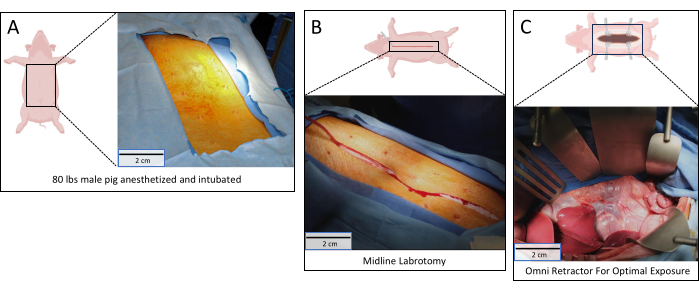
Figure 1: Operative setup. (A) Pig positioned in supine position with the abdomen sterilely prepped; (B) Midline laparotomy made from xiphoid to pubis; (C) Retroperitoneum exposed with Omni retractor in place. Scale bars = 2 cm. Please click here to view a larger version of this figure.
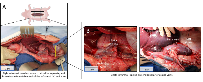
Figure 2: Retroperitoneal dissection. (A) Retroperitoneum dissection complete with circumferential control of the infrarenal IVC and renal veins. The yellow rectangle is magnified in Figure 2B; (B) Magnified image of infrarenal IVC and renal veins; (C) Renal veins tied off. Scale bars = 2 cm. Abbreviation: IVC = inferior vena cava. Please click here to view a larger version of this figure.
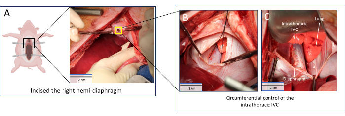
Figure 3: Obtaining supraceliac control. (A) Right hemidiaphragm incised. The yellow square is magnified in Figure 3B; (B) Extension of right hemidiaphragm incision to expose intrathoracic IVC and descending thoracic aorta; (C) Circumferential control of intrathoracic IVC obtained. Scale bars = 2 cm. Abbreviation: IVC = inferior vena cava. Please click here to view a larger version of this figure.
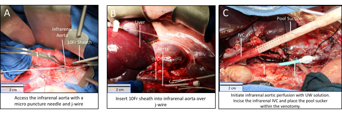
Figure 4: Infrarenal aortic cannulation, visceral isolation, and perfusion. (A) Proximal and distal control of the infrarenal aorta obtained; (B) 10Fr sheath placed in the infrarenal aorta by Seldinger technique; (C) Visceral perfusion via the infrarenal aortic sheath and drainage via venotomy and pool sucker in infrarenal IVC. Scale bars = 2 cm. Abbreviation: IVC = inferior vena cava. Please click here to view a larger version of this figure.
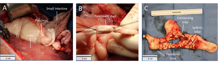
Figure 5: Total pancreatectomy. (A) Viscera becoming paler during perfusion; (B) pancreatic duct isolated; (C) Explanted porcine pancreas. Scale bars = 2 cm. Please click here to view a larger version of this figure.
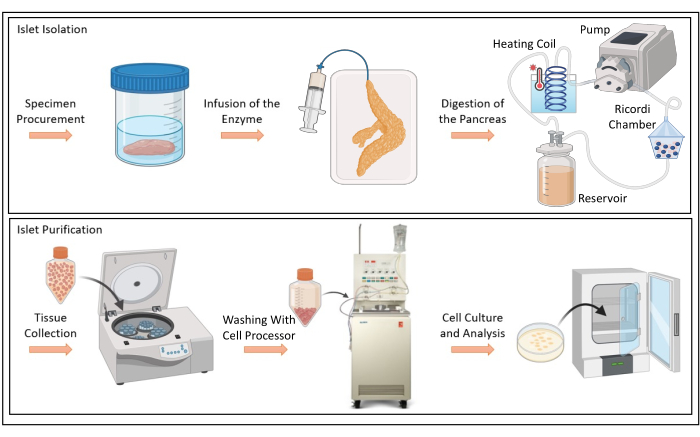
Figure 6: Islet isolation and purification. Schematic depiction of infusion of enzyme solution, digestion of porcine pancreas, tissue collection, cell washing with Cell Processor, and cell culture and analysis. Please click here to view a larger version of this figure.
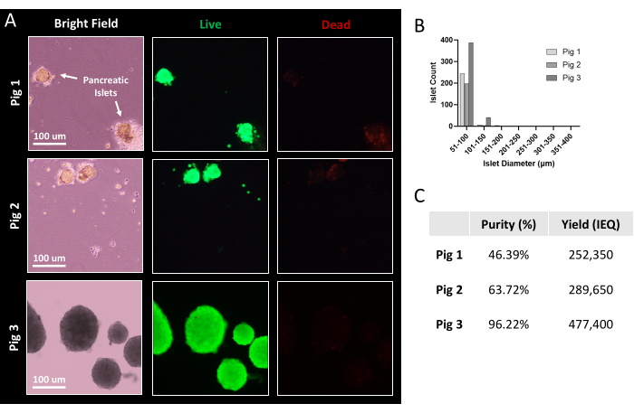
Figure 7: Representative islet viability and yield after purification. (A) Porcine islets after magnified bright-field imaging and live/dead staining. Green demonstrates live cells; red demonstrates dead cells. Scale bars = 100µm (B) Histogram distribution of porcine islet size following purification. (C) Table indicating islet purity and yield from three porcine pancreatic islet isolations. Abbreviation: IEQ = islet equivalent, the standard unit for islet quantification. Please click here to view a larger version of this figure.
Pancreatic islet xenotransplantation using porcine donors is a promising strategy for the treatment of Type I diabetes. Islet isolation is challenging, and final islet viability and yield are highly susceptible to hypotension and tissue ischemia encountered during organ procurement16,17. To optimize pancreas procurement and preservation, this protocol presents a new method for aortic cannulation, visceral isolation, and perfusion during the procurement of the porcine pancreas prior to islet isolation.
Porcine islet isolation success is dependent on optimal procurement and preservation of the donor pancreas. The removal of all blood from the pancreas tissue is crucial for optimizing islet isolation as residual blood in the tissue leads to complement activation and thrombosis of pancreatic microvasculature, which can deteriorate islet yield and viability4,18. Previous techniques also include cannulation of the infrarenal aorta with intra-arterial flushing13,14,15,19. However, a major advantage of the technique presented here is the use of Seldinger technique for aortic cannulation to maintain a bloodless operative field. This not only minimizes hemodynamic instability before aortic cross clamping but also keeps the dissection planes pristine for the pancreatectomy, enabling faster organ recovery and minimizing WIT. This technique is also adaptable based on the size of the pig, as a smaller-caliber sheath could be used for mini-pigs with smaller-diameter aortas.
An additional strength of this technique is the significant reduction of WIT. Prior reports suffer from extended periods of WIT given pancreatectomy following the induction of cardiac arrest or exsanguination. Even when combined with ex vivo intraarterial and intraductal flushing, these procurement techniques are hindered by WIT, to which the porcine pancreas is particularly sensitive due to species-specific abundance of proteolytic enzymes and high fat content20. Prolonged WIT can also alter pancreatic tissue distention, likely due to an increase in resistance in the pancreatic ducts. This can significantly reduce the amount of enzyme reaching the distal pancreatic ducts, the overall efficiency of the digestion, and the yield of the islet isolation5.
Some limitations and pitfalls may be encountered during the procurement, but the majority can be avoided with careful planning and clearly defining personnel roles at the start of the procedure. Although the operative procurement can be carried out by two operators, a third assistant, who is not sterile, is needed to prepare the UW solution and container for the pancreas. When performing the intrathoracic portion of the procedure, it is important for the liver to be handled with gentle retraction. Injury and fracture of the liver can lead to persistent bleeding throughout the procedure. If this occurs, the liver can be packed with multiple lap pads to limit bleeding and limit blood obscuring the pancreatic procurement site. Of note, the identification and isolation of the pancreatic duct are critical for this procedure.
For the purification to go smoothly, two well-trained laboratory personnel should be available to perform the experiment. We recommend that one person maintain sterility during the procedure, while the other can be nonsterile. This will help with the handoff of critical materials during the procedure while maintaining sterility during pancreatic isolation.
During the perfusion procedure, the lumbar arteries that emerge from the infrarenal aorta are typically not identified and individually ligated. This may lead to a negligible volume of UW solution to disperse into non-viscera. However, we believe that minimizing the aortic dissection time and the risk of vascular injury during lumbar vessel manipulation outweigh the slight inefficiency of the UW perfusion not reaching the pancreatic tissue.
Once the pancreatic duct is identified, a catheter must be cannulated through the duct to proceed with the infusion of the pancreas. If the pancreatic duct is damaged, the parenchyma will not distend during subsequent perfusion of digestive solution during islet isolation. In the case of poor distention, islet isolation should be aborted. If the distention is favorable, the remaining enzyme can be injected into the parenchyma. In summary, this protocol provides a method for visceral isolation and perfusion during procurement of the porcine pancreas for islet isolation and purification. The use of the Seldinger technique for aortic cannulation minimizes hemodynamic changes and blood loss during procurement. This method can be used in combination with islet isolation to provide porcine islets for future studies and, ultimately, xenotransplantation.
The authors have nothing to disclose.
This manuscript was supported by a clinical innovation grant from the Mid-America Transplant Foundation.
1 M NaOH, 100 mL | Millipore Sigma | 1310-73-2 | |
10x HBSS, 500 mL (15 to 30 °C) | Thermo Fisher | 14065056 | |
1x D-PBS, 500 mL | Thermo Fisher | 14200075 | |
1x HBSS, 1 L | Thermo Fisher | 14025076 | |
250 mL Conical Tubes | Millipore Sigma | CLS430236-6EA | |
Amphotericin B [50 mg] | Millipore Sigma | PHR1662 | |
Antiseptic Povidone Iodine 10% | Millipore Sigma | 25655-41-8 | |
BioRad Gradient Former | BioRad | 395 | |
Calcium Chloride dihydrate | Thermo Fisher | 10035-04-8 | |
COBE Cell Processor | Ebay | 317690 | |
Digestion circuit reservoir, Gibco bottle, 1 L | Thermo Fisher | 10341001 | |
Dimethylsulfoxide (DMSO) | Thermo Fisher | 85190 | |
Dithizone | Thermo Fisher | 60-10-6 | |
Gradient Former Connection Kit | BioRad | 1652008 | |
Heparin [1000 U/mL] | Emergency Medical Products | 0409-2720-01 | |
HEPES Buffer (1 M), 100 mL | Thermo Fisher | 15630080 | |
Human Serum Albumin 25%, 100 mL | Celprogen | HSA2001-25-1 | |
Liberase (100 mg) | Millipore Sigma | 5401020001 | |
LIVE/DEAD Viability/Cytotoxicity Kit | Thermo Fisher | L3224 | |
Lympholyte 1.1 Media 500 mL | Cedarlane Labs | CL5020 | |
Masterflex LS 16 Tubing | Masterfle | 96419-16 | |
Masterflex Peristaltic Pump | Masterflex | 07522-30 | |
PenStrep (2 mg/mL) | Celprogen | PS-30-002 | |
Pulmozyme (1 mg/mL, 2.5 mL/vial) | Dornase Alfa | 8931278 | |
Recovery Medium, PIM(R) | Prodo Laboratories, Inc. | PIM-R001GMP | |
RPMI 1640 | Millipore Sigma | R8758-1L | |
Standard Culture Medium, PIM(S) | Prodo Laboratories, Inc. | PIM-S001GMP | |
University of Wisconsin (UW) Solution, 3 L | Global Transplant Solutions | 1000-0046-06 | |
Waterbath | Sigma Alderich | CLS6783-1EA |
.