Sensing of Barrier Tissue Disruption with an Organic Electrochemical Transistor
Summary
The Organic Electrochemical Transistor is integrated with live cells and used to monitor ion flux across the gastrointestinal epithelial barrier. In this study, an increase in ion flux, related to disruption of tight junctions, induced by the presence of the calcium chelator EGTA (ethylene glycol-bis(beta-aminoethyl ether)-N,N,N’,N’-tetra acetic acid), is measured.
Abstract
The gastrointestinal tract is an example of barrier tissue that provides a physical barrier against entry of pathogens and toxins, while allowing the passage of necessary ions and molecules. A breach in this barrier can be caused by a reduction in the extracellular calcium concentration. This reduction in calcium concentration causes a conformational change in proteins involved in the sealing of the barrier, leading to an increase of the paracellular flux. To mimic this effect the calcium chelator ethylene glycol-bis(beta-aminoethyl ether)-N,N,N',N'-tetra acetic acid (EGTA) was used on a monolayer of cells known to be representative of the gastrointestinal tract. Different methods to detect the disruption of the barrier tissue already exist, such as immunofluorescence and permeability assays. However, these methods are time-consuming and costly and not suited to dynamic or high-throughput measurements. Electronic methods for measuring barrier tissue integrity also exist for measurement of the transepithelial resistance (TER), however these are often costly and complex. The development of rapid, cheap, and sensitive methods is urgently needed as the integrity of barrier tissue is a key parameter in drug discovery and pathogen/toxin diagnostics. The organic electrochemical transistor (OECT) integrated with barrier tissue forming cells has been shown as a new device capable of dynamically monitoring barrier tissue integrity. The device is able to measure minute variations in ionic flux with unprecedented temporal resolution and sensitivity, in real time, as an indicator of barrier tissue integrity. This new method is based on a simple device that can be compatible with high throughput screening applications and fabricated at low cost.
Introduction
The gastrointestinal epithelium is an example of barrier tissue, which controls the passage of molecules between different compartments of the body. The epithelium consists of elongated columnar cells joined together by complexes of proteins that provide a physical barrier1 against pathogens and toxins, while allowing the passage of water and nutrients required to sustain the body. This selectivity is due to the polarization of the epithelial cells, which creates two different membrane domains: the apical side of the cells exposed to the lumen and the basal side of the cells anchored on the underlying tissues2,3. Tight junctions (TJ) are complexes of proteins present at the apical portion of epithelial cells and are part of a larger complex known as the apical junction4. Ion flow across the barrier tissue may either go via the transcellular (through the cell) or via a paracellular (between two adjacent cells) pathway. The sum of the transport through both pathways is known as the transepithelial resistance. The apical junction is responsible for the regulation of ions and molecules passing across the barrier5,6 via a specific opening and closing function. A dysfunction or disruption of these protein complexes is often related to disease7-11. In addition, many enteric pathogens/toxins are known to specifically target this complex, thereby entering the body and leading to diarrhea, most likely as a consequence of massive dysregulation of ion/water flow across the barrier12-14. Barrier tissue may also be modified by changing the extracellular microenvironment. Cadherin is a critical protein for cell-cell adhesion, and is involved in the formation of the apical junction. Calcium is required for the correct structural conformation of Cadherin, and a decrease in extracellular calcium has been shown to result in the destruction of the cell-cell junction and a subsequent opening of the paracellular pathway between the cells15. In this study, EGTA (Ethylene glycol-bis(beta-aminoethyl ether)-N,N,N',N'-tetra acetic acid), a specific calcium chelator, was used to induce a breach in barrier tissue, as it has been shown to have a rapid and drastic effect on paracellular ion flow16,17. This calcium chelator was used on a confluent and differentiated monolayer of the Caco-2 cell-line. Cultured in cell culture inserts, this cell line is known to develop the characteristics of the gastrointestinal tract and is widely used by the pharmaceutical industry to test the absorption of drugs18,19.
Methods to monitor barrier tissue integrity are plentiful. These methods are often optical, relying on immunofluorescence staining of particular proteins known to be at the apical junction20, or relying on the quantification of a fluorescent tracer molecule that is normally impermeable to the barrier tissue21,22. However, label-free methods (i.e. without a fluorophore/chromophore) are preferable as the use of a label can incur artifacts, and often increases cost and assay time. Electrical, label-free monitoring of barrier tissue has recently emerged as a dynamic monitoring method23. For example recent technological advances in electrical impedance spectroscopy have allowed the development of a commercially available scanning apparatus24,25 that can measure transepithelial resistance (TER), a measurement of the ion conductance across the cell layer.
Organic electronics has created a unique opportunity to interface the world of electronics and biology26,27 28,29 by using conducting polymers that can conduct both electronic and ionic carriers. A new technique to detect breaches in barrier tissue using the OECT30-32 was recently introduced. This device was validated against existing techniques used to assess barrier tissue integrity, including immunofluorescence, permeability assays using Lucifer yellow, and impedance spectroscopy using the Cellzscope. In the case of all toxic compounds tested, the OECT was found to operate with equal or better sensitivity, and with increased temporal resolution, compared to the above techniques. In this device, PEDOT:PSS, a conducting polymer that has been shown to be stable and biocompatible33,34, is used as the active material in the transistor channel. The OECT is composed of drain and source electrodes on either side of a conducting polymer channel. This is then placed in contact with an electrolyte, which forms an integral part of the device. A gate electrode is immersed in the electrolyte (Figure 1), and when a positive gate voltage is applied at the gate, cations from the electrolyte are forced into the channel, thus dedoping the conducting polymer and resulting in a change in the source-drain current. The device is thus extremely sensitive to minute changes in ionic flux due to amplification by the transistor. A cell layer grown on a cell culture insert was placed between the gate electrode and the conducting polymer channel. The presence of an intact cell layer acts as barrier for the cations entering into the conducting polymer, therefore, in the presence of an intact monolayer, the drain current decreases (Figure 2: transition from region a to b). In the presence of a toxic compound, the barrier tissue will progressively lose its integrity, letting the cations enter into the polymer film and increasing the drain current (Figure 2: region c). With this technique, the breach in barrier tissue is seen by the modulation of the drain current, corresponding to the modulation of the flux across the monolayer. This device is able to measure minute variations in ionic flux with unprecedented temporal resolution and sensitivity in real time. This technology will be of interest in the domain of toxicology for drug testing, disease diagnostics or basic research as the barrier model can be easily adapted. This method will also help to reduce animal experimentation, as it allows the validation of in vitro models to replace in vivo testing.
Protocol
1. PEDOT:PSS Solution Preparation
- To 50 ml of PEDOT:PSS, add ethylene glycol (increases conductivity) in a volume ratio of 1:4 (ethylene glycol to PEDOT:PSS), 0.5 μl/ml of Dodecylbenzenesulfonic acid (DBSA) as a surfactant, and 10 mg/ml 3-glycidoxypropyltrimethoxysilane (GOPS) as a cross-linker to promote adhesion of the conducting polymer to the glass slide.
2. OECT Fabrication (Figure 3)
- Define thermally evaporated gold source and drain contacts via lift-off lithography:
- Spin coat photoresist on a normal clean glass slide at 3,000 rpm for 30 sec. Glass dimensions are 3 in x 1 in.
- Define patterns by photolithography. Dimensions may be altered, however the dimensions listed here were shown to lead to the optimal sensitivity. Then use a developer.
- Evaporate 5 nm and 100 nm of chromium and gold, respectively.
- Lift-off the photoresist in an acetone bath for 1 hr, leaving the substrate with the source and drain Au contacts area only.
- The desired length of the PEDOT:PSS channel is 1 mm. This is achieved by patterning using a parylene-C (Pa-C) peel-off technique:
- Load 3.5 g of Pa-C in the coating setup. Uniformly evaporate 2 µm of parylene-C on top of the substrate with Au contacts.
- Pattern the channel by photolithography:
- Spin coat photoresist at 3,000 rpm for 30 sec.
- Use a mask to illuminate channel and gold contact 20 sec to UV, the exposed photoresist will become soluble in the developer.
- Use developer to open the channel area on the photoresist.
- Etch the Pa-C in the channel area by a 15 min plasma step (O2 (50 sccm) and CHF4 (3 sccm) plasma at 160 W) in order to open the channel and the gold contact.
- Deposit the PEDOT:PSS mixture solution by spin coating at 500 rpm for 45 sec. Bake for 30 sec at 110 °C.
- Peel-off the Pa-C to reveal the PEDOT:PSS channel of the substrate underneath. This channel should slightly overlap with the Au contacts made in Protocol 1.
- Bake samples for 1 hr at 140 °C under atmospheric conditions.
3. Device Assembly
- Make PDMS by mixing two solutions (usually supplied together in a kit) at a ratio 1:10 of curing solution in base solution. Bake it for 1 hr at 120 °C.
- Design well by using a hole punch and cut a square around the desired area.
- Glue a PDMS well on top of the channel, to result in a channel area of approximately 6 mm2. Ensure that the source and drain contacts are not covered.
- Make a plastic support with a hole in it (can use the holder for the cell culture insert) and then glue it on top of the PDMS. Let it dry overnight.
- Test the well for leaking by prefilling with water.
- Solder electric wires on the source and drain contact with tin.
4. Cell Culture
- Prepare cell culture media as follows:
- In DMEM, add 1% Glutamine, 10% Fetal Bovine Serum, 0.5% Penicillin-streptomycin, and 0.1% Gentamicin.
- Sterilize the cell culture media using a sterile filter device.
- Maintain Caco-2 cells between passage 49 and 68 at 37 °C in a humidified atmosphere of 5% CO2, in cell culture media.
- Divide cells once a week using trypsin and seed at 1.5 x 10⁴ cells/insert.
- Change cell culture media twice a week over 3 weeks.
5. Measurements with OECT
- Connect an Ag/AgCl wire to a sourcemeter for use as the gate electrode. Immerse the tip in cell culture media, which is used as the electrolyte, as illustrated in Figure 1a.
- Connect wires from source and drain to the sourcemeter (dual channel setup).
- Apply a square pulse positive voltage (VGS) between the gate and the source (used as reference electrode: ground). A negative constant voltage between drain and source (VDS) is applied and corresponding current (IDS) is measured.
- Use a PC running program data collection. OECT parameters entered in a customized acquisition program should be as follows: VDS = -0.2 V, VGS = 0.3 V, VGS on time = 2 sec, off time = 28 sec.
- Read out source-drain current (IDS) and gate current (IGS). Carry out the measurement for several minutes to ensure a stable baseline signal.
6. Integrating Cells with OECT for Measurement
Note: Prior to the experiment, the integrity of the cell layer may be verified by measuring TER with an Impedance spectroscopy device. TER of each 3-week old Caco-2 cell insert should be above 400 Ω.cm2.
- During off time of OECT measurement: Remove the gate electrode from the electrolyte, incorporate the cell culture insert, and replace the gate electrode inside the cell culture insert.
- Carry out a baseline measurement with the cells for several minutes to ensure a stable signal. This baseline will be used as for calculation of the normalized value corresponding to an intact monolayer (0).
7. Preparation and Introduction of Toxic Compound
- Prepare a 1 M solution of EGTA in DI water, first adjusting the pH of the solution to 7.4 with Tris base.
- Add EGTA solution in the appropriate volume to obtain the desired concentration (e.g. between 5 mM and 100 mM) taking into account the volume. The EGTA should be added in the basal chamber during the off time.
- Measure continuously for 90 min as in Protocol 5. If the measurement is being carried out at room temperature, the stability of the cells will not remain constant after 90 min.
- At the end of the run, scratch the cell layer to result in a complete destruction of the barrier layer and measure for 15 min, this point could be done by removing the filter and immersing the gate electrode in the media. This baseline will be used as for calculation of the normalized value corresponding to a destroyed monolayer.
Representative Results
During the first step of measurement, the drain current may vary somewhat, but in most cases it should remain stable (Figure 2, section a). If the signal is not stable, the transistor should be discarded and replaced. This stability check also ensures that any initial losses in conductivity of the device do not affect the subsequent measurement. After several minutes of measurement, the insert with cells forming barrier tissue is placed on top of the channel. The drain current should immediately decrease (Figure 2, section b.). If the drain current does not decrease, this could imply that the cells have not correctly formed a barrier, or were damaged during manipulation, and the filter should be discarded and replaced. By adding a toxic compound to the barrier tissue, the modulation of the drain current should increase progressively or immediately, depending on the compound used and the concentration (Figure 2, section c.).
Data were collected using a sourcemeter and a customized acquisition program. From this program different parameters were obtained, which are run in a data analysis program to obtain the mid-modulation corresponding to each gate pulse. The mid modulation corresponds to the value of the current at the middle of the peak (Figure 4). The mid modulation values obtained during sensing of an intact monolayer were normalized to 0 while the mid modulation obtained after the scratch were normalized to 1. Normalized results are used to compare data from different devices (Figure 5). From this data the dose response of difference concentrations of toxic compound over time can be seen.
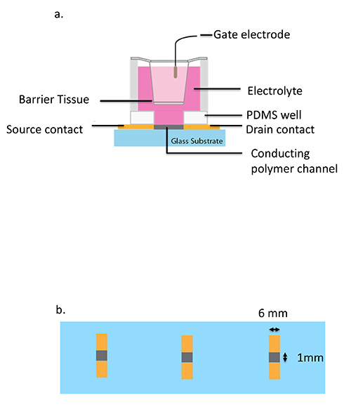
Figure 1. Schematic of an Organic Electrochemical Transistor integrated with cell culture insert a) from a side view. b) Top view of channel (grey) and contact (yellow) dimensions on a glass slide with a channel length of 6mm and channel width of 1 mm. This figure has been modified from Jimison30.
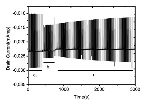
Figure 2. Overview of in situ measurement of drain current response to square gate pulses over time. Measurement conditions are: VDS = -0.2 V, VGS = 0.3 V, VGS on time = 2 sec, off time = 28 sec. a) Corresponds to the OECT operated before integration with cells, ensuring the stability of the device. b) Corresponds to the integration of the barrier tissue forming cells, again ensuring a stable signal before commencing testing of toxic compound. c) Corresponds to the addition of EGTA to the barrier tissue cells, note the evolution of the signal over time.
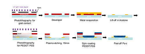
Figure 3. Fabrication processes of the OECT. Click here to view larger image.
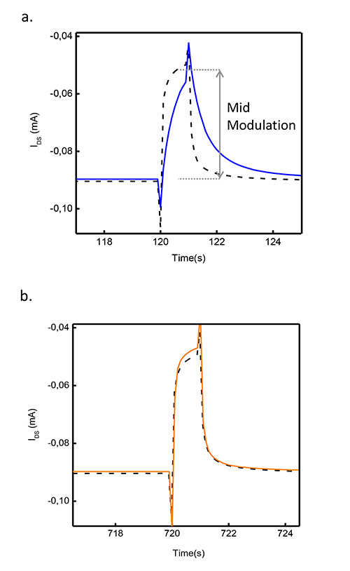
Figure 4. OECT drain current response to a single square gate pulse. Measurement conditions are as follows: VDS = -0.2 V, VGS = 0.3 V, VGS on time = 2 sec, off time = 28 sec. a) OECT transient response before (dashed line) and after (blue line) the addition of barrier tissue forming cells grown on a filter. b) OECT transient response after the addition of toxic compound on a barrier tissue (orange line) and without barrier tissue (dashed line).
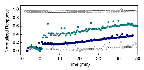
Figure 5. Typical normalized result From the OECT. The normalized response of the OECT on introduction of device (empty circle), cell layer (black cross), 5 mM EGTA (dark blue circle) and 10 mM EGTA (cyan circle) introduced at t = 0, were measured over a period of 50 min. Click here to view larger image.
Discussion
This technique provides a novel method to integrate an organic electrochemical transistor with live cells to measure barrier tissue integrity. The main advantages of this technique are the rapidity and sensitivity, but also the low cost of the device for dynamic monitoring of barrier tissue.
As this method uses live cells, a critical point is to be sure to use a monolayer, which represents an intact barrier layer. The parameters of the barrier should be defined during the characterization of the cell line. It is therefore crucial not to damage the cell layer when the gate electrode is immersed in the electrolyte on top of the cell layer.
Another important point is the fabrication of the device; no leakage should occur between the edge of the PDMS and the plastic holder to avoid liquid volume variation. In order to achieve reproducible results, attention should be also given to the soldering of the wire, as burning the gold electrode will result in poor/no contact with the conducting polymer channel and so poor/no signal.
To facilitate the analysis and to obtain better results a few minutes delay between each step of the experiment should be allowed for the device to stabilize between steps. As a variation of the signal from device to device is observed, normalization of results is required to be able to compare the results of each experiment. However, in the future larger scale production of the device will allow greater device-device reproducibility.
The main limitation of the technique is the format, as for the moment only one sample can be measured. This will be solved soon by the development of a multiacquisition sensor and in the future the creation of a 96-well plate format. This will open the way for this technique for use in high throughput screening of barrier tissue. In parallel, planar devices are also under development to allow simultaneous optical and electronic measurements.
In summary, a novel device that can be fabricated at low cost that is capable of label-free monitoring of barrier tissue has been developed. This device shows greater sensitivity and higher temporal resolution than existing methods. The integration of in vitro models with devices such as the OECT can be a great alternative to animal testing for drug discover and toxicology.
Disclosures
The authors have nothing to disclose.
Acknowledgements
The authors of this paper do not have any competing financial interests.
Materials
| CLEVIOS PH 1000 | HERAUS CLEVIOS | ||
| AZ9260 resin | CIPEC SPECIALITIES | ||
| Dodecylbenzenesulfonic acid (DBSA) | Acros Organic | ||
| 3-glycidoxypropyltrimethoxysilane (GOPS) | Sigma Aldrich | ||
| 24-well Suspended cell Culture insert Millicell PET 0.4 μm Millipore | Dominique dutscher | 51705 | |
| 24-well cell culture plate BD Falcon | Dominique dutscher | 51705 | |
| STERICUP-GP PES 0.22 μM | Dominique dutscher | 51246 | |
| ADVANCED DMEM Marque GIBCO | Fisher scientific | E3434T | |
| FBS HEAT INACT. S.AMERICAN | Fisher scientific | E3387M | |
| PENICILLIN STREPTOMYCIN | Fisher scientific | E3470C | |
| GLUTAMAX | Fisher scientific | E3524T | |
| TRYPSIN 0.05% EDTA | Fisher scientific | E3513N | |
| EGTA (Ethylene glycol-bis(2-aminoethylether)-N,N,N′,N′-tetraacetic acid) | Sigma Aldrich | E4378 | |
| ETHYLENE GLYCOL, ANHYDROUS, 99.8%, | Sigma aldrich | ||
| Caco-2 cells | ATCC | ||
| PDMS | Dow corning | SYLGARD 184 SILICONE ELASTOMER | |
| Au (99.99%) | NEYCO | AU3X6 | |
| Chromium (99.95%) | NEYCO | ||
| Parylene C | Specialty Coating Systems | ||
| Ag/AgCl wire | HARVARD APPARATUS | ||
| Photoresist | CIPEC SPECIALITIES | Résine AZ9260 |
References
- Farquhar, M. G., Palade, G. E. Junctional complexes in various epithelia. J. Cell Biol. 17, 375-412 (1963).
- Gaillard, J. L., Finlay, B. B. Effect of cell polarization and differentiation on entry of Listeria monocytogenes into the enterocyte-like Caco-2 cell line. Infect. Immun. 64, 1299-1308 (1996).
- Anderson, J. M., Balda, M. S., Fanning, A. S. The structure and regulation of tight junctions. Curr. Opin. Cell Biol. 5, 772-778 (1993).
- Guttman, J. A., Finlay, B. B. Tight junctions as targets of infectious agents. Biochim. Biophys. Acta. 1788, 832-841 (2009).
- Anderson, J. M. Molecular structure of tight junctions and their role in epithelial transport. News. Physiol. Sci. 16, 126-130 (2001).
- Anderson, J. M., Van Itallie, C. M. Tight junctions: Closing in on the seal. Curr. Biol. 9, (1999).
- Ma, T. Y., Boivin, M. A., Ye, D., Pedram, A., Said, H. M. Mechanism of TNF-{alpha} modulation of Caco-2 intestinal epithelial tight junction barrier: role of myosin light-chain kinase protein expression. Am. J. Physiol. Gastrointest. Liver Physiol. 288, 422-430 (2005).
- Schulzke, J. D., et al. Epithelial tight junctions in intestinal inflammation. Ann. N.Y. Acad. Sci. 1165, 294-300 (2009).
- Fisher, S. J., Swaan, P. W., Eddington, N. D. The ethanol metabolite acetaldehyde increases paracellular drug permeability in vitro and oral bioavailability in vivo. The J. Pharmacol. Exp. Therap. 332, 326-333 (2010).
- Ma, T. Y., Nguyen, D., Bui, V., Nguyen, H., Hoa, N. Ethanol modulation of intestinal epithelial tight junction barrier. Am. J. Physiol. 276, 965-974 (1999).
- Nemeth, E., Halasz, A., Barath, A., Domokos, M., Galfi, P. Effect of hydrogen peroxide on interleukin-8 synthesis and death of Caco-2 cells. Immunopharmacol. Immunotoxicol. 29, 297-310 (2007).
- Vogelmann, R., Amieva, M. R., Falkow, S., Nelson, W. J. Breaking into the epithelial apical-junctional complex–news from pathogen hackers. Curr. Opin. Cell Biol. 16, 86-93 (2004).
- Nusrat, A., et al. Clostridium difficile toxins disrupt epithelial barrier function by altering membrane microdomain localization of tight junction proteins. Infect. Immun. 69, 1329-1336 (2001).
- Obert, G., Peiffer, I., Servin, A. L. Rotavirus-induced structural and functional alterations in tight junctions of polarized intestinal Caco-2 cell monolayers. J. Virol. 74, 4645-4651 (2000).
- Nagar, B., Overduin, M., Ikura, M., Rini, J. M. Structural basis of calcium-induced E-cadherin rigidification and dimerization. Nature. 380, 360-364 (1996).
- Boulenc, X., et al. Importance of the paracellular pathway for the transport of a new bisphosphonate using the human Caco-2 monolayers model. Biochem. Pharmacol. 46, 1591-1600 (1993).
- Artursson, P., Magnusson, C. Epithelial transport of drugs in cell culture. II: Effect of extracellular calcium concentration on the paracellular transport of drugs of different lipophilicities across monolayers of intestinal epithelial (Caco-2) cells. J. Pharm. Sci. 79, 595-600 (1990).
- Artursson, P. Epithelial transport of drugs in cell culture. I: A model for studying the passive diffusion of drugs over intestinal absorptive (Caco-2) cells. J. Pharm. Sci. 79, 476-482 (1990).
- Artursson, P., Karlsson, J. Correlation between oral drug absorption in humans and apparent drug permeability coefficients in human intestinal epithelial (Caco-2) cells. Biochem. Biophys. Res. Commun. 175, 880-885 (1991).
- Balda, M. S., et al. Functional dissociation of paracellular permeability and transepithelial electrical resistance and disruption of the apical-basolateral intramembrane diffusion barrier by expression of a mutant tight junction membrane protein. J. Cell Biol. 134, 1031-1049 (1996).
- Hubatsch, I., Ragnarsson, E. G. E., Artursson, P. Determination of drug permeability and prediction of drug absorption in Caco-2 monolayers. Nat. Protoc. 2, 2111-2119 (2007).
- Uchida, M., Fukazawa, T., Yamazaki, Y., Hashimoto, H., Miyamoto, Y. A modified fast (4 day) 96-well plate Caco-2 permeability assay. J. Pharmacol. Toxicol. Methods. 59, 39-43 (2008).
- Krug, S. M., Fromm, M., Gunzel, D. Two-Path Impedance Spectroscopy for Measuring Paracellular and Transcellular Epithelial Resistance. Biophys. J. 97, 2202-2211 (2009).
- Wegener, J., Abrams, D., Willenbrink, W., Galla, H. J., Janshoff, A. Automated multi-well device to measure transepithelial electrical resistances under physiological conditions. BioTechniques. 37, 592-594 (2004).
- Weber, C. R., Shen, L., Wu, L., Wang, Y., Turner, J. R. Occludin is Required for Tumor Necrosis Factor (TNF)-Mediated Regulation of Tight Junction (TJ) Barrier Function. Gastroenterology. 140, (2011).
- Owens, R. M., Malliaras, G. G. Organic electronics at the interface with biology. MRS Bull. , (2010).
- Lin, P., Yan, F., Yu, J. J., Chan, H. L. W., Yang, M. The Application of Organic Electrochemical Transistors in Cell-Based Biosensors. Adv. Mater. 22, 3655-3660 (2010).
- White, H. S., Kittlesen, G. P., Wrighton, M. S. Chemical Derivatization of an Array of 3 Gold Microelectrodes with Polypyrrole – Fabrication of a Molecule-Based Transistor. J. Am. Chem. Soc. 106, 5375-5377 (1984).
- Bernards, D. A., Malliaras, G. G. Steady-state and transient behavior of organic electrochemical transistors. Adv. Funct. Mater. 17, 3538-3544 (2007).
- Jimison, L. H., et al. Measurement of Barrier Tissue Integrity with an Organic Electrochemical Transistor. Adv. Mater. 24, 5919-5923 (2012).
- Tria, S., Jimison, L. H., Hama, A., Bongo, M., Owens, R. M. Sensing of EGTA Mediated Barrier Tissue Disruption with an Organic Transistor. Biosensors. 3, 44-57 (2013).
- Tria, S. A., Jimison, L. H., Hama, A., Bongo, M., Owens, R. M. Validation of the organic electrochemical transistor for in vitro toxicology. Biochim. Biophys. Acta. 1830, 4381-4390 (2013).
- Zhu, Z. T., et al. A simple poly(3,4-ethylene dioxythiophene)/poly(styrene sulfonic acid) transistor for glucose sensing at neutral pH. Chem. Commun. , 1556-1557 (2004).
- Lin, P., Yan, F., Yu, J., Chan, H. L., Yang, M. The application of organic electrochemical transistors in cell-based biosensors. Adv. Mater. 22, 3655-3660 (2010).
