Cancer cells are embedded in a collagen gel and then sandwiched in an acellular fibrin gel to generate a 3D culture system in which the invasiveness and formation of satellite tumors may be monitored.
Mammalian cell culture in monolayers is widely used to study various physiological and molecular processes. However, this approach to study growing cells often generates unwanted artifacts. Therefore, cell culture in a three-dimensional (3D) environment, often using extracellular matrix components, emerged as an interesting alternative due to its close similarity to the native in vivo tissue or organ. We developed a 3D cell culture system using two compartments, namely (i) a central compartment containing cancer cells embedded in a collagen gel acting as a pseudo-primary macrospherical tumor and (ii) a peripheral cell-free compartment made of a fibrin gel, i.e. an extracellular matrix component different from that used in the center, in which cancer cells can migrate (invasion front) and/or form microspherical tumors representing secondary or satellite tumors. The formation of satellite tumors in the peripheral compartment is remarkably correlated to the known aggressiveness or metastatic origin of the native tumor cells, which makes this 3D culture system unique. This cell culture approach might be considered to assess cancer cell invasiveness and motility, cell-extracellular matrix interactions and as a method to evaluate anti-cancer drug properties.
Investigating the fundamental and biomedical characteristics of cancer cell invasion/migration and subsequent metastasis establishment is the subject of an intense research1,2. Metastasis is the ultimate stage of cancer and its clinical management remains elusive. A better understanding of metastasis at the cellular and molecular levels will enable the development of more efficient therapies3.
Several properties of metastatic cells can be explored in vitro4 including their stemness and potential to acquire a transition state (e.g., epithelioid-mesenchymal transition) to migrate and invade within and from the primary tumor5. However, the in vitro assessment of invasion/metastasis processes has been a challenge since it virtually excludes the contribution of the blood/lymphatic circulation. Organotypic cultures that embed tumor fragments in collagen gels have previously been used to monitor cancer aggressiveness. Although the complexity of tumors is preserved (e.g., the presence of non-cancerous cells), tumor fragments are exposed to limited medium diffusion, to sampling variation, and to an overgrowth of stromal cells6. An alternative method consists in growing cancer cells within components of the extracellular matrix (ECM), which mimics the three-dimensional (3D) cell environment. The proliferation of breast cancer cell lines in a collagen gel and/or a basement membrane-derived matrix is amongst the best-characterized examples of 3D cell culture. By using specific 3D cell culture environments, the disorganized assembly observed for breast cancer cells grown under standard conditions can be reversed to the spontaneous formation of mammary acini and tubular structures7-10. Furthermore, the formation of multicellular tumor spheroids derived from adenocarcinoma cancer cells congregated using different techniques (e.g., hanging drops, floating spheroids, agar embedment) now constitutes the most commonly used 3D cell culture assay11-13. However, this assay is limited by the restricted set of cancer cell lines that can form spheroids and by the short period available to study cells in these conditions.
In this visualized technique, we herein introduce a sophisticated 3D cell culture assay where cancer cells of interest are embedded in a collagen gel to allow the in vitro formation of a pseudo-primary tumor that can be alternatively coated with a basement membrane-derived matrix. Once formed, the pseudo-primary tumor is then sandwiched in an acellular matrix (fibrin gel in the present case), which allows the cancer cells to cross the interface between the two matrix compartments (see Figure 1). Interestingly, secondary tumor-like structures originating from the pseudo-primary tumor along with aggressive cancer cells appear in the fibrin gel. Such a 3D culture system offers the flexibility required to investigate, for example, anticancer drugs, gene expression and cell-cell and/or cell-ECM interactions14-16.
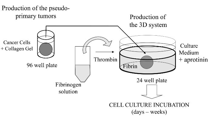
Figure 1: Overview of the Method. Schematic summary of the method to generate the 3D cell culture system as a model for cancer studies. Please click here to view a larger version of this figure.
NOTE: No ethics consideration since animal and human cancer cells were purchased or kindly provided to us.
1. Making Collagen Plugs (Pseudo-primary Tumor)
2. First Layer of Fibrin Gel
3. Second Layer of Fibrin Gel and Sandwiched Collagen Plug
4. Cell Culture Medium Conditions
As previously mentioned, an interesting feature of this 3D cell culture assay is that cancer cells can not only migrate from the collagen plug to the adjacent fibrin gel, but also establish secondary tumors (e.g., satellite tumor-like structures). This can be directly observed with an inverted phase contrast microscope at low and high magnifications through the gel thickness, especially with a long working distance condenser (Figure 2). Using this 3D cell culture method, the behavior of known metastatic cells can be readily compared to that found in non-metastatic cells, as demonstrated repeatedly with different experimental setups15. For example, numerous satellite tumors are found randomly dispersed in the fibrin gel with the metastatic cell line B16F10 while only very few tiny satellite tumors can so be detected with its non-metastatic counterpart, B16F0 cell line (Figure 2).
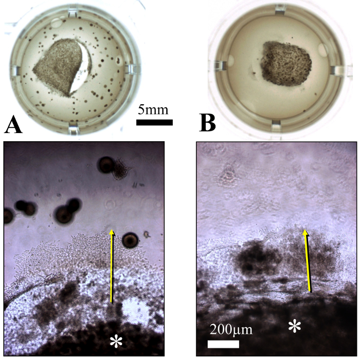
Figure 2: Behavior of Mouse Melanoma B16F10 (A) and B16F0 (B) Cells in the 3D Cell Culture System. The B16F10 and B16F0 cell lines are known to be metastatic and weakly metastatic, respectively. An upper view of the composite gel is shown after 15 days in culture in 24-well plate. The collagen plug (*) is adjacent to a fibrin gel layer in which cells have migrated (arrows) and formed satellite tumors as seen at higher magnification. Quite conveniently, these cell lines express a high level of melanin that allows for direct visualization (i.e. without staining). Please click here to view a larger version of this figure.
The shape of the migration front and satellite tumors may vary as a function of cell line characteristics. For instance, the murine mammary gland carcinoma 4T07 cells, which are poorly metastatic but are very invasive locally, form a migration front that extends radially into the fibrin gel (Figure 3A). After 14 days of culture, those cells have pulled away from the migratory front to form stellar-shape structures closely resembling mammary gland structures (Figure 3B-D).
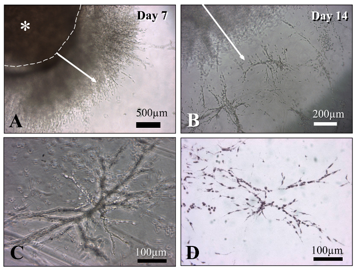
Figure 3: Mouse Mammary Gland Carcinoma 4T07 Cells. These cells demonstrate a strong capacity to migrate in the fibrin gel (A). The dashed line delineates the collagen plug and the arrows show the direction of the migration front. The cells evenly invade the fibrin matrix (A), and well-organized, stellar-shape tubular structures can be observed in 3D (B and C). Views under phase contrast microscopy (A–C) and histological sections stained by hematoxylin and eosin (D) are shown. Please click here to view a larger version of this figure.
The cell structures observed with this 3D culture system, particularly the satellite tumors, can be quantified by planar projection after staining the gels with a methylene blue solution as shown previously16. Staining gel samples with Hoechst 33258 at various stages of the cell culture enables to visualize the cell nuclei under epifluorescence inverted microscopy (Figure 4).
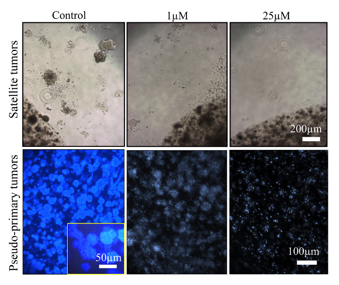
Figure 4: Hormone-Responsive Breast Cancer Cells. Human Breast Cancer MCF-7 Cells were Exposed to Different Concentrations of Tamoxifen. Satellite tumors in fibrin gels (upper row) were observed by phase contrast microscopy. DNA from the collagen gels (indicating the presence of cells in pseudo-primary tumors) (lower row), was stained with Hoechst 33258 and visualized using epifluorescence microscopy. As expected, tamoxifen induced DNA fragmentation as a result of apoptosis. Please click here to view a larger version of this figure.
Reagents | Volume |
DMEM 5x (no bicarbonate) Prepare from powder | 1 ml |
FBS | 0.5 ml |
0.26 M NaHCO3 | 0.5 ml |
1 N NaOH | 20 µl |
ddH2O | 100 µl |
Keep this solution on ice | |
Cell suspension | 880 µl |
Well mix on ice and at last quickly add: | |
RTT Collagen (3.5 mg/ml) | 2 ml |
Total volume | 5 ml |
Table 1: Collagen Plugs. Reagents required and preparation of a collagen solution for 18-19 plugs. Mix well immediately after addition of the collagen (by inverting the container up and down), while avoiding air bubbles, and then promptly distribute 200 µl to each well.
Steps | Problems | Possible reasons | Solutions |
Step 1. Collagen plug |
Air bubbles | Inappropriate pipetting* | Mix well while avoiding excessive bubbles. |
Draw each 200 µl with pipette piston all way down and release only 200 µl (reverse pipetting).* | |||
Collagen plug contraction | Collagen plug shrinks with certain cell lines, particularly when incubated overnight. | Check if cells induce collagen contraction before starting experiment. It is recommended to monitor contraction over a few days. Otherwise, plugs may contract in the sandwiched gel. | |
Gelation does not occur | Check reagents for freshness, molarity, pH, etc.); make sure no reagent has been omitted. | Start again from beginning. | |
Step 2. First layer of fibrin gel |
Air bubbles | Same comments*; need practice. | Same solution.* Air bubbles may remain in the fibrin gel but they usually disappear over time at 37 °C. |
Gelation does not occur | Wrong quantity of fibrinogen used; check thrombin concentration or degradation. | Start again from beginning. | |
Assume the thrombin solution is inadequate; slightly increase the concentration and/or volume of thrombin (e.g., twice). | |||
Residual liquid phase after gelation | Fibrinogen solution and thrombin not well mixed. | Start again from beginning. | |
Fibrin is not homogenous (i.e. fibrillar structures, agglomerates) | Thrombin has inadequately diffused in the gel; excessive or insufficient agitation | Need practice; start again from beginning. | |
Step 3. Sandwich/2nd fibrin layer |
Same comments as in step 2. | ||
Collagen plug is rapidly (few hours) surrounded by empty areas in fibrin | Fibrin has not polymerized adequately in contact with collagen gel | Start again from beginning | |
Collagen plugs may be progressively (days-weeks) surrounded by empty areas in fibrin | Local lysis; | If it is restricted to a few places around the plug, the experiment can go on. Otherwise consider to start from beginning. Could be due to cells secreting excessive amount of plasminogen activator. | |
Do not forget the antifibrinolytic agent! | |||
Step 4 and 5. Cell culture and follow-up |
Fibrinolysis | Problem with the antifibrinolytic agent (degradation, suboptimal dose, etc.). | If extensive amount of satellite tumors or invasion, increase the dose of antifibrinolytic agent. |
Acidification | Excessive cell growth. | Change cell culture medium more frequently. |
Table 2: Troubleshooting. Possible issues and proposed solutions.
As an important technical footnote, it is essential that no gap is present at the interface between the central and the peripheral gels. Otherwise, it might reduce the capacity of the cells to migrate/invade the fibrin gel. A space between the collagen and the fibrin gels may form during the first 24 hr of culture if thrombin has not been appropriately diluted. It is also possible that the cell line tested might lead the collagen gel to contract during culture, thereby causing a relatively large space to form between both gels. This is especially expected when stromal cells are mixed in the collagen gel along with cancer cells due to myofibroblastic-like activity18. In order to avoid this artifact, the collagen plug should be incubated at 37 °C for a longer period (≥48 hr) to induce gel contraction prior to assembling the fibrin sandwich.
A problem also encountered with the assay is the presence of discernible fibrin fibrils or agglomerates. This confers a cloudy instead of translucent aspect to the fibrin gel, which complicates data interpretation due to perturbation of the cancer cell migratory pathways. Such issues may arise from inadequate or excessive shaking of the fibrinogen solution during the polymerization process in the culture plates. It may also be due to the improper addition of thrombin into the fibrinogen solution (step 2.3). An improperly formed or cloudy fibrin gel cannot be fixed once solidified. Collagen or fibrin gels may contain air bubbles that, in most cases, will be spontaneously freed out after a few days in the incubator; nonetheless, proper pipetting minimizes their occurrence. It is recommended to introduce an anti-fibrinolytic agent at the onset of the cell culture period. Fibrinolysis may occur after a long period of cell culture due to cellular activity and the activation of proteolytic enzymes (i.e., plasminogen activator) within the collagen plug and the satellite tumors, depending on the cancer cell types and their aggressiveness19.The release of the collagen plug from the degraded fibrin gel and cell attachment to the bottom of the plates are two consequences of excessive fibrinolysis (see troubleshooting section in Table 2). A limitation of the present technique is the thickness of the fibrin gel, which can make it difficult to observe cells through the entire gel. However, the use a microscope with a long working distance condenser helps to circumvent this limitation and to monitor cells along the entire Z-axis. Confocal or intravital microscopy may also provide an alternative to visualize the cells in the fibrin gel.
One of the major objectives for developing this 3D cell culture model was to provide an extracellular matrix compartment through which cancer cells are able to freely migrate and develop. Fibrin was selected because of its presence in tumors, which owes to increasing vascular permeability induced by inflammation, necrosis and altered angiogenesis, and since fibrin is also known to facilitate tumor cell invasion20,21. The interface between the two compartments presents much interest as it mimics the production of new supportive matrix, such as fibrin, in tumors. Moreover, oxygen and nutrient diffusion, and in the longer run, pH stability, are possibly dysregulated in the collagen compartment compared to the peripheral fibrin compartment in which diffusion is facilitated. Due to the large number of cancer cells initially aggregated in a central collagen plug, signs of necrosis/apoptosis are observed within the primary tumor during culture, simulating the phenomenon observed during the expansion of primary tumors in patients22. Moreover, the formation of satellite tumors is remarkably related to the aggressiveness of cancer cells as well as their metastatic behavior. Depending on the cell line and type of experiment, the incubation period required for observing relevant metastatic processes may be as long as 30 days. This is usually longer than for other 3D assays, but the added delay brings the advantage of studying a model more representative of in situ tumors.
It is well recognized that cells grown in 3D culture models display differences in cell signaling and gene expression compared to monolayer cell cultures5,23. Growing cancer cells in 3D also affects their sensitivity to chemotherapeutic agents24-26. An important and attractive feature of this 3D cell culture model resides in the possibility to modify the ECM concentration, and thus the stiffness of the gel, which may be of interest to those working on the biomechanical properties of the cell-cell and cell-ECM interactions, as well as the effect of matrix properties on cell invasion/migration processes27. The 3D cell culture assay presented here can be adapted to suit different settings and/or experimental conditions. For example, the assay can be scaled up to accommodate studies requiring a large number of cells; by doubling the amount of material and the number of cells, collagen plugs can be prepared in a 48-well plate and layered onto a fibrin gel in a 6-well plate. On the other hand, it may be difficult to miniaturize the culture system for high-throughput studies and measurements because of inconsistent homogeneity of the fibrin gel.
The introduction of non-cancerous cells in one of the two gel compartments may modify the behavior of the cancer cells. For instance, the formation of satellite tumors is exacerbated when the prostate cancer cell line PC3 is mixed with fibroblasts and endothelial cells14. Alternatively, fluorescently-labeled cell lines can be introduced in the gel compartments to investigate cell-cell interactions and/or to track the cells during the culture period. Furthermore, histological sections can be prepared after gel fixation; however, the quality of the immunohistochemistry results may vary between antibodies since cross-reactions with fibrin may occur. Routine staining of histological sections such as with hematoxylin and eosin (H&E) can reveal cell organization inside the gel matrices (Figure 3D).
It is relatively easy to remove the collagen plug from the fibrin gel (e.g., by pulling out the plug with a glass pipette). Thus, cells and satellite tumors present in the fibrin gel can be grown separately from those found in the collagen plug. Cells from the satellite colonies can be harvested by extracting the colonies with surgical scissors or a scalpel and growing them in culture medium without aprotinin to free cells from the fibrin gel. Collecting cells from the different compartments of the composite gel can allow various further studies, including gene and protein expression analysis. It can also be used to isolate cell populations with aggressive phenotypes for further in vitro or in vivo studies. For instance, this 3D culture model was used to identify genes involved in melanoma metastasis16. All those representative applications clearly demonstrate the flexibility allowed by a bi-compartmental 3D cell culture system in which cell co-culture can be carried out.
In conclusion, the design of our 3D culture system is flexible and can offer new avenues to investigate biological and molecular events during cancer cell apoptosis, migration and metastasis as well as for anticancer drug evaluation.
The authors have nothing to disclose.
Work partially funded by Prostate Cancer Canada (grant # D2014-4 to SG and CJD) and the Canadian Institutes of Health Research (grant # MOP-111069 to SG). We would like to thank Dr. Richard Poulin for editorial assistance and Mrs. Chanel Dupont for technical assistance.
Freeze-dried collagen | Sigma-Aldrich | C7661 | from rat tail tendon (soluble dispersion) or home-made (see Rajan et al., ref.#14) |
Fibrinogen (freeze-dried) | Sigma-Aldrich | F8630 | Type I-S, 65-85% protein with ≥75% of protein is clottable |
Thrombin | EMD Chemicals Inc. | 605157 | Gibbstown, NJ; NIH units/mg dry weight |
Growth factor-reduced Matrigel | Corning | 356234 | Previously from BD Biosciences |
Aprotinin | Sigma-Aldrich | A6279 | solution at 5-10TIU/ml (Trypsin Inhibitor Unit) |
Micro-spoons | Fisher Scientific | 2140115 | Fisherbrand Handi-Hold Microspatula |
96 well plate, round base | Sarstedt | 3925500 | |
24 well plate | Sarstedt | 3922 | |
Dulbecco's modified Eagle's Medium | Sigma Chemical, Co. | D5546 | DMEM |
Fetal Bovine Serum | VWR | CAA15-701 | FBS, Canadian origin. |
Trypsin-EDTA | Sigma Chemical, Co. | T4049 | |
Hank’s Balanced Salt Solution | Sigma Chemical, Co. | H8264 | HBSS |