Autophagy activation is beneficial in the prevention of a number of diseases. One of the physiological approaches to induce autophagy in vivo is physical exercise. Here we show how to activate autophagy by aerobic exercise and measure autophagy levels in mice.
Autophagy is a lysosomal degradation pathway essential for cell homeostasis, function and differentiation. Under stress conditions, autophagy is induced and targets various cargos, such as bulk cytosol, damaged organelles and misfolded proteins, for degradation in lysosomes. Resulting nutrient molecules are recycled back to the cytosol for new protein synthesis and ATP production. Upregulation of autophagy has beneficial effects against the pathogenesis of many diseases, and pharmacological and physiological strategies to activate autophagy have been reported. Aerobic exercise is recently identified as an efficient autophagy inducer in multiple organs in mice, including muscle, liver, heart and brain. Here we show procedures to induce autophagy in vivo by either forced treadmill exercise or voluntary wheel running. We also demonstrate microscopic and biochemical methods to quantitatively analyze autophagy levels in mouse tissues, using the marker proteins LC3 and p62 that are transported to and degraded in lysosomes along with autophagosomes.
Autophagy is an evolutionarily conserved degradation pathway, which is induced in response to various stress conditions such as starvation and hypoxia1,2. During autophagy, double-membrane vesicles, called autophagosomes, incorporate unnecessary or damaged subcellular components and transport them into lysosomes for degradation3. Basal autophagy is essential for cellular function and organism development, and impaired basal autophagy is implicated in many disorders, including neurodegeneration, tumorigenesis and type 2 diabetes4,5,6.
The best-known physiological autophagy inducer is starvation. However, it has two major limitations. First, starvation takes a long period to effectively induce autophagy in animals, e.g., 48 hr of food restriction in mice in most organs. Second, starvation barely induces brain autophagy, due to a relatively stable nutrient supply in the brain. In fact, it is also difficult to detect autophagy induction by small-molecule inducers, as many drugs cannot pass the blood brain barrier. Thus, to better analyze the function of autophagy activation in disease pathogenesis, we recently discovered that exercise is a more potent physiological method to induce autophagy in a short period of time7,8,9. Compared with starvation, autophagy is effectively induced by treadmill running as fast as 30 min. Thus, exercise is a convenient and potent physiological approach to study the mechanism of autophagy in mediating health benefits and preventing diseases.
There are several protein markers for the detection of autophagy activity, including LC3 and p62. LC3 (microtubule-associated protein 1A/1B-light chain 3) is a cytosolic protein (LC3-I form) that is conjugated to PE (phosphatidylethanolamine) upon autophagy induction. PE-lipidated LC3 (LC3-II form) is recruited onto autophagosomal membranes and can be used to visualize autophagosomes when labeled with GFP. Its translocation from the cytosol to punctate structures of autophagosomes under microscopy is an indication of autophagy induction. p62 is a cargo receptor for autophagy substrates (such as ubiquitination proteins), and is incorporated into autophagosomes as well. Since the protein is degraded in lysosomes along with autophagosomes, its levels can be used to measure the autophagy flux. Here we show how to use these markers to quantify autophagy in different mouse tissues induced by aerobic exercise, including forced exercise (treadmill) and voluntary exercise (running wheels). The same procedures can also be applied to in vivo measurement of autophagy after treatment of other inducers.
All procedures involving animals were performed according to guidelines approved by the Northwestern University Institutional Animal Care and Use Committee (IACUC).
1. Mouse Models
2. Exercise-induced Autophagy
3. Autophagy Flux Evaluation
4. Tissue Harvest and Fixation
5. Imaging Analysis of GFP-LC3 Puncta
6. Western Blot Analysis on Autophagy Markers
This protocol describes two different methods to induce autophagy in mouse tissues by aerobic exercise: a total of 90 min of forced exercise on a multi-lane treadmill proceeded by two days of acclimation; or two weeks of voluntary exercise on a running wheel used by single-housed mice. In each exercise protocol, we can measure the autophagy flux by fluorescence microscopy and western blot analysis in various organs.
We used a transgenic mouse line expressing GFP-tagged LC3 as a reporter system to monitor autophagy by exercise1. Upon autophagy induction, LC3 translocates from the cytosol to the autophagosome in punctate structures. After sectioning, formation of GFP-LC3 puncta can be directly visualized by fluorescence microscopy (Figure 1A). Alternatively, autophagosome structures can also be immunostained by an LC3 antibody for imaging. Either 90 min of treadmill exercise or 2 weeks of voluntary running increased the number of GFP-LC3 puncta in both skeletal muscle (vastus lateralis) and cerebral cortex, compared to the resting condition (Figure 1B). It should be noted that the frontal cortex region has been the major region in the brain where autophagy is clearly induced by either method so far. Exercise also induced the conversion of LC3 from the cytosolic form (LC3-I) to the lipid-conjugated form (LC3-II), which can be detected by western blot analysis (Figure 1C).
Exercise-induced increment of autophagosomes (represented by LC3-II and GFP-LC3 puncta) is due to an elevated autophagic flux, rather than a block in autophagosome degradation, assessed by the use of inhibitors of lysosomal degradation, such as bafilomycin A1 or chloroquine. Here we measured the degradation of the autophagic cargo receptor p62 as an example. Exercise (90 min of treadmill) caused a higher degradation of p62 in skeletal muscle than the resting condition, which was rescued by injection with chloroquine prior to exercise (Figure 2). The similar results are also observed with voluntary exercise by running wheels. Thus, aerobic exercise by treadmill or running wheels induces autophagy in vivo, measured by the steady-state level of LC3 and the degradation of p62.
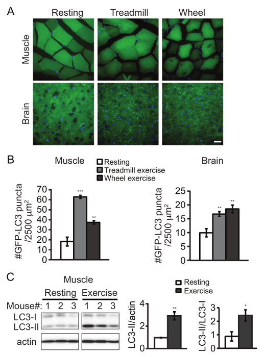
Figure 1. Aerobic Exercise Induces Autophagy in Mouse Tissues. Representative images (A) and quantification (B) of GFP-LC3 puncta in skeletal muscle (vastus lateralis) and brain (frontal cortex) of GFP-LC3 transgenic mice under the control condition (resting), after 90 min of forced exercise (treadmill) or after 2 weeks of voluntary exercise (wheel). Results represent mean ± s.e.m of 10 pictures per mouse. N = 5 mice. The following emission wavelengths were used: GFP – 525 nm; DAPI – 490 nm. (C) Western blot detection of LC3 in skeletal muscle (vastus lateralis) from rested and exercised (by treadmill) mice. Quantification data represent the level of LC3-II normalized to actin (left) and the ratio of LC3-II to LC3-I (right). N = 3 mice. Statistic is comparing each value to the control sample. Results represent mean ± s.e.m. *, P < 0.05; **, P < 0.01; ***, P < 0.001, t-test. Scale bar, 25 µm. Please click here to view a larger version of this figure.
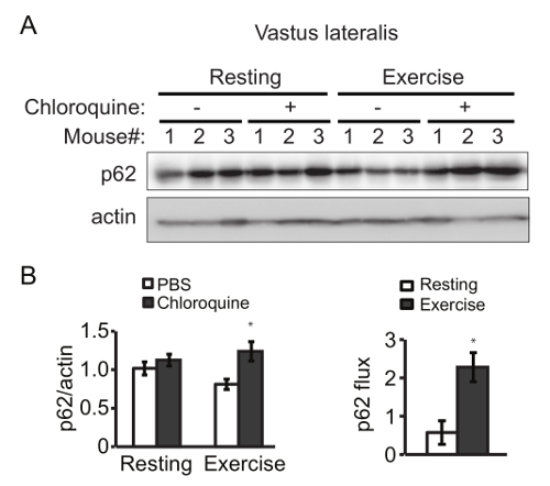
Figure 2. Exercise Increases Autophagy Flux in Mouse Skeletal Muscle. (A) Western blot detection of p62 in vastus lateralis from rested and exercised mice in the presence or absence of the lysosomal inhibitors chloroquine. (B) (Left) Quantification analysis of p62 normalized to the corresponding actin band. (Right) The p62 flux is determined by subtracting the normalized densitometric value of PBS-treated p62 from that of chloroquine-treated p62. Results represent mean ± s.e.m. N = 3 mice. *, P < 0.05, t-test. Please click here to view a larger version of this figure.
Autophagy is a catabolic process that provides energy and reduces cytotoxicity by lysosomal degradation of cytoplasm components or damaged organelles. Studying autophagy is important to understand the regulation of cellular homeostasis and the mechanisms of stress response. New models and methodologies are emerging in the research field15, to study how impaired autophagy contributes to numerous pathological processes16,17.
Nutrient deprivation (starvation) and pharmacological induction are commonly used approaches to induce autophagy in vitro and in vivo. These methods, especially for animal models, may show adverse effects that can influence the overall results. For example, since it requires at least 48 hr of starvation to induce a detectable level of autophagy, the animals may not have energy needed for regular motor activity, which affects the outcome of many subsequent behavioral studies. Yet pharmacological inducers may also lead to side effects due to their lack of specificity to the autophagy pathway. The major autophagy inducer rapamycin and its derivatives suppress mTOR activity and cause metabolic dysfunction and immunosuppression18,19,20, which should be taken into consideration in the experimental design for long-term treatment. Thus, we have been working on more physiological and robust ways for autophagy activation in animal models.
Recently we and others have identified exercise as an effective, faster and safer autophagy inducer in vivo7,8,9. Both forced exercise by treadmill and voluntary exercise by running wheel have been used to analyze the effects of exercise on autophagy activation, with variations in exercise duration and intensity21,22. For example, an hour of treadmill running (starting at speed of 10 m/min to a maximum of 40 m/min) induces abundant LC3 lipidation in skeletal muscle21, and longer-term voluntary wheel running for 3 months or 4 – 5 weeks also increases basal autophagy and enhances the expression of autophagy proteins in skeletal muscle21,22.
Here we described and compared the two methods (treadmill and running wheel) to exercise mice, and presented optimized shorter protocols with high efficiency in autophagy induction. Each approach has pros and cons. A single bout of 90 min of forced exercise on treadmill is sufficient to induce autophagy in skeletal muscle and brain. We have done a time course of treadmill exercise7, and found that the exercise conditions described here induce the maximal level of autophagy in mouse skeletal muscle, which is also validated by others23. Under these conditions, mice undergo aerobic exercise; as previously reported, aerobic exercise is induced by prolonged running with gradual increases in speeds on a treadmill 24,25,26, whereas the anaerobic condition is obtained by a short duration of high intensity exercise, which increases muscle mass but does not necessarily enhance exercise endurance27,28. Furthermore, the running speed reported here in this protocol positively correlates with oxygen consumption capacity, by indirect methods to assess the aerobic capacity29. Therefore, aerobic exercise on a treadmill is a fast and effective way to induce autophagy.
However, forced running in an enclosed space may exert stress to mice. Thus, to avoid giving additional stress to animals, we use finger nudges or wire tassels as a method to keep mice running, instead of the built-in electric shock. Compared to the treadmill, the use of running wheel has many advantages. It is less stressful, does not require researchers' observation time, and is convenient to study long-term effects of autophagy activation. Yet exercise on a running wheel is voluntary and thus generates variability in terms of running distance and speed among different mice or the same mouse on different days. Therefore, it is necessary to run mice on a wheel for an extended period of time (e.g., several weeks) to minimize the individual variability. Importantly, the approach also requires using an odometer at night to verify that the mouse is actually running. We measured the average running distance of wild-type C57BL/6 mice over a period of 2 weeks, and found that they run approximately 1 km/night at the beginning of training (day 1) and can run up to 8 – 10 km/night at day 14. Overall, the running wheel is an effective method to stimulate autophagy in most organs, although, it is harder to capture autophagosome accumulation in the skeletal muscle compared to using the treadmill. The reason is that skeletal muscle has a high autophagic flux and a fast autophagosome degradation rate, which requires immediate tissue harvesting after running to detect the autophagy induction by GFP-LC3 puncta measurement. In either protocol, rapid PFA perfusion and fixation of the tissues after animal euthanasia is a critical step of the procedure to preserve the autophagosome structures that are otherwise easily degraded during dissection.
This protocol demonstrates how to use aerobic exercise to stimulate autophagy in mouse tissues, including brain and skeletal muscle. These methods can be used to study the mechanisms of the autophagy pathway in the maintenance of tissue function, such as mitochondrial maintenance23, and to study the long-term effects of autophagy induction on the regulation of behavior and health of animal disease models, such as enhancing the analgesic effects of cannabinoids9. Both the treadmill and running wheel methods induce a good level of autophagy; yet it is important to consider their differences when choosing the best approach to meet different research goals.
The authors have nothing to disclose.
We thank the Northwestern University Mouse Histology and Phenotyping Laboratoryfor technical support and assistance, and Noboru Mizushima (University of Tokyo) for providing GFP-LC3 transgenic mice. A. R. and C. H. were supported by the startup funds from Northwestern University and the grant from National Institutes of Health (DK094980).
Treadmill | Columbus Instruments | 150-RM Exer 3/6 | |
Mouse running wheel | Super Pet | 100079365 | diameter 11.4 cm |
Odometer | Bell | DASHBOARD 100 | |
Syringe pump | KD Scientific | KDS100 | |
Fluorescence microscope | Nikon | Model: inverted microscope ECLIPSE | |
Cryostat | Leica | CM 1850UV | |
Homogenizer | IKA | 003737001 / Model: T10 Basic S1 | |
Chloroquine | CAYMAN CHEMICAL COMPANY | 14194 | |
Parafolmaldehye | SIGMA-ALDRICH | P6148 | Personal protection equipment required. This product may release formaldehyde gas, a chemical known to cause cancer |
Mounting media | Vector Laboratories | H-1200 | |
p62 antibody | BD Biosciences | 610833 | |
LC3 antibody | Novus Biologicals | NB100-2220 | |
2X Laemmli Sample Buffer | Bio-Rad Laboratories | 161-0737 | |
ImageJ | NIH |