This manuscript describes the efficient, non-viral delivery of miR to endothelial cells by a PEI/MNP vector and their magnetization. Thus, in addition to genetic modification, this approach allows for magnetic cell guidance and MRI detectability. The technique can be used to improve the characteristics of therapeutic cell products.
To date, the available surgical and pharmacological treatments for cardiovascular diseases (CVD) are limited and often palliative. At the same time, gene and cell therapies are highly promising alternative approaches for CVD treatment. However, the broad clinical application of gene therapy is greatly limited by the lack of suitable gene delivery systems. The development of appropriate gene delivery vectors can provide a solution to current challenges in cell therapy. In particular, existing drawbacks, such as limited efficiency and low cell retention in the injured organ, could be overcome by appropriate cell engineering (i.e., genetic) prior to transplantation. The presented protocol describes the efficient and safe transient modification of endothelial cells using a polyethyleneimine superparamagnetic magnetic nanoparticle (PEI/MNP)-based delivery vector. Also, the algorithm and methods for cell characterization are defined. The successful intracellular delivery of microRNA (miR) into human umbilical vein endothelial cells (HUVECs) has been achieved without affecting cell viability, functionality, or intercellular communication. Moreover, this approach was proven to cause a strong functional effect in introduced exogenous miR. Importantly, the application of this MNP-based vector ensures cell magnetization, with accompanying possibilities of magnetic targeting and non-invasive MRI tracing. This may provide a basis for magnetically guided, genetically engineered cell therapeutics that can be monitored non-invasively with MRI.
Gene and cell therapy are powerful tools that have the potential to solve current challenges in CVD treatment. Despite the fact that both of these approaches are currently being tested in clinical trials, they are not yet ready for wide clinical application1. Notably, a common approach to tackle the challenges of gene and cell therapy is to develop multifunctional gene delivery vectors suitable for clinical application. The lack of safe and efficient gene delivery systems is the main concern of gene therapy. At the same time, the genetic engineering of cellular products prior to transplantation could overcome the serious challenges of cell therapy, such as low efficiency (e.g., in the cardiac field, only ~ 5% of functional improvement is achieved post-stem cell transplantation1) and poor retention/engraftment at the site of injury (i.e.,cell retention drops below 5 – 10% within minutes to hours post-application, regardless of the administration route2,3,4).
To date, viral vectors greatly exceed non-viral systems in terms of efficiency, which has resulted in their wider application in clinical trials (~ 67%)5. However, viral vehicles carry serious risks, such as immunogenicity (and the subsequent inflammatory response, with severe complications), oncogenicity, and limitations in the size of the carried genetic material6. Due to these safety concerns and the high costs of viral vector production, the use of non-viral systems is preferable in certain cases7,8. It is particularly suitable for disorders that require transient genetic correction, such as the expression of growth factors controlling angiogenesis (e.g., for CVD treatment) or the delivery of vaccines.
In our group, a delivery system was designed by combining branched 25-kDa polyethyleneimine (PEI) and superparamagnetic iron oxide nanoparticles (MNP) bound together by biotin-streptavidin interaction9. This vector is a potential tool for the genetic engineering of cells, allowing for their simultaneous magnetization prior to transplantation. The latter provides a basis for magnetic guidance/retention, which is particularly promising nowadays, as advanced magnetic targeting techniques are being successfully developed10. Moreover, the resulting magnetically responsive cells have the potential to be non-invasively monitored by magnetic resonance imaging (MRI) or magnetic particle imaging11,12.
In the case of the PEI/MNP vector, polyamine ensures nucleic acid condensation and thus protection from degrading factors, vector internalization in cells, and endosomal escape5. The MNPs complement the properties of PEI, not only in terms of magnetic guidance, but also by reducing the known PEI toxicity7,13,14. Previously, PEI/MNP vector properties were adjusted in terms of delivery efficiency (i.e., pDNA and miRNA) and safety by using fibroblasts and human mesenchymal stem cells15,16.
In this manuscript, a detailed protocol on the application of PEI/MNPs for the generation of miRNA-modified cells is described17. For this purpose, HUVECs are used and represent an established model for in vitro angiogenesis. They are challenging to transfect and are susceptible to toxic influence18,19,20. In addition, we provide an algorithm to evaluate such cells in vitro, including their targeting, intercellular communication, and MRI detection.
Human umbilical cords for cell isolation were obtained postpartum from informed, healthy women who gave their written consent to the use of this material for research according to the Declaration of Helsinki. The ethical committee of the University of Rostock has approved the presented study (reg. No. A 2011 06, prolonged 23 September, 2013).
1. Preparation of Transfection Complexes
2. Cell Preparation
3. Transfection
4. Analysis of Vector Safety
5. Testing of Transfection Efficiency
6. Cell Targeting Evaluation and Magnetic Cell Separation
7. Defining MRI Detection Limits
The main purpose of the proposed protocol is to produce magnetically responsive miR-modified cells and to conduct their accurate characterization (Figure 1). As a result, efficiently transfected cells, responsive to magnetic selection and guidance and detectable with MRI, should be obtained.
First, the identities of isolated HUVECs were confirmed by typical staining with the endothelial marker CD31 (PECAM) (Figure 2A) and by their ability to form tubes on the appropriate basement membrane matrix (Figure 2B). The obtained cells took up the miR introduced by PEI/MNP very efficiently; microscopy demonstrated that all cells were positive for a signal of tagged miR (Cy3) 24 h post-transfection (Figure 3A). Importantly, no cell death was recorded compared to untreated cells (Figure 3B), and normal appearance was maintained (Figure 3A). As observed with confocal laser scanning microscopy, the signal of Cy3-miR was primarily located inside the cells. Moreover, a more detailed investigation of the intracellular localization of all parts of the transfection vector and miR was carried out with higher resolution using 3-color labeled complexes. SIM, miR, PEI, and MNP signals were all detected in the cytoplasm in the perinuclear region (Figure 3C).
Furthermore, a detailed investigation of transfection vector safety has proven that miR/PEI/MNP-HUVECs are capable of forming tubes on the appropriate basement membrane matrix and that the resulting network is comparable to that of untreated cells used as a positive control (Figure 4A). Moreover, transfected cells maintained GJIC, as 3D-FRAP experiments revealed. In particular, when cells are loaded with fluorescent GJ-permeable dye and one of them is bleached, its fluorescence recovers due to the communication with neighboring cells and the resulting dye transfer (Figure 4B).
Importantly, due to the presence of the superparamagnetic compound40 (Figure 5A), PEI/MNP application allows not only cell transfection, but also the simultaneous magnetization. The resulting amount of intracellular iron was measured using MPS (Figure 5B) for all applied MNP concentrations within the optimal range (2.5 pmol/cm2 miR; NP 25 complemented with 5-25 µg/mL MNP): 0.37 ± 0.079 to 0.7 ± 0.150 pg iron/cell17. As the use of magnetic separation columns demonstrated, for all these MNP concentrations, the amount of magnetically responsive cells in the total volume of transfected cells is ~70% (Figure 5C). In addition, transfected HUVECs are visible with MRI (Figure 5D) inside of in vitro agarose phantoms, mimicking mouse tissue susceptibility. Furthermore, the magnetic targeting of transfected HUVECs in dynamic conditions simulated in vitro was proven to be efficient (Figure 6). In this experimental setup, even the constant vigorous movement of culture medium did not prevent prevailing cell growth in the area near magnet application.
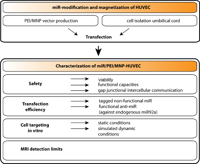
Figure 1. Schematic Illustration of the Production of Magnetized miR-modified HUVECs and Their Analysis. The PEI/MNP (polyethyleneimine/superparamagnetic nanoparticle-based vector) is applied to deliver microRNA (miR) to human umbilical vein endothelial cells (HUVECs). The obtained cell product is characterized by the following parameters: safety (including cell viability, functionality, and capacity for intercellular communication); transfection efficiency (i.e., miR uptake efficiency and the functionality afterwards); magnetic targeting in vitro in static and simulated dynamic conditions; magnetic resonance imaging (MRI) detection. Please click here to view a larger version of this figure.
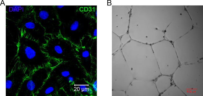
Figure 2. Characterization of Isolated HUVEC. A. Representative image of isolated and cultured HUVECs stained with endothelial CD31 marker. The nuclei are counterstained with DAPI (blue). The scale bar is 20 µm. B. Representative result of the tube formation assay performed on isolated HUVECs; the image was recorded using a laser scanning confocal microscope (differential interference contrast) 18 h after cell seeding on the basement membrane matrix. The scale bar is 100 µm. Please click here to view a larger version of this figure.
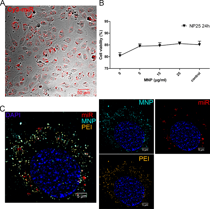
Figure 3. miR Delivery to HUVECs using PEI/MNP. A. The uptake of tagged miR (Cy3, red) 24 h post-transfection (2.5 pmol/cm2 of Cy3-tagged miR, NP 25 and MNP 25 µg/mL) and the maintenance of normal cell appearance are illustrated. Images were taken by confocal laser scanning microscopy using a 514-nm excitation laser (Cy3, red) and differential interference contrast. The scale bar is 50 µm. B. Cell viability 24 h post-transfection was assessed by flow cytometry. The plot represents the results obtained for HUVECs treated with the following complex compositions: 2.5 pmol/cm2 of Cy3-miR; NP ratio 25 complemented with 5 to 25 µg/mL of MNPs. The bars depict the ratio between the average number of viable cells and the whole cell population (n=6; error bars: SEM). C. Structured illumination microscopy (SIM) imaging of 3-color labeled miR/PEI/MNP complexes taken up by HUVECs. The cells were transfected as described above, incubated, and fixed 24 h post-treatment; the nuclei were counterstained with DAPI. Raw SIM data were recorded in z-stacks and processed; the representative images are the result of the maximum intensity projection. The following excitation lasers were applied: 405 nm (DAPI, blue), 488 nm (Atto488-PEI, orange), 565 nm (Atto565-MNP, cyan), and 633 nm (Cy5-miR, red). The scale bar is 5 µm. Please click here to view a larger version of this figure.
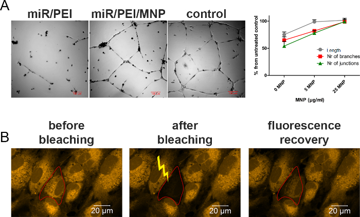
Figure 4. The Safety of HUVECs Modification with miR/PEI/MNP. A. A tube formation assay was performed with transfected cells (2.5 pmol/cm2 of scr-miR; NP 25 and MNP 25 µg/mL) 48 h post-treatment. Images were acquired with a laser scanning confocal microscope (i.e., differential interference contrast) 18 h after cell seeding on basement membrane matrix. HUVECs transfected with miR/PEI were used as a toxicity control and untreated cells as a positive control. The scale bar is 100 µm. The plot reflects the ratio between the transfected HUVECs and the untreated cells using several parameters: tube length, number of branches, and junctions (n= 3, error bars: SEM). B. The gap junction-mediated intercellular communication of miR/PEI/MNP-modified HUVECs was evaluated 24 h post-transfection. For this purpose, cells were loaded with the GJ-permeable dye calcein (orange); fluorescence recovery after photobleaching (FRAP) was examined in 3D by scanning test cells in z-stacks. Representative images of calcein-loaded cells before the bleaching, directly after bleaching, and 15 min post-bleaching (with recovered fluorescence) are depicted. The scale bar is 20 µm. Please click here to view a larger version of this figure.
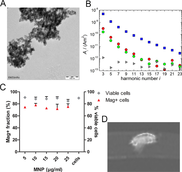
Figure 5. Cell Magnetization Resulting from Transfection with PEI/MNP and its Application. A. Representative image of filtered MNPs, taken with TEM, illustrating their clustered morphology. A small size (~ 5 – 15 nm) of single iron oxide particles (resulting in superparamagnetism) is thereby confirmed. The scale bar is 100 nm. B. The intracellular loading of transfected cells was evaluated by magnetic particle spectroscopy (MPS) 24 h post-transfection. The representative panel depicts the MPS spectrum of examined samples. In particular, pure MNP suspension (50 µL) served as a reference (blue squares); the circles are the MPS spectra of two transfected cell samples (red circles: MNP 25 µg/mL; green circles: MNP 5 µg/mL), while the gray triangles show the detected background spectrum obtained by a measurement without any sample. Note the logarithmic scale of the amplitude (A). C. The amount of magnetically modified cell was quantified by the application of transfected HUVECs (2.5 pmol/cm2 miR; NP 25 complemented with 5-25 µg/mL of MNP), collected by trypsinization 24 h post-treatment, to a magnetic cell separation column. The resulting magnetically positive fraction (i.e., that remained in the column) was counted, and its percentage out of the whole amount of transfected cells is reflected on the plot for each MNP concentration (red triangles). Cell viability was subsequently examined by Trypan blue exclusion assay (gray rhombuses). The data are presented as the mean ± SEM (n= 3). D. An illustrative MRI image of transfected HUVECs (300,000 cells; 2.5 pmol/cm2 miR; NP 25 & 25 µg/mL of MNP) embedded into an agarose phantom was recorded with a 7.1 T animal MRI system. Please click here to view a larger version of this figure.
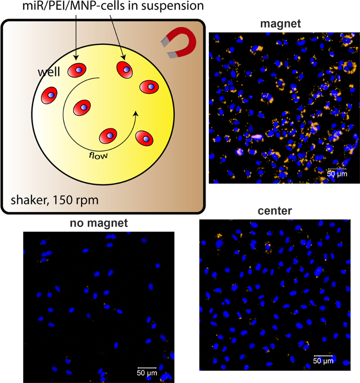
Figure 6. In Vitro Magnetic Targeting of miR/PEI/MNP-HUVECs in Simulated Dynamic Conditions. HUVECs were collected 24 h post-transfection (2.5 pmol/cm2 of Cy3-miR; NP 25 & MNP 25 µg/mL) and re-seeded in fresh culture medium in the wells, with a small magnet locally attached to the side wall. In turn, this culture plate was fixed to a shaker rotating at 150 rpm. Cell attachment and growth were evaluated 12 h later by fixing cells with PFA, staining their nuclei with DAPI, and performing laser scanning confocal microscopy (excitation lasers: 405 nm, DAPI, blue; 514 nm, Cy3, orange). Representative images depict cell growth in the area with magnet application; without magnet application; and in the center of the culture well, where the flow of liquid was concentrating the cells. The scale bars are 50 µm. Please click here to view a larger version of this figure.
The production of genetically engineered cells loaded with superparamagnetic nanoparticles for their further magnetically controlled guidance is presented in the current protocol. The successful application of this strategy allows for the resolution of some difficulties of cell therapy, such as low retention and poor engraftment in the injured area2,3,4, by providing a targetable cell product for transplantation. Moreover, the simultaneous introduction of properly selected genetic material could promote cell properties and could thus increase functional benefits41.
The current protocol has been developed on HUVECs as a model. These cells are known to be difficult to transfect and susceptible to toxic influence18,19,20. The demonstrated level of fluorophore-labeled miR uptake over 95% after PEI/MNP transfection is typically obtained with PEI-based vectors and commonly used cationic lipid-based transfection reagents (e.g., Lipofectamine)42. In addition, the optimal NP ratio of 25 corresponds with previously published values of 20 – 40 for the NP. Nevertheless, the highly efficient uptake of tested fluorophore-tagged miR does not necessarily guarantee proper miR processing and function after delivery43. This point is particularly relevant in the case of PEI, with its tight electrostatic condensation of NA. Thus, the resulting functional capacity of delivered miR must also be tested. Here, a functional miR endogenously expressed in HUVEC, miR-92a31, was selected, and anti-miR against it was delivered using PEI and PEI/MNP. As a result, an almost complete knockdown of the target miR-92a proved the efficient release of the anti-miR from the transfection complexes.
Importantly, in previous works, the successful application of the PEI/MNP vector was demonstrated for the delivery of plasmid DNA (pDNA) and miR to other cell types, both adherent (e.g., cos7, human mesenchymal stem cells15,16) and in suspension (e.g., bone marrow-derived CD133+ hematopoietic stem cells44). All these experiments have confirmed the known fact that the transfection efficiency and toxicity are cell type-dependent45. Thus, the selection of the optimal vector composition (i.e., miR amount, NP ratio, and amount of MNP iron) should be carried out for every particular cell type, using previously established optimal conditions as reference points.
Moreover, the transient character of the achieved genetic modification should be accounted for. On the one hand, it is very suitable for certain function, such as the short-term delivery of growth factors. On the other hand, the duration of expression may not last long enough for a variety of purposes requiring long-term expression. Nevertheless, the proven safety of the PEI/MNP vector supports its possible repeated administration once the conditions are further optimized.
PEI and its derivatives are commonly used due to their high efficiency compared to other non-viral vehicles and their flexibility to modification7. However, despite PEI success in vitro and in vivo, its application in clinical trials is very limited (i.e., few phase 1 and 2 trials or cancer patients)7,14 due to PEI toxicity7,13. As previous works of our group16, as well as this protocol, have demonstrated, MNPs are able to decrease PEI toxicity significantly. In particular, miR/PEI transfection has damaged the ability of HUVECs to form tubes on their matrix, thereby failing a main in vitro functional test for these cells. In contrast, tube formation performance of miR/PEI/MNP-HUVECs was comparable to untreated cells. Notably, this decrease of PEI toxicity was achieved by introducing low levels of components such as iron oxide, which are biocompatible and routinely used as a contrast reagent in human subjects40.
Apart from maintaining functional properties, it is important that modified cells are capable of establishing intercellular communication, as this feature is necessary for the proper integration of cell therapeutics post-transplantation46. In addition, modified cells can be exploited as carriers for the delivery of therapeutic miR to injured tissues47. In this case, their capacity to exchange molecules with neighboring cells defines their success as a vehicle. Since PEI/MNP transfection allows GJIC in modified HUVECs, they have the potential for integration in vivo and to provide miR delivery to damaged areas.
Notably, previously published works on cell magnetization for subsequent targeting have reported the cell loading of ~4 and 30 pg of iron/cell48,49,50,51. These numbers significantly exceed the values ~0.16-0.7 pg/cell, which were sufficient for the in vitro targeting of NA/PEI/MNP-HUVECs in static and simulated dynamic conditions. Such low iron amounts are beneficial in terms of safety: a commonly known mechanism of iron oxide nanoparticle-associated toxicity (i.e., the production of reactive oxygen species (ROS)) is known to be directly associated with the amount of internalized nanoparticles40. In addition, several studies have demonstrated that high intracellular levels of iron oxide nanoparticles can lead to dose-dependent cytoskeletal disorganization and damage52,53. Importantly, most reported cases of cell magnetization for targeting purposes do not include genetic cell manipulation. In contrast, the miR/PEI/MNP vector provides an opportunity to simultaneously modify cells and to add magnetic properties.
However, low MNP loading might be not sufficient for magnetic targeting in a turbulent environment with blood flow. In order to come closer to the challenging in vivo situation, dynamic conditions were simulated in vitro: culture plates with magnetized miR/PEI/MNP-HUVECs in suspension were placed on the rotating shaker34. This experiment clearly cannot cover all the interactions that occur in vivo. However, it led to a better understanding of the targeting potential of PEI/MNP-modified cells. As a result, highly efficient cell targeting was demonstrated when even active shaking of the culture medium did not interfere with cell movement towards the magnet54. For this purpose, miR-modified HUVECs were loaded with at least ~ 0.16 pg of iron/cell. Moreover, magnetically responsive cells can be sorted. A magnet-based cell separation procedure applied to the current study has not compromised the viability of the miR/PEI/MNP-cells. Importantly, the application of a static magnetic field (i.e., targeting experiments that involved the application of a magnetic field for 12 – 24 h) did not have a damaging effect on targeted cells. Therefore, the demonstrated production of magnetized genetically modified cells provides the basis for further in vivo studies addressing the efficiency of cell retention ensured by magnetic force. Notably, the most successful in vivo studies employing the targeting of magnetized cells51,54,55 used cell retention after local administration instead of cell guidance after intravenous injection. Therefore, magnet-based retention at the site of injection appears desirable for further in vivo applications of miR/PEI/MNP-modified cells.
The authors have nothing to disclose.
We would like to thank G. Fulda (Electron Microscopy Center, Rostock University, Germany) for the technical support in acquiring TEM images of filtered superparamagnetic nanoparticles and in performing their X-ray analysis. The work carried out at the RTC Rostock was supported by the Federal Ministry of Education and Research Germany (FKZ 0312138A, FKZ 316159 and VIP+03VP00241) and the State Mecklenburg-Western Pomerania with EU Structural Funds (ESF/IV-WM-B34-0030/10 and ESF/IV-BM-B35-0010/12) and by the DFG (DA 1296-1), the Damp-Foundation, and the German Heart Foundation (F/01/12). Frank Wiekhorst was supported by the EU FP7 research program “Nanomag” FP7-NMP-2013-LARGE-7.
PEI 25 kDa | Sigma Aldrich | 408727 | |
EZ-Link Sulfo-NHS-LC-Biotin | Thermo Scientific | 21335 | |
PD-10 Desalting Columns | GE Healthcare | 17085101 | Containing Sephadex G-25 Medium |
Ninhydrin Reagent solution 2% | Sigma Aldrich | 7285 | |
Glycine | Sigma Aldrich | 410225 | |
Pierce Biotin Quantitation Kit | Thermo Scientific | 28005 | |
Microplate reader Model 680 | Bio-Rad | ||
Streptavidin MagneSphere Paramagnetic Particles | Promega | Z5481 | |
Millex-HV PVDF Filter | Merck | SLHV013SL | 0.45µm |
Libra 120 transmission electron microscope | Zeiss | Acceleration Voltage 120KV | |
Sapphire X-ray detector | EDAX-Amatek | ||
Cell culture plastic | TPP | ||
NHS-Esther Atto 565 | ATTO-TEC GmbH | AD 565-31 | |
NHS-Esther Atto 488 | ATTO-TEC GmbH | AD 488-31 | |
Cy5 miRNA Label IT kit | Mirus Bio | MIR 9650 | |
Biotin Atto 565 | ATTO-TEC GmbH | AD 565-71 | |
Collagense Type IV Gibco | Thermo Scientific | 17104019 | |
Endothelial growth medium, EGM-2 | Lonza | CC-3156 & CC-4176 | |
Penicillin/Streptomycin | Thermo Scientific | 15140122 | 100 U/ml, 100µg/ml |
Matrigel | BD Biosciences | 356234 | |
anti-PECAM-1 antibody | Santa Cruz | sc-1506 | |
MS MACS columns | Miltenyi Biotec | 130-042-201 | |
Near-IR Live/Dead Cell Stain Kit | Thermo Scientific | L10119 | |
Cy3 Dye-Labeled Pre-miR Negative Control | Thermo Scientific | AM17120 | "Cy3-miR" or "Cyanine-miR3" in the manuscript |
Pre-miR miRNA Precursor Molecules – Negative Control | Thermo Scientific | AM17110 | "scr-miR" in the manuscript |
Anti-hsa-miR92a-3p synthetic Inhibitor | Thermo Scientific | AM10916 | |
LSM 780 ELYRA PS.1 system | Zeiss | ||
Paraformaldehyde | Sigma Aldrich | 158127 | 4% solution in PBS |
DAPI nuclear stain | Thermo Scientific | D1306 | |
NucleoSpin RNA isolation Kit | Machery-Nagel | 740955 | |
mirVana miRNA Isolation Kit | Thermo Scientific | AM1560 | |
TaqMan MicroRNA Reverse Transcription Kit | Thermo Scientific | 4366596 | |
StepOnePlus Real-Time PCR System | Applied Biosystems | ||
High-Capacity cDNA Reverse Transcription Kit | Thermo Scientific | 4368814 | |
hsa-miR-92a TaqMan assay | Thermo Scientific | 000431 | Mature miRNA Sequence: UAUUGCACUUGUCCCGGCCUGU |
FastGene Taq Ready Mix | Nippon Genetics | LS27 | |
ITGA5 TaqMan assay | Thermo Scientific | Hs01547673_m1 | |
RNU6B TaqMan assay | Thermo Scientific | 001093 | |
18S rRNA Endogenous Control | Thermo Scientific | 4333760F | |
Gelatin | Sigma Aldrich | G7041 | |
CellTrace Calcein Red-Orange | Thermo Scientific | C34851 | |
PBS | Pan Biotech | P04-53500 | |
BSA | Sigma Aldrich | ||
MACS buffer | Miltenyi Biotec | 130-091-221 | |
Agarose | Sigma Aldrich | A9539 | |
7.1 Tesla animal MRI system | Bruker Corporation | A7906 | |
ImageJ software | National Institutes of Health | upgraded with an AngiogenesisAnalyzer (NIH) | |
MPS device | Bruker Biospin | ||
Matlab software | Mathworks | ||
Ring Neodym Magnet | magnets4you GmbH | RM-10x04x05-G | ø 10 mm; remanescence is ~1.3T, coercivity ≥ 955 kA/m |
Click-iT EdU Alexa Fluor 647 Imaging Kit | Thermo Scientific | C10340 | |
FluorSave Reagent | Merck | 345789 | |
Ultrasonic bath | Bandelin electronic | Type: RK 100 SH |