Tightly-focused femtosecond laser can deliver precise stimulation to cells by being coupled into a confocal microscopy enabling the real-time observation and photostimulation. The photostimulation can activate cell molecular events including ERK signaling pathway and mitochondrial flashes of reactive oxygen species.
Direct control of cellular defined molecular events is important to life science. Recently, studies have demonstrated that femtosecond laser stimulation can simultaneously activate multiple cellular molecular signaling pathways. In this protocol, we show that through coupling femtosecond laser into a confocal microscope, cells can be stimulated precisely by the tightly-focused laser. Some molecular processes that can be simultaneously observed are subsequently activated. We present detailed protocols of the photostimulation to activate extracellular signal regulated kinase (ERK) signaling pathway in Hela cells. Mitochondrial flashes of reactive oxygen species (ROS) and other mitochondrial events can be also stimulated if focusing the femtosecond laser pulse on a certain mitochondrial tubular structure. This protocol includes pretreating cells before photostimulation, delivering the photostimulation by a femtosecond laser flash onto the target, and observing/identifying molecular changes afterwards. This protocol represents an all-optical tool for related biological researches.
The technology of controlling cellular signaling molecules is an important part of the development of life science. Traditionally, the most commonly-used method is biochemical treatment by drugs or biological materials1,2,3. Over the past decade, the invention of optogenetics opens a new era for cellular molecular signal modulation. Transfection with light sensitive proteins by gene engineering makes light become a powerful tool to modulate various protein activities in target cell. This technology has made encouraging progresses such as excitation and inhibition of neural signal, promoting gene expression, manipulating cellular signal patterns, leading different cell fates and pathological investigation4,5,6,7,8,9. However, light can only work by transfecting cells with optogenetic proteins. In current stage, there exist rare methods that enable light to control cellular molecules directly besides optogenetics.
Femtosecond laser has advanced biological researches by providing efficient multiphoton excitation while maintaining good biological safety. By deploying diverse photo processing strategies, it has realized numerous achievements such as multiphoton microscopy, microsurgeries and multiphoton optogenetic applications10,11,12,13,14,15,16. Recent investigations show that femtosecond laser stimulation has been demonstrated as a highly efficient optical method to directly induce molecular signaling events. It has been found that tightly-focused femtosecond laser irradiation on endoplasmic reticulum (ER) is able to deplete calcium in ER and activate calcium-release-activated calcium (CRAC) channels to form calcium signals in cells17. This photo-activated calcium signal can spread between multiple types of cells18,19,20. Furthermore, it also has the ability to activate cell signaling pathways such as nuclear factor of activated T cells (NFAT) and ERK signaling pathway21,22. By adjusting the intensity and localization of femtosecond laser exposure in cells, for example, focusing the laser on mitochondria, it can influence mitochondrial morphology and molecular events23,24,25. Specifically, bursts of mitochondrial ROS generation can be excited by photostimulation, which is remarked as fluorescent flashes in mitochondria (mitoflashes).
Hence, the photostimulation technology is of good potential to be widely applied in related biological research. It is also a good chance to extend femtosecond laser applications in controlling of cellular signaling molecules and functions besides microscopy. Here, we provide the technical details of photostimulation. The photostimulation is achieved by coupling a femtosecond laser to a confocal microscope to provide single target cell with a short flash photostimulation. It can initiate efficient and controllable ERK activation in the cell. If the photostimulation is located on the mitochondrial tubular structure, the mitochondrial membrane potential, morphology, ROS, and permeability transition pores, can all be controlled by the photostimulation. Based on this photostimulation scheme, we provide a detailed method of activating ERK signaling pathway and influencing multiple mitochondrial events in Hela cells. This protocol elucidates the process of delivering femtosecond laser stimulation into target cells.
The photostimulation system is established on a confocal microscope with a femtosecond laser coupling into it for simultaneous stimulation and continuous microscopy. The femtosecond laser (wavelength: 1,040 nm, repetition rate: 50 MHz, pulse width: 120 fs, maximum output average power: 1 W) is split into two beams before coupling. One is guided through a relay telescope consisting of a pair of lenses. It is then directly reflected into the back-aperture of an objective (60x, N.A. = 1.2, water immersion) to form a diffraction-limit focus (Stim-A). The other is reflected to the scanning optical path of confocal microscope to work as a two-photon scanning mode (Stim-B). Stim-A presents a fixed focus point in the center of field of view (FOV). Stim-B is a pre-designed partial confocal scanning area in the FOV. Stim-A and Stim-B are shown in Figure 1A. A CCD camera under the dichroic mirror (DM) provides bright-field imaging for monitoring the focus of femtosecond laser.
There are some crucial essentials for the following experiments. In this protocol, a femtosecond fiber laser source (1040 nm, 50 MHz, 120 fs) is used as an example. In practice, most commercial femtosecond oscillators can be used as long as the pulse width is shorter than 200 fs and the peak power density should be above the level of 1011 ~ 1012 W/cm2. For example, a Ti: Sapphire laser usually used for multiphoton microscopy is able to replace the femtosecond laser showed in Figure 1B. The laser power and some other photostimulation parameters need to be tuned because the optical parameters (pulse width, wavelength, and repetition rate) vary a lot in different femtosecond lasers that thus induce different multiphoton excitation efficiencies.
Along with femtosecond laser stimulation, the confocal microscopy provides continuous cell imaging to monitor molecular dynamics in real time in both Stim-A and Stim-B modes. Both photostimulation schemes (Stim-A and Stim-B) are controlled by a mechanical shutter with millisecond resolution (Figure 1).
In Stim-A mode, the position of laser focus is fixed in the center of FOV. A relay telescope is used to ensure the focus of femtosecond laser to be located on confocal imaging plane by tuning the distance between two lenses in the vertical direction (the laser propagation direction, vertical to the confocal imaging plane, as shown in Figure 1). By the bright-field imaging of CCD camera, the diameter of laser focus can be measured (~2 µm, Figure 2B). The stimulation durations and exposure times are controlled by a shutter during the confocal imaging process.
In Stim-B mode, the stimulation area can be pre-assigned manually in the confocal imaging controlling software to any form like line, polygon, or circle. The shutter is synchronized with the confocal scanning process. It opens at the pre-designed time which is set through the confocal imaging controlling software. Then, the stimulation area is scanned by femtosecond laser as confocal microscopy. Thus, the sample is only stimulated by femtosecond laser when confocal scanning process enters a given imaging frame.
The photostimulation system can be established on both inverted and upright metallurgical microscopes according to the experiment subjects. In vitro cells cultured in Petri dishes are better to work with inverted microscopes. Animals, especially brains of live animals, are more suitable with upright microscopes. In this study, we take the inverted microscope as an example. It should be noted that the cover of Petri dish is not opened during the whole experiments.
CAUTION: The protocol presented below involves using NIR femtosecond laser and toxic chemicals. Please pay attention to all possible damages induced by experiment procedures. Please read safety data sheets of all relevant chemicals or other materials before use. Please follow the safety instructions of the laser facilities or consult professionals for guidance before operate laser source.
1. Experimental Preparation
2. Cell Culture and Transfection
NOTE: Hela cell (cell line derived from cervical cancer cells taken on February 8, 1951 from Henrietta Lacks)26 is used as an example in this protocol.
3. Activation of ERK2 by Photostimulation of Femtosecond Laser
4. Activation of eIF4E (Substrate of ERK) by Femtosecond Laser Simulation
5. Activation of Mitoflashes and Other Mitochondrial Events by Photostimulation
NOTE: To observe mitochondrial morphological dynamics, Hela cells are transfected with Mito-GFP in step 5.1 to fluorescently indicate mitochondria. To observe mitoflashes, Hela cells are transfected with mt-cpYFP in step 5.1.
6. Oscillation of Mitochondrial Membrane Potential on Target Mitochondria in Hela Cells by Femtosecond Laser Stimulation
7. Changes of Bax and Cytochrome C on the Target Mitochondria in Hela Cells by Femtosecond Laser Stimulation
NOTE: In this experiment, seed the cells in Petri dishes with cell location grids (Figure 3B) to localize the cell which is treated by femtosecond laser. Stain the cells with TMRM to localize the mitochondrion which is selected to be stimulated by femtosecond laser.
The photostimulation can be performed simultaneously along with continuous confocal scanning microscopy. The photostimulation can start at any pre-defined time slot in the time-lapse confocal microscopy sequence. The confocal microscopy can monitor cellular molecules by fluorescent imaging. The molecular responses to photostimulation and other dynamics can be identified in this way. Theoretically, if ERK is activated, it will be phosphorylated move from the cytoplasm to cell nucleus27. The specific cell fate can be regulated by the certain patterns of this ERK signal28. In recent studies, optical modulation based on optogentics has provided a high precise control of the ERK signal in duration and magnitude and revealed that perturbed ERK signal transmission dynamics drives improper proliferation in cancer cells7,8. Here, we demonstrate the ERK2 translocation into nucleus after treating cell with a short flash of femtosecond laser by using the method presented in this protocol. As shown in Figure 5A, ERK2-GFP fluorescence reaches the maximum after several minutes of femtosecond laser simulation. The ERK2 molecules will be dephosphorylated after activating downstream substrates in the nucleus, and then the ERK2 comes back to the cytoplasm indicated by decreasing of nuclear GFP fluorescence. ERK2 can be activated for multiple times by multiple photostimulations (Figure 5B). Therefore, it is able to manipulate the ERK2 signal pattern precisely by controlling interval time between multiple stimuli. In addition, ERK2 can be activated in adjacent cells around the stimulated cell occasionally (Figure 5C). This observation indicates that some diffusible molecules may be released by the cell treated with femtosecond laser to activate ERK2 in the adjacent cells. Phosphorylation of ERK2 downstream protein eIF4E can be confirmed and visualized by immunofluorescence microscopy (Figure 5D). This result indicates that femtosecond laser stimulation can successfully activate ERK signaling pathway. More detailed results are in Wang S., et al.22.
Mitochondrial oxidative flashes (mitoflashes) are oxidative bursts in mitochondria that root from complex mitochondrial molecular dynamics. In last past decade, mitoflashes are realized to be an elemental mitochondria signaling event and take an important part in multitudinous cell functions29,30,31. Traditionally, mitoflashes are usually observed by chance when treating cells with chemicals to provide indirect stress to mitochondria29,30. By implementing this photostimulation scheme, we achieve a controllable and precise manner to excite mitoflashes at single mitochondrial tubular level. The successful mitoflash excitation is shown in Figure 6A. Interestingly, the properties of mitoflashes such as pulse peak, width and response durations32 are closely related to the femtosecond laser power. More detailed quantitative analysis of mitoflashes excited by femtosecond laser stimulation is in Wang S., et al.32. This photostimulation to mitochondria also shows varieties of mitochondrial molecular dynamics, including fragmentation and restoration of mitochondrial morphology (Figure 6B), and oscillation of mitochondrial membrane potential (Figure 6C). Similar to photo-activated mitoflashes, these mitochondria events have different performance with different power intensities of photostimulation. It is different from activation of ERK signaling pathway. The influence of femtosecond laser is highly restricted on a single mitochondrial tube. More detailed results are in Wang Y., et al.24 and Shi F., et al.25.
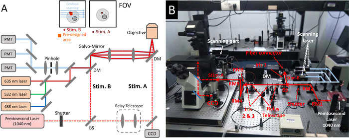
Figure 1: The photostimulation scheme established on a femtosecond laser coupling into a confocal microscope. (A) Optical paths of (B) the photostimulation and confocal imaging system. The femtosecond laser is at first split by a 50/50 beam splitter into two beams. The transmission is expanded by a relay telescope and then reflected into the objective to form Stim-A. The reflection beam is aligned through the microscope scanning system to form Stim-B. A CCD camera is used to provide a bright-felid imaging to monitor cells and focus of femtosecond laser in Stim-A mode. Stim-A = a fixed focus in the center of FOV; Stim-B = a special scanning frame at pre-designed area. DM = dichroic mirror, BS = beam splitter, RM = reflective mirror. The wavelength of confocal scanning laser is 488 nm/532 nm/635 nm, and the typical collection wavelength interval of fluorescence is <560 nm/560-625 nm/> 625 nm. The fiber femtosecond laser (1,040 nm, 120 fs, 50 MHz, 1 W) can be replaced by a Ti = Sapphire laser (810 nm, 80 MHz, 65 fs, 1 W) or other commercial femtosecond laser oscillators. Please click here to view a larger version of this figure.
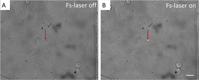
Figure 2: In Stim-A mode, the localization and size of femtosecond laser focus on the target cell at confocal imaging plane under bright-felid imaging. (A) Femtosecond laser is blocked by the shutter. (B) Femtosecond laser is on. Arrow: the reference arrow is set at the center of FOV to confirm that the femtosecond laser focus locates the correct position. Scale bar = 10 µm. Please click here to view a larger version of this figure.
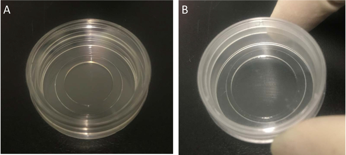
Figure 3: The Petri dish used for cell culture, transfection and photostimulation experiments. (A) The 35 mm Petri dish with a 15 mm diameter and 0.17 mm thickness glass bottom. (B) The 35 mm Petri dish with a glass bottom and an imprinted 500 µm cell location grid. Please click here to view a larger version of this figure.
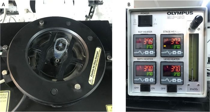
Figure 4: The CO2 incubation system. (A) The CO2 incubator stage and (B) the control panel. Please click here to view a larger version of this figure.
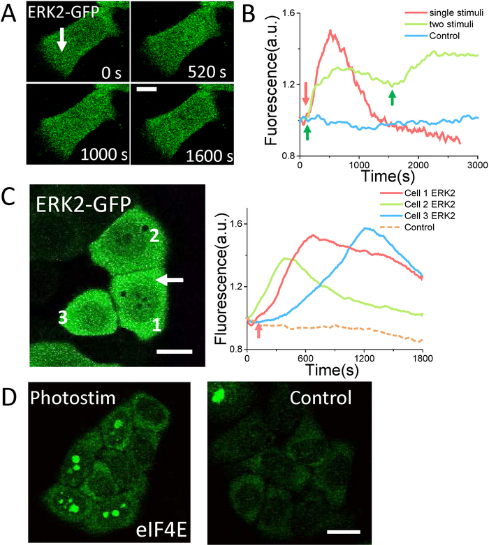
Figure 5: ERK photoactivation by using Stim-A mode. (A) Single photo stimulus (1,040 nm, 40 mW, 0.1 s) induces ERK2-GFP translocation into nucleus and then back to cytoplasm. (B) ERK2 signal pattern mediated by single femtosecond laser exposure (red arrow, 1,040 nm, 40 mW, 0.1 s) and two photostimulations (green arrows, 810 nm, 30 mW, 0.1 s). (C) ERK activation in surrounding cells by single short femtosecond laser exposure in target cell (white arrow, 810 nm, 24 mW, 0.2 s). (D) The Immunofluorescence of eIF4E-P shows a significant increase 24 h after single photosimulation (810 nm, 25 mW, 0.2 s). Scale bar = 10 µm (A), and 20 µm (B, C). The fluorescence of ERK2-GFP and eIF4E-P is excited by 488 nm excitation laser and collected in <560 nm channel. Please click here to view a larger version of this figure.
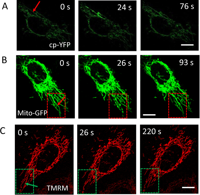
Figure 6: Multiple mitochondria events induced by photostimulation under Stim-A mode. (A) Excitation of mitoflash on the stimulated mitochondrial tubule by single stimulus (810 nm, 16 mW, 0.1 s). (B) Mitochondria morphological fragmentation and restoration induced by single photostimulation (1,040 nm, 30 mW, 0.1 s). (C) Oscillation of mitochondrial membrane potential by single femtosecond laser stimulation (1040 nm, 20 mW, 0.1 s). Arrows in (A) (B) and (C) indicate the position of photostimulations.Scale bar = 10 µm. The fluorescence of Mito-GFP or mt-cpYFP is excited by 488 nm excitation laser and collected in <560 nm channel. The fluorescence of TMRM is excited by 532 nm excitation laser and collected in 560-625 nm channel. Please click here to view a larger version of this figure.
0.05 s | 0.1 s | 0.2 s | 0.5 s | |
810 nm, 65 fs, 80 MHz | 20 – 65 mW | 10 – 60 mW | 5 – 50 mW | 5 – 40 mW |
1040 nm, 120 fs, 50 MHz | 30 – 100 mW | 20 – 80 mW | 15 – 70 mW | 10 – 60 mW |
Table 1: Recommended stimulation duration and average power of femtosecond laser in Stim-A mode.
0.05 s | 0.1 s | 0.2 s | 0.5 s | |
810 nm, 65 fs, 80 MHz | 25 – 40 mW | 20 – 30 mW | 15 – 25 mW | 10 – 25 mW |
1040 nm, 120 fs, 50 MHz | 40 – 60 mW | 30 – 50 mW | 25 – 40 mW | 20 – 30 mW |
Table 2: Recommended stimulation duration and average power of femtosecond laser in Stim-A mode at 2 x 2-3 x 3 µm2 in cell.
We demonstrate a photostimulation strategy by combining a femtosecond laser with a laser scanning confocal microscope system. The photostimulation can directly work as a two-photon microscopy by defining Stim-B accordingly. We provide a detailed protocol for utilizing a short flash of femtosecond laser to trigger ERK signaling or mitoflashes in target cells. The different stimulation modes can be performed according to different experimental purposes and systems. Stim-A can be easily set up based on a confocal microscope. The femtosecond laser can be focused at diffraction-limit level at the FOV center. The subcellular target needs to be moved to the femtosecond laser focus position manually and can be stimulated for arbitrary durations, totally independent from confocal microscopy. Therefore, Stim-A can protect the sample that is out of the focus area perfectly and is suitable for long-duration photostimulation. The stimulation region of Stim-B can be pre-defined as any area in the FOV. It can be performed automatically. But the stimulation duration and exposure time can only follow along with the confocal scanning. After setting the dwell time of each pixel and the total frame size, the stimulation duration in a single microscopy frame is actually fixed. The photostimulation can also be performed periodically frame by frame. It can also be used to activate photosensitizers and optogenetic proteins that requires less exposure duration but large area.
A critical consideration in this protocol is how to determine the parameters of femtosecond laser stimulation. According to our previous studies, the cellular molecular activity is mostly induced by multiphoton exaction17,21,22,23,24,25. Generally multiphoton exaction efficiency is related with all femtosecond laser parameters including wavelength, pulse width, repetition rate. The cellular response is further related with the laser power, stimulation position/region, and exposure duration simultaneously. In principle, the stimulation efficiency conflicts with cell safety. Stronger photostimulation such as higher average power, longer stimulation duration and larger total incident laser energy contributes to higher stimulation efficiencies but brings higher risks of cell damage. Since those parameters are also relative with each other and all contribute to the stimulation and damage effect, it is impossible to use only one parameter, like the total incident laser energy, or laser average power, to define it is safe or not. Considering both signal activation efficiency and cell safety, here, we provide recommended average power and stimulation duration of two femtosecond laser source for reference in Table 1 and Table 2. Besides, the parameters of femtosecond laser stimulation should be changed according to the actual optical system and biological sample.
According to the discussions above, we intend to present more detailed effective ranges of photostimulation parameters. The wavelength of femtosecond laser can be changed at NIR range used for photostimulation. The pulse width should be short to provide a high peak power and high nonlinear exaction efficiency. Long pulse duration is not recommended for this protocol. Usually, the typical pulse width for two-photon microscopy (<200 fs) is the proper choice. The repetition rate of the laser is not a key factor. The repetition rate up to MHz is suitable for this protocol. The most critical factor for cellular molecular responses is the laser power and stimulation duration of femtosecond laser. For activating ERK, according to our previous work22, the average power at 15 mW (810 nm) or 20 mW (1,040 nm) and exposure duration at 0.2 s are enough to provide sufficient stimulation to activate ERK signaling while maintaining ultrahigh cell viability in Hela cells. On the contrary, average power over 80 mW (810 nm) or 120 mW (1,040 nm) and exposure duration longer than 1 s can induce irreversible damage to cells. Mitochondria are generally more sensitive to photostimulation. The stimulation with an average power higher than 40 mW (810 nm) or 80 mW (1,040 nm) and exposure duration over 0.2 s may induce significant damage like irreversible fragmentation in stimulated mitochondrion or even in all mitochondria across the whole cell. It should be noted that the photosensitivity of mitochondria to photostimulation varies a lot in different cell types. For example, in HeLa cells, the laser power needs only around 6 mW (810 nm, 0.1 s duration). All mitochondria can response to the photostimulation in the form of fragmentation, mitoflashes, and MMP oscillations. But in human mesenchymal stem cells, the power needs to be increased to around 15 mW (810 nm, 0.1 s duration), and still around half stimulated mitochondria show no response to photostimulation. In Stim-B mode, another factor may affect the photo activation efficiency and cell damage is the stimulation area. The photostimulation may cause cell damage by delivering on a small stimulation area. But the same stimulation may fail to activate ERK by delivering on a larger stimulation area. We recommend that the stimulation area in target cell is around 2 x 2-3 x 3 µm2 and does not exceed 25 µm2.
The localization of stimulation region is also important for applying this protocol. According to previous works17,22, the photostimulation in that area can deplete the Ca2+ store in ER effectively and activate the CRAC channel. Then, the Ca2+ influx can activate ERK signaling pathway subsequently. Therefore, we recommend delivering the photostimulation on the ER region in Hela cells to achieve a high ERK activation efficiency. The ER region can be easily distinguished from cytoplasm or nucleus under bright-field microscopy by a phase-contrast objective. In order to introduce mitochondrial signaling events, the focus of femtosecond laser can be easily mounted on the selected mitochondrial structure following the related procedures.
Photostimulation methods described in this protocol are also able to be used to affect other multiple cellular events such as inducing Ca2+ and ROS signals17,20,21. It should be noted all those previous works have provided the evaluation of cell safety after photostimulation. For example, significant morphological changes of cells, bubbling, mitochondrial fragmentation and swelling of the whole cell, decreased proliferation rate of cells, and some other unusual changes of the fluorescence during confocal microscopy all imply high cell damage. It should be very careful to monitor the cell status. Nevertheless, with well control of the laser power and stimulation parameters, the cell viability could be maintained in a very high level simultaneously with high stimulation efficiency. Hence, this photostimualtion method of femtosecond laser is of good potential to further extend to more areas and applied in relevant applications.
The authors have nothing to disclose.
The work was supported by National Natural Science Foundation of China (81571719 and 81661168014, 81673014, and 81870055), Shanghai Municipal Science and Technology Committee (18QA1402300 and 16XD1403100), and National Key R&D Plan (2017YFA0104600).
inverted microscope | Olympus | ||
femtosecond laser | Fianium | ||
CO2 incubation system | Olympus | MIU-IBC | |
petri dish | NEST | 801002 | |
petri dish with imprinted grid | Ibidi | 81148 | |
ERK-GFP | addgene | 37145 | A gift from Rony Seger's lab |
mt-cpYFP | A gift from Heping Cheng's lab | ||
mito-GFP | Invitrogen | C10508 | |
Tetramethylrhodamine (TMRM) | Invitrogen | T668 | Dilute in DMSO |
polyethylenimine (PEI) | Sigma-Aldrich | 9002-98-6 | Dilute in PBS |
paraformaldehyde | Solarbio | P8430 | Dilute in PBS |
Triton X-100 | Solarbio | T8200 | Dilute in PBS |
Bovine Serum Albumin (BSA) | Sigma-Aldrich | 9048-46-8 | Dilute in PBS |
Tween20 | Sigma-Aldrich | 9005-64-5 | |
anti-eIF4E antibody | abcam | ab76256 | |
anti-Bax antibody | abcam | ab53154 | |
anti-cytochrome C antibody | abcam | ab90529 | |
secondary antibody (anti-Rabbit IgG H&L, Alexa Fluor 488) | abcam | ab150077 |