Here, we present a protocol that demonstrates the technique and necessary controls for Laser Doppler perfusion imaging to measure blood flow in the mouse hindlimb.
Blood flow recovery is a critical outcome measure after experimental hindlimb ischemia or ischemia-reperfusion. Laser Doppler perfusion imaging (LDPI) is a common, noninvasive, repeatable method for assessing blood flow recovery. The technique calculates overall blood flow in the sampled tissue from the Doppler shift in frequency caused when a laser hits moving red blood cells. Measurements are expressed in arbitrary perfusion units, so the contralateral non-intervened upon leg is usually used to help control measurements. Measurement depth is in the range of 0.3-1 mm; for hindlimb ischemia, this means that dermal perfusion is assessed. Dermal perfusion is dependent on several factors—most importantly skin temperature and anesthetic agent, which must be carefully controlled to result in reliable readings. Furthermore, hair and skin pigmentation can alter the ability of the laser to either reach or penetrate to the dermis. This article demonstrates the technique of LDPI in the mouse hindlimb.
Skin ulceration with inadequate wound healing is a leading cause of amputations in human patients1. Adequate wound healing requires higher levels of arterial perfusion than are needed to maintain intact skin, which is compromised in patients with peripheral arterial disease2,3,4. Several other rheumatologic conditions and diabetes can also lead to disturbed and inadequate skin microcirculation to heal wounds5,6. Many diabetic patients have concomitant peripheral arterial disease, placing them at especially high risk for amputation. Laser Doppler perfusion imaging (LDPI) is used in clinical situations to evaluate the skin microcirculation, as well as in research situations to evaluate blood flow and blood flow recovery after experimental hindlimb ischemia, ischemia-reperfusion, and microsurgical flaps7.
The LDPI system projects a low power laser beam that is deflected by a scanning mirror to move over a region of interest. This differs from Laser Doppler flowmetry, which provides a perfusion measurement for the small area of tissue in direct contact with the flowmetry probe8. When the laser beam interacts with moving blood in the microvasculature, it undergoes a Doppler frequency shift, which is photodetected by the scanner and converted to arbitrary perfusion units. Because LDPI is a light-based technique, it is limited in terms of depth of penetration to 0.3-1 mm, meaning that for the most part dermal perfusion is assessed7. Dermal flow can be altered by skin temperature and the sympathetic nervous system, which may be affected by various anesthetic agents9. Measurements from the optical laser are also affected by ambient lighting conditions, skin pigmentation, and can be blocked by overlying fur or hair7.
LDPI is the most commonly used research technique to monitor perfusion recovery after ischemia because it is noninvasive, does not require contrast administration, and has quick scan times allowing data collection on multiple animals. This makes it ideal to help determine whether treatments aimed at therapeutic arteriogenesis or angiogenesis are effective in small animal models. Blood flow recovery after hindlimb ischemia as measured by LDPI correlates well with collateral artery development when assessed by other means such as Microfil casting or micro-CT10,11. The goal of this protocol is to demonstrate the assessment of hindlimb perfusion using LDPI.
Animal experiments were performed according to a protocol approved by the University of Washington Institutional Animal Care and Use Committee.
1. Scanner preparation
2. Mouse pre-scanning preparation
3. Scanning
4. Capturing LDPI data (Figure 3)
5. Analysis
Successful LDPI should result in consistent repeated measures scans, with no more than 100-150 perfusion unit variation (corresponding to about 10% of the usual mean perfusion for the mouse footpad) between the three scans (Figure 2). As demonstrated in Figure 2, repeat scans help determine that the mouse has been appropriately equilibrated so that the ischemic/control ratio best reflects the underlying blood flow as opposed to variation in dermal perfusion caused by temperature variation. Using single scans for datapoints will increase the variability leading to the need for more experimental mice. When used for hindlimb ischemia, the surgical hindlimb should have decreased global perfusion when compared to the control hindlimb. Results are expressed as a ratio of surgical hindlimb perfusion/control hindlimb perfusion. As mice initially vasodilate and develop their intrinsic collateral network over time, blood flow recovery by LDPI should be seen over a postoperative time course (Figure 4). The degree of recovery is dependent on the mouse strain and severity of the hindlimb ischemia model.
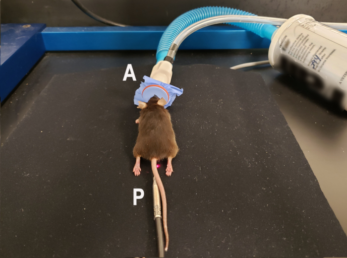
Figure 1. Mouse positioning for laser Doppler perfusion imaging of the ventral footpads. Anesthetized mouse using isoflurane nose cone (A) is placed in the prone position with hindlimbs extended to allow scanning of the ventral footpads. Rectal temperature probe (P) for homeothermic blanket is in place to maintain consistent body temperature during the scan. The homeothermic blanket pad is underneath the nonreflective neoprene material used to provide background for the scan. Laser indicating the middle of the scanning region is visible next to the mouse's tail. Please click here to view a larger version of this figure.
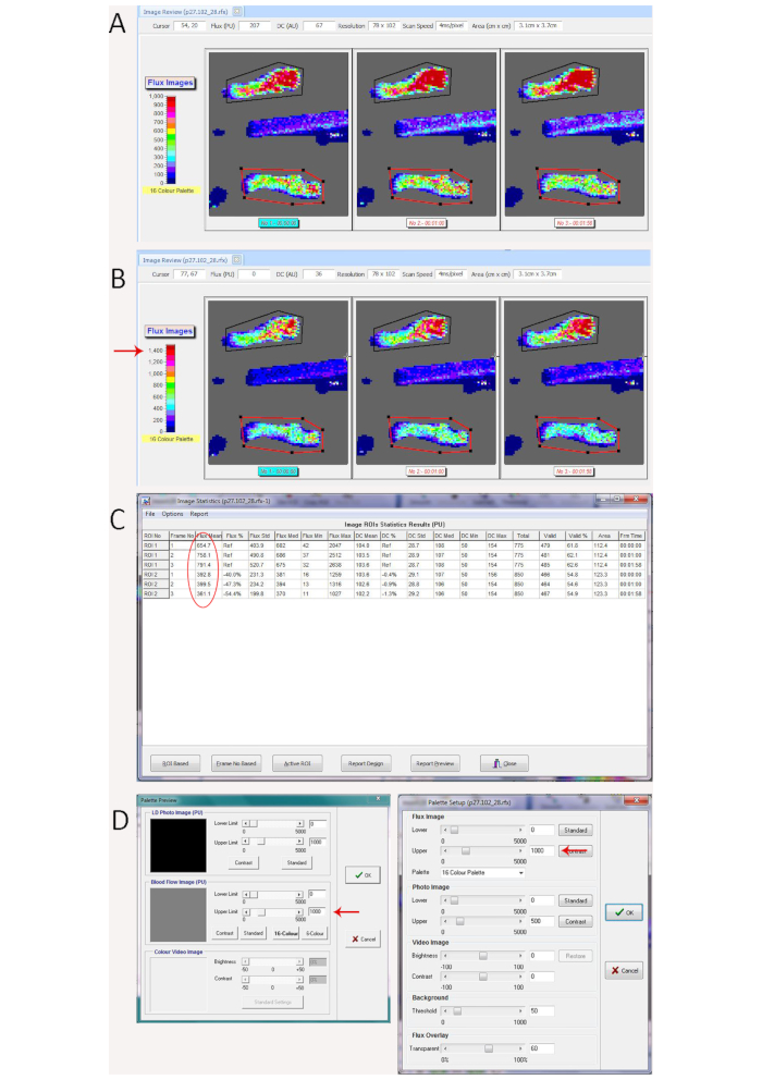
Figure 2. Demonstration of scan variation from mouse temperature change better seen with color palette adjustment. (A) Laser Doppler repeated scans with significant variation caused by mouse core temperature variation during the repeated scans, which is visible based on the perfusion units translated to color on the repeated flux scans. (B) Changing the dynamic range on the color palette (shown on the left of the scan window) from 0-1000 in A to 0-1500 (red arrow) in B makes the variation more obvious. (C) Statistics showing mean perfusion values for the region of interest (circled in red) for the control hindlimb (Polygon 1 in black on the rfx image in A and B) ranges between 655 for the 1st scan to 791 on the 3rd scan and mean perfusion for the region of interest for the ischemic hindlimb (Polygon 2 in red on the rfx image in A and B) showed less variation (361 to 400), leading to significant differences in the ischemic/control ratio between the repeated scans (0.60, 0.53, and 0.46). (D) Window to change the dynamic range of the color palette in the measurement software (left panel) and image review software (right panel). Red arrows show where to increase or decrease the upper range. Please click here to view a larger version of this figure.
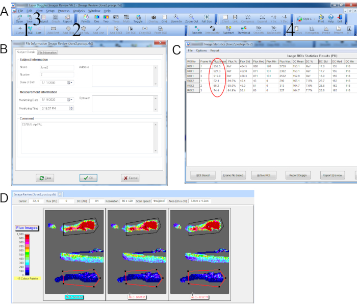
Figure 3. Data capture for laser Doppler perfusion imaging with repeated scans. (A) Top toolbar with 1. Add ROI icon. 2. Add Polygon icon. 3. Subject details icon (accesses window pop-up in B). 4. Statistics icon (accesses window pop-up in C). (B) Subject details window. (C) Statistics window showing the mean perfusion values (circled in red) for each ROI. (D) Repeated scan with polygon ROI traced around control hindpaw (black) and ischemic hindpaw (red). Please click here to view a larger version of this figure.
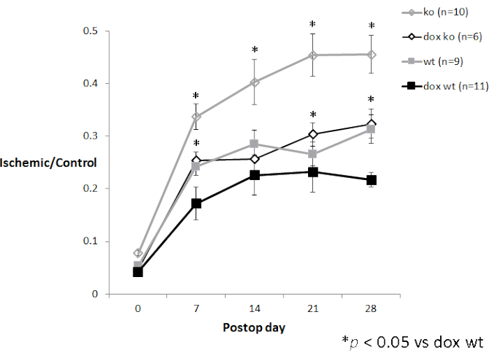
Figure 4. Time course experiment with LDPI data. P27 knockout mice (3-5 month old female CDKN1b-/- mice on a C57Bl/6 background) after femoral artery ligation with (n=6) and without (n=10) oral doxycycline treatment compared to age-matched female wildtype C57Bl/6 mice with (n=11) and without (n=9) oral doxycycline treatment (unpublished data from the author). Error bars represent standard error of the mean (SEM). Please click here to view a larger version of this figure.
Consistent technique is critical for obtaining reliable results with LDPI. The same anesthetic, temperature settings, mouse position, and region of interest should be used throughout the entire time course. Different anesthetic agents will result in higher or lower perfusion values9. Isoflurane anesthesia is convenient because of its rapid onset and emergence as well as overall safety. A consistent percentage of isoflurane should be used as depth of anesthesia with this vasodilatory agent may alter skin perfusion. If the region of interest includes fur, then the same method of hair removal should be used each time, as chemically depilated mice will have higher perfusion values than mice whose fur was removed with electric clippers7. Mouse temperature has a large effect on perfusion imaging, with mice at 36 °C having significantly lower perfusion values than mice at 38 °C7,12. The ischemic hindlimb may also react differently to regional temperature variation than the control hindlimb (Figure 2). In this protocol, a homeothermic blanket is used to maintain mouse temperature during scanning, which provides more consistent temperature control during the scanning process than pre-equilibrating the mouse on a 37 °C warming plate for five minutes and then scanning on a non-warmed surface as shown by Niiyama et al.13
If only the footpads are chosen as the region of interest, then prone positioning is preferred because of reproducibility in the scanned region of interest. The advantage of this approach is that it studies the area furthest from the heart, and the most clinically relevant area corresponding to where ischemic foot ulcers are common. The footpads are hairless, so clipping or depilation is not necessary, simplifying preparation and time for measurement. Also, non-white mice may have patches of pigmentation in the skin of the calf or thigh, which can interfere with LDPI measurement. If the chosen region of interest includes the calf and thigh, then supine positioning is preferred because the femoral and saphenous artery run along the ventral surface of the hindlimb and can be imaged using LDPI7. From the supine position, consistent imaging of the foot is difficult to capture, as the side and top surfaces may be variably imaged.
Collateralization and blood flow recovery after hindlimb ischemia is dependent on a number of different factors including hindlimb ischemia model, mouse strain, gender, and age. Certain strains of mice such as C57Bl/6 have robust baseline collaterals, with a less dramatic drop in perfusion after induction of acute hindlimb ischemia, whereas others such as BALB/c have poor collaterals14,15. Female mice have worse recovery than male mice. Older mice also have worse blood flow recovery than younger mice16. Therefore, mice need to be strain, age, and gender matched for reliable conclusions to be drawn regarding blood flow recovery using LDPI data. Even with rigorous matching and using inbred strains of mice, there is a certain amount of biologic variability to the mouse response to hindlimb ischemia, so adequate mouse numbers (usually 8-10 mice per timepoint) are required for valid data. Furthermore, normalization of LDPI does not necessarily mean restoration of normal levels of arterial flow as the measurement is done in anesthetized mice who do not have any skeletal muscle demand. Finally, because of the limitations in depth of penetration, detailed anatomic studies of collateral pathways that may run through the deeper musculature of the thigh and calf are not possible with LDPI.
Several other methods have been used to assess blood flow recovery including perfusion-based imaging of the skin such as laser speckle imaging17,18,19 or deeper structures such as contrast-enhanced ultrasound of skeletal muscle20, MRI21, and (13)N-ammonia PET22. Also used are anatomic-based imaging of collateral vessels such as micro-CT10, OCT23, and contrast-enhanced ultrasound with intravital microscopy24. Because of fast scan time, relative ease of data capture and analysis, and avoidance of the need for intravenous contrast, LDPI is the predominant method used by most groups in the literature. Weaknesses include that the technique measures blood flow velocities and provides data in arbitrary perfusion units rather than measuring absolute tissue perfusion, the scanning depth is relatively shallow, and it provides relatively poor anatomic detail.
LDPI is most commonly used to assess recovery after various hindlimb ischemia models7. It has also been used in ischemia-reperfusion research both in the hindlimb25 as well as in deeper splanchnic organs or the spinal cord26,27,28. Assessment of deep structures however requires surgical exposure of the structure to be scanned, making repeated measurements more difficult because of scarring. A further application is assessment of flap reperfusion after microsurgery29.
In conclusion, LDPI is an effective, easily performed, and repeatable method for measuring hindlimb dermal perfusion as a reflection of overall arterial perfusion. Consistent technique is required when using LDPI to obtain reliable data.
The authors have nothing to disclose.
This work was carried out with the use of facilities and resources at the VA Puget Sound Health Care Center. The work is that of the author and does not necessarily reflect the position or policy of the Department of Veterans Affairs or the United States government. Dr Tang is currently funded via the VA (Merit 5 I01 BX004975-02).
Black nonreflective material | Fabric store, black neoprene recommended by company | ||
F/air cannister | A.M. Bickford Inc | 80120 | |
Homeothermic blanket with rigid metal probe | Harvard Apparatus | Also comes with flexible probe, but this is less durable | |
Isoflurane Anesthesia machine | Drager | Multiple manufacturers | |
Isoflurane induction chamber | VetEquip | 941444 | 2 L chamber |
Moor laser Doppler perfusion imager | Moor Instruments | MoorLDI2-IR | Higher resolution imager (MoorLDI2-HIR) |
Mouse Anesthesia nose cone | Multiple manufacturers | ||
Nair | Nair | ||
Oxygen tank | Multiple manufacturers | ||
Surgilube | Multiple distributors |