Here we present a protocol for combining two sample processing techniques, high-pressure freezing and microwave-assisted sample processing, followed by minimal resin embedding for acquiring data with a focused ion beam scanning electron microscope (FIB-SEM). This is demonstrated using a mouse tibial nerve sample and Caenorhabditis elegans.
The described sample preparation technique is designed to combine the best quality of ultrastructural preservation with the most suitable contrast for the imaging modality in a focused ion beam scanning electron microscope (FIB-SEM), which is used to obtain stacks of sequential images for 3D reconstruction and modelling. High-pressure freezing (HPF) allows close to native structural preservation, but the subsequent freeze substitution often does not provide sufficient contrast, especially for a bigger specimen, which is needed for high-quality imaging in the SEM required for 3D reconstruction. Therefore, in this protocol, after the freeze substitution, additional contrasting steps are carried out at room temperature. Although these steps are performed in a microwave, it is also possible to follow traditional bench processing, which requires longer incubation times.The subsequent embedding in minimal amounts of resin allows for faster and more precise targeting and preparation inside the FIB-SEM. This protocol is especially useful for samples that require preparation by high-pressure freezing for a reliable ultrastructural preservation but do not gain enough contrast during the freeze substitution for volume imaging using FIB-SEM. In combination with the minimal resin embedding, this protocol provides an efficient workflow for the acquisition of high-quality volume data.
High-pressure freezing is the sample preparation method of choice for obtaining high-quality ultrastructural preservation, which represents the native state of a sample much better than conventional preparation methods using chemical fixation1. This cryo-preparation method is useful for samples such as myelinated mouse tissue2 and a strict requirement for use of the model organism Caenorhabditis elegans3. After freeze substitution and resin embedding, these samples are usually analyzed by transmission electron microscopy (TEM) or electron tomography (ET). If bigger volumes must be imaged using FIB-SEM or serial block-face imaging for high-resolution large-scale 3D reconstructions, in our experience proper imaging by SEM is often hampered by the lack of contrast. In the FIB-SEM, the image is usually recorded by the detection of backscattered electrons from the primary electron beam. The yield of backscattered electrons is proportional to the content of heavy metals in the sample. Therefore, protocols were especially designed for volume imaging to enhance the contrast by additional heavy metal impregnation. Such methods are based on chemically fixed samples and apply a combination of osmium tetroxide-thiocarbohydrazide-osmium tetroxide4, as described by Knott et al.5, for serial block-face and focused ion beam scanning electron microscopy. Modifications including the use of formamide and pyrogallol6 or lead aspartate7 have been successfully applied for different imaging techniques.
The protocol provided here combines the cryo-preparation of specimens by HPF and freeze substitution with subsequent microwave-assisted processing for enhanced contrast using thiocarbohydrazide/osmium tetroxide in acetone at room temperature. We demonstrate this on the myelinated nervus tibialis of mice and in Caenorhabditis elegans, which represent samples that require high-pressure freezing for high-quality ultrastructural preservation. In addition, it is shown how, after dehydration and infiltration, the samples are embedded with as little resin as possible. This minimal resin embedding8 allows for faster targeting of the structure of interest and reduces the time spent on sample processing, including less time required for exposing the region of interest with the ion beam. After performing further sample preparation steps inside the microscope, imaging and milling of the sample is carried out continuously to acquire a stack of images. For 3D visualization, image processing software (IMOD) is used to reconstruct parts of the dataset.
Our workflow describes how the most suitable contrasting of samples for volume imaging can be combined with the best ultrastructural preservation by HPF and freeze substitution. This is useful for samples that strictly require cryo-preparation. Applications are limited to small samples that can be prepared by HPF. In samples of different nature, such as plant material or microorganisms, this protocol requires adaptation.
All experiments including samples from animals described here have been approved by the Institutional Animal Care and the Lower Saxony State Office for Customer Protection and Food Safety (LAVES).
1. High-pressure freezing and freeze substitution
2. Microwave-assisted processing
NOTE: Perform all of the following steps at room temperature12 using a temperature control unit to keep the temperature stable (see Table of Materials).
3. Minimal resin embedding8
4. Preparation for FIB-SEM
5. Data acquisition inside the FIB-SEM
6. Data visualization
The workflow starts with a sample (here, a freshly dissected mouse nervus tibialis) being placed in metal carriers for high-pressure freezing (Figure 1a). The carriers are recovered from liquid nitrogen (Figure 1b) and placed in a freeze substitution unit on top of the frozen first chemical cocktail (Figure 1c). After a long freeze substitution protocol including 2% osmium tetroxide and 0.1% uranyl acetate, the samples are removed from the carriers at room temperature (Figure 1d). To further enhance the contrast, the samples are transferred to plastic tubes to be processed in the microwave (Figure 1e). The vacuum chamber and temperature control unit are used to optimize the process (Figure 1f).
To be able to perform the minimal resin embedding, toothpicks and sheets of plastic film are needed (Figure 2a). After infiltrating the samples with resin using the microwave, they are placed on pieces of plastic film and moved around until no resin is left on the sample surface. A halogen lamp is used to help drain the remaining resin and leave the sample minimally embedded (Figure 2b) on the plastic film. It should be noted that more resin removed from the top of the sample is good. There should still be a small amount left underneath the sample to keep it attached to the substrate. The sample being polymerized on the plastic film is cut and mounted on top of SEM stub with silver conductive resin (Figure 2c). The stub is polymerized for at least 4 h at 60 °C (Figure 2d). The components should be mixed thoroughly or the mixture may not polymerize correctly. To avoid charging inside the scanning electron microscope, the stub is sputter coated with gold or platinum/palladium (Figure 2e).
The samples are placed in the FIB-SEM and imaged with a secondary electron detector to target the region of interest (Figure 3a,e). An ion beam is used to remove material directly in front of the region of interest to expose a cross section (Figure 3b-d, 3f-h). Standard protocols often suffer from a lack of membrane contrast (Figure 3b,f), whereas the enhanced protocol provides a strong membrane contrast (Figure 3c-d, 3g-h).
The EM data (after post-processing) are visualized using IMOD, an image processing and modelling program. To achieve a better understanding of the 3D information, virtual reslicing of the data is used (Figure 4a). Different structures of the dataset are segmented manually (Figure 4b-d).
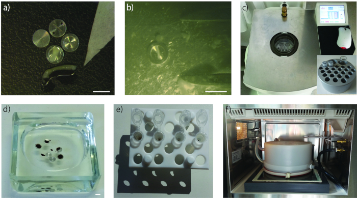
Figure 1: High-pressure freezing, freeze-substitution, and microwave-assisted processing. (a) Sample carrier containing the mouse nervus tibialis, scale bar 3 mm. (b) Sample carrier containing the mouse nervus tibialis after high-pressure freezing, scale bar 3 mm. (c) Automatic freeze substitution (AFS) unit with samples. Inset: custom-made metal container for up to 23 sample vials and two large vials containing chemicals in the AFS. (d) Samples being removed from carriers in a glass dish in acetone, scale bar 3 mm. (e) Samples in reaction tubes to be put in the microwave for processing. (f) Vacuum chamber and temperature control unit of the microwave. Please click here to view a larger version of this figure.
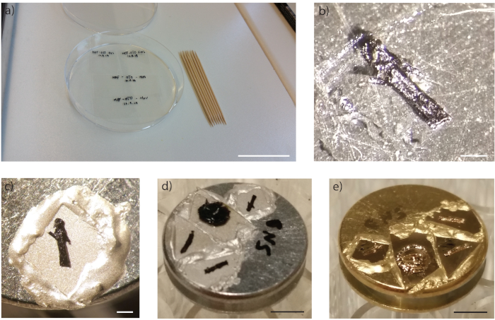
Figure 2: Minimal resin embedding and preparation for the FIB-SEM. (a) Plastic film and toothpicks that are used for the minimal resin embedding, scale bar 4 cm. (b) Nervus tibialis drained of resin on top of the plastic film, scale bar 250 µm. (c) Nervus tibialis polymerized on plastic film, then cut and mounted on top of the SEM stub with silver conductive resin, scale bar 250 µm. (d) Samples polymerized on top of the SEM stub, scale bar 3 mm. (e) Samples coated with gold on the SEM stub, scale bar 3 mm. Please click here to view a larger version of this figure.
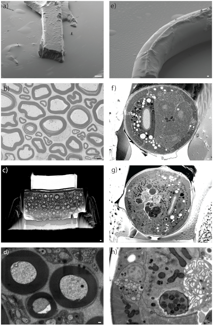
Figure 3: Preparation of the sample inside the FIB-SEM. (a and e) Secondary electron image inside the FIB-SEM of the sample surface (a) Nervus tibialis, scale bar 100 µm. (e) C. elegans, scale bar 2 µm. (b-d) and (f-h) Cross-section through sample using ESB detector for imaging. (b and c) Nervus tibialis, scale bar 2 µm. (f and g) C. elegans, scale bar 1 µm and 200 nm. (b and f) Shown are the results of high-pressure freezing and freeze substitution without enhancement, whereas all other images show results of enhanced freeze substitution. (d and h) Detailed image of the sample using the ESB detector. (d) Nervus tibialis, scale bar 200 nm. (h) C. elegans, scale bar 200 nm. Please click here to view a larger version of this figure.
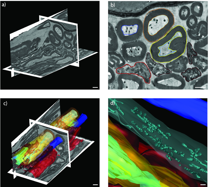
Figure 4: Image acquisition and visualization. (a) EM data shown in IMOD with virtually resliced x/z- and y/z-planes, scale bar 2 µm. (b) Segmented axons on EM data (blue), Remak bundles (red), myelin sheaths (yellow and orange), and mitochondria (turquoise), scale bar 2 µm. (c and d) 3D model, scale bar 2 µm and 500 nm. Please click here to view a larger version of this figure.
The protocol was developed to illustrate the optimal preservation and contrast to perform serial block-face imaging with a FIB-SEM. Therefore, we chose to apply cryo-immobilization followed by post-staining using freeze substitution and microwave-assisted processing. Therefore, this protocol is limited to samples that are small enough for high-pressure freezing. The size limitations of 3 to 6 mm in width and thickness of ~200 µm are established by the size of the sample carrier, which matches the sample size that can be properly frozen with this technique. This is relevant for the mouse nerve sample, since the sciatic nerve is too large in diameter to fit into the 0.2 mm carriers that are required to ensure proper freezing. Therefore, careful dissection of a smaller nerve such as the tibial nerve or other thin nerve such as the femoral nerve is recommended. Since the myelin sheath is sensitive to stretching, great care must be taken during dissection of the fresh and viable nerve to avoid handling artifacts. In general, only viable samples should be used for electron microscopic studies.
Microwave-assisted processing and minimal resin embedding are designed for speeding up the preparation and targeting process. The microwave-assisted processing applying a modified OTO protocol4 is used for room temperature chemical fixation. A household microwave will not yield the same results, since there is no homogenous distribution of microwaves, its temperature is not controlled, and there is no vacuum that can be applied. The smaller a sample, the better penetration of the chemicals; therefore, the best results are achieved by smaller samples. To avoid damage to the sample by overheating, temperature control and application of the minimally required microwave power are critical. The microwave-assisted processing steps can be performed on the bench if there is no microwave available, which will lead to longer processing times. To directly target structures in the SEM, it is crucial to remove as much resin as possible from the top of the sample. After recording a dataset, post-processing of the raw data is necessary to reduce file size and improve the signal-to-noise ratio. Modern volume imaging techniques produce large amounts of data. Therefore, to perform the data processing in a fast and sufficient manner, sufficient RAM on the workstation is needed. For alignment operations at least two times as much RAM as the dataset size is required.
This protocol has been tested on the nervus tibialis of the mouse as well as in C. elegans. Hall et al.12 used a similar enhancement step after their freeze substitution on the bench for preparation of C. elegans. For any other model organism such as the zebrafish, adjustments of the protocol are likely necessary. One possible modification is to change the composition of the freeze substitution cocktail, such as by the addition of water that is used for contrast enhancement18. Moreover, the duration of the freeze substitution has to be adapted to the sample and can be considerably shortened according to the quick freeze substitution protocol19. One possibility is the application of agitation for accelerating the freeze substitution process20. After the freeze substitution, further modifications are possible, such as repeated application of enhancing chemicals and osmium tetroxide21. During microwave processing, the temperature, incubation times, and power settings can be varied to optimize results for the respective sample.
This protocol shows that such an enhancement can be combined with other freeze substitution protocols and different types of samples as described by Hall12 that are imaged in a FIB-SEM or by serial block-face scanning electron microscopy. These imaging techniques require enhanced contrast, which is less important for transmission electron microscopy.
The authors have nothing to disclose.
The FIB-SEM and A.S. (the position of the FIB-SEM operator) are funded by the Cluster of Excellence and Deutsche Forschungsgemeinschaft (DFG) Research Center Nanoscale Microscopy and Molecular Physiology of the Brain (CNMPB). We thank the lab of Thomas Müller-Reichert for providing the C. elegans samples. We thank Ulrich Weikert for participating in the movie.
Instrumentation | |||
Leica HPM100 | Leica | ||
Automatic Freeze Substitution | Leica | ||
Laboratory microwave with temperature control unit | Ted Pella | ||
EM ACE600 with gold target | Leica | ||
Crossbeam 540 | Zeiss | ||
Halogen lamp 12 V/ 20 W | Osram | ||
Oven | VWR | ||
Freezing | |||
Bovine Serum Albumin | Sigma-Aldrich | A2153 | |
M9 | Homemade | According to C. Elegans- A practical approach I.A.Hope | |
Hexadecene | Sigma-Aldrich | 52276 | |
Polyvinylpyrrolidone | Sigma-Aldrich | P2307 | |
A type carrier | Wohlwend GmbH | #241 | |
B type carrier | Wohlwend GmbH | #242 | |
Slit carrier | Wohlwend GmbH | #446 | |
Plastic Pasteur pipettes | VWR | 612-1684 | |
Forceps | FST | 11200-10 | |
Freeze substitution | |||
Acetone | science services | 10015 | |
Tannic acid | Sigma-Aldrich | 403040 | |
Osmium tetroxide | EMS | 19100 | |
Uranyl acetate | SPI-Chem | 02624-AB | |
Acetone | EMS | 10015 | |
Thiocarbohydrazide | Sigma-Aldrich | 223220 | |
Nunc CryoTubes | Sigma-Aldrich | V7884-450EA | |
Watch glass dishes, 150 mm | VWR | 216-2189H | |
Eppendorf tubes | Eppendorf | 0030 120.094 | |
Durcupan resin | Sigma-Aldrich | 44610 | |
Mounting | |||
SEM stubs | Science Services | E75200 | |
Aclar | Science Services | 50425-10 | |
Toothpicks | |||
filter paper | VWR | 512-3618 | |
conductive silver resin | EMS | 12670-EE | EPO-TEK EE 129-4 |
Software | |||
Image acquisition | Zeiss | SmartSEM | |
Image acquisition | Zeiss | Atlas5 A3D | |
Image processing | Open source | Fiji | http://fiji.sc/#download |
Image visualization | Open source | IMOD | http://bio3d.colorado.edu/imod/ |