1. Water/Oil/Water Double Emulsion Microsphere Production
- First prepare 2% and 0.5% w/v solutions of polyvinyl alcohol (PVA) in deionized water. Stir the solutions at 50 °C until clear, this may take several hours. Prepare a solution of 2% v/v Isopropyl Alcohol in deionized water.
- Prepare an aqueous solution of desired hydrophilic protein. The table below provides example formulations.
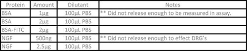
Table 1: Example Protein Solutions. The following protein solutions have been successfully encapsulated and electrospun using this protocol. Other hydrophilic protein solutions can be used as needed.
- Place 40 ml of the 0.5% PVA solution into a 50 ml centrifuge tube and set aside.
- In a 15 ml centrifuge tube dissolve 300 mg of 65:35 poly(lactic-co-glycolic acid) (PLGA) in 3 ml of Dichloromethane. A vortex mixer can be used to accelerate PLGA dissolution.
- Combine 200 µl of protein solution and 4 µl of 2% PVA solution. Pour the protein mixture into the PLGA solution (step 1.4). The solutions will remain mostly separate.
- Place the tube into a beaker of ice water. Using a wand sonicator at ~10 Watts (RMS), agitate the solution for a few seconds (5-10) until a uniform creamy white emulsion is created.
- Pour the emulsion into the 50 ml tube containing 0.5% PVA (step 1.3). Mix the solution at high speed on a vortex mixer for ~20 sec. The solution will develop a cloudy appearance.
- Transfer the emulsion to a 200 ml beaker and place on a stir plate at 350 rpm for 2 min. Add 50 ml of 2% isopropyl alcohol to the beaker on the stir plate. Allow the mixture to continue stirring for a minimum of 1 hr to allow the DCM to evaporate and the PLGA to harden.
- Transfer the microsphere solution into centrifuge tubes.
- Centrifuge at 425 x g for 3 min. The microspheres will collect at the bottom of the tube and appear white. Carefully remove the supernatant from the tube, above the microspheres, and store in a 500 ml bottle.
- Rinse the microspheres with deionized water by filling the tube three quarters full and shaking it to redistribute the microspheres in the liquid.
- Repeat steps 1.10 & 1.11 four times.
- Following the final rinse, remove the supernatant again and place in the 500 ml bottle with the other samples. Freeze the microspheres collected in the centrifuge tube, then lyophilize for at least 24 hr.
- Visualize the microspheres under a light microscope or with a Scanning Electron Microscope. Microspheres no larger than 60 µm are for effective electrospinning. If microspheres are too large, longer sonicating or vortexing times may be required in step 1.6 or 1.7.
- Store the dried microspheres in a -20 °C freezer.
- Optional: Use a protein assay, per manufacturer instructions, to test the amount of protein in the 500 ml bottle from step 1.1030. This is used to calculate the percent of protein encapsulated in the microspheres, by subtracting the amount in the solution from what was used in the production process.
Note: To visualize the protein location in the microsphere add Rhodamine 2 µg/ml to the PLGA solution31 and encapsulate a FITC conjugated protein. Figure 1 shows an example.
2. Electrospinning with Microspheres
- Prior to preparing electrospinning solution, create a 0.5% w/v solution of photoinitiator, in deionized water by dissolving at 37 °C. This process can take several hours.
- Create a 2% w/v methacrylated hyaluronic acid (MeHA) (see Burdick et al. for synthesis)29, 3% w/v 900 kD poly(ethylene oxide) (PEO) and 0.05% w/v photo initiator solution in deionized water.
- Calculate the correct amount of MeHA and PEO for the desired volume. For example, 10 ml of electrospinning solution requires 200 mg of MeHA and 300 mg of PEO.
- Dissolve the PEO in deionized water at 90% of the final volume desired (9 ml for this example). This may take several hours, a heated stir plate or water bath at 37 °C can be used to accelerate the process.
- Next add the MeHA and use a vortex mixer to stir the solution until clear. This will only take a few minutes.
- Finally add the 0.5% photo initiator solution to fill the remaining 10% volume (1 ml for this example).
- Add microspheres at the desired concentration up to 400 mg/ml. Mix the solution on a vortex mixer until the microspheres are evenly distributed in the solution.
- Transfer the solution to a syringe and attach a 6 inch 18 gauge blunt tip needle.
- Place the syringe in a syringe pump and set it to dispense at 1.2 ml/hr.
- Tape a layer of aluminum foil on the collection plate or mandrel. This allows for easy clean up and storage of the finished scaffold. A rotating mandrel is used to create aligned fibers. A flat plate or stationary mandrel will result in randomly arranged fibers.
- Connect the ground wire from a high voltage power source to the collection apparatus. Connect the positive lead to the needle.
- Adjust the syringe pump and collection surface so that there is 15 cm between the needle tip and collection surface.
- Start the polymer pumping, when the solution is visible at the end of the syringe, turn on the voltage source and set the voltage to 24 kV. CAUTION: Once the voltage is turned on do not touch any metal part of the system. Charge may also jump short distances from electrified parts to skin.
- Run the solution until desired scaffold thickness is achieved. When complete turn off voltage source and syringe pump.
- Remove foil with scaffold attached. Completed scaffolds containing protein are stored in a -20 °C freezer.
3. Protein Bioactivity Testing
- Prepare cell culture media. Add 10% v/v Fetal Bovine Serum, 1% v/v L-Glutamine, and 1% v/v Penicillin-Streptomycin to Dulbecco’s Modified Eagle’s Medium.
- Select glass coverslips that fit completely into a well plate.
- Use 3-(Trimethoxysilyl)propyl methacrylate to treat the coverslips as described by the manufacturer. Methacrylation enhances scaffold adherence to the coverslips.
- Attach methacrylated coverslips to the collection area of the electrospinner with removable double sided tape before electrospinning. Spinning onto the coverslips eases handling and viewing.
- Electrospin to desired thickness as described above.
- After electrospinning carefully remove coverslips from mandrel. Place the scaffold coated coverslips into a clear nitrogen chamber and ensure that all oxygen is purged.
- Place the chamber and scaffold under a 10 mW/cm2 365 nm light for 15 min. After crosslinking place into appropriately sized well plate. Ensure that the scaffold side is facing up.
- Place scaffolds under a germicidal lamp for 30 min to sterilize. If desired fibronectin or other protein is used as a coating to enhance cell adhesion. Follow manufacturer’s instructions to coat scaffolds.
- Harvest Dorsal Root Ganglia (DRG) as previously described by Hollenbeck32. One DRG will be needed for each scaffold covered coverslip tested.
- Place 100-200 µl of media on each scaffold in the well plate. Carefully place one DRG on each scaffold in the media droplet. For thick scaffold more media may be needed; DRG need to be fully submerged and not floating.
- Incubate the scaffold and DRG at 37 °C for 4 hr to allow the cell to adhere to the scaffold.
- Fill the media to the appropriate level for the well and place back into the incubator. Continue incubating for 4-6 days.
- After the incubation period carefully remove the media from each well and gently wash once with PBS. Fix cells for 30 min using 4% w/v paraformaldehyde.
- Stain cells using an antibody stain for neurofilament. This will allow visualizing of neurite outgrowth for quantification. DAPI can also be used to view nuclei of the cells. An example staining protocol was described by Sundararaghavan and colleagues14.
- Visualize the cells using a fluorescent microscope.
- Place well plate on the stage of the microscope.
- Locate the cell mass using the filter and excitation settings for DAPI.
- Once the cell is switch the filter to FITC to visualize the extended neurites. Using the stitch function on the microscope collect and combine as many images as necessary to see the entire structure. Repeat for DAPI, FITC and bright field.
Microspheres 50±14 µm in diameter with an over 85% protein encapsulation have been consistently produced and electrospun into scaffolds. Size was determined by imaging samples of microspheres from three separate production batches. The images where captured on an optical microscope and lengths where measured using commercial lab software. Figure 1 shows a histogram of the size distribution. Encapsulation rate was also tested from three separate microsphere batches, by quantifying the protein that escaped during the production process.
Figure 2 shows representative microspheres with Rhodamine (2 µg/ml) in the PLGA shell and bovine serum albumin (BSA)-FITC encapsulated within. The images were taken using a fluorescent microscope. The shell can be clearly seen (red) with the protein concentrated on the interior (green).
To evaluate encapsulation, the microspheres were filled with BSA and tested for protein release with a Bradford Protein Assay. PLGA breaks down by hydrolysis of ester bonds, creating larger gaps for protein escape. The microspheres continue releasing for over 60 days (Figure 3). The release begins with an initial burst then continues as microspheres break down.
After electrospinning the microspheres can be seen throughout the 3D fibrous structure. Figure 4 shows an SEM image of the complete scaffold where the spheres can be seen in multiple layers (indicated by arrows). Distribution of microspheres in the scaffolds was measured to be 15.3 ± 3.6 spheres/mm2. The scaffold holds the microspheres in place preventing migration away from the target site. As the microspheres break down they release NGF into the scaffold to encourage neurite growth33,34. This creates a continual delivery of protein throughout the scaffold to support the growth of cells and encourage cells to infiltrate into the scaffold.
Finally, Dorsal Root Ganglia (DRG) were used to test the viability of NGF in the microspheres, within the scaffold. Figure 5 shows a DRG seeded on a scaffold containing microspheres loaded with NGF next to one containing microspheres with no protein in them. There are longer neurite extensions visible extending from the DRG on the NGF scaffold, indicating that the NGF remained viable and is stimulating growth.
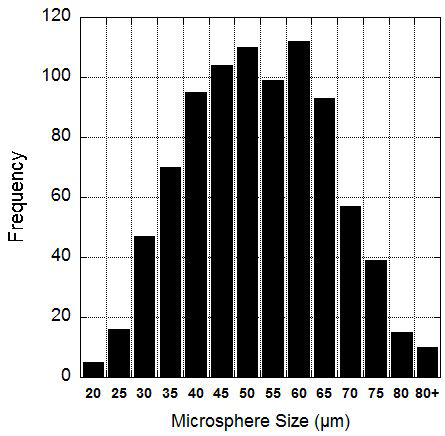
Figure 1. Microsphere size distribution. Microspheres produced by this protocol are 50±14 µm in diameter. The graph shows a histogram of the microsphere diameter in 5 µm bins.
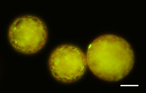
Figure 2. Fluorescent Image of Microspheres. 2 µg/ml Rhodamine was included in the PLGA solution to visualize the microsphere shell (red) and BSA-FITC was encapsulated to visualize the protein (green). Protein can clearly be seen within the sphere. Scale bar = 20 µm. Please click here to view a larger version of this figure.
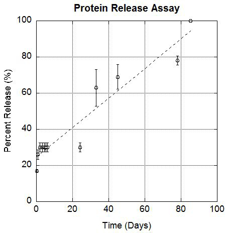
Figure 3. Protein Release Curve. BSA release from the microspheres over 60 days was measured using a Bradford Protein Assay. The initial burst is likely due to BSA that was attached to the outer surface. As the PLGA breaks down, protein is released over time.
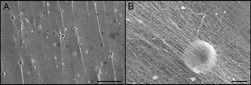
Figure 4. Scaffold containing Microspheres. The electrospinning process allows microspheres to be incorporated throughout the depth of the scaffold. (A) Arrows indicating the location of microspheres. Scale bar = 500 µm. (B) High magnification image of one microsphere with the nanofibers from the scaffold above it. Scale bar = 20 µm. Please click here to view a larger version of this figure.
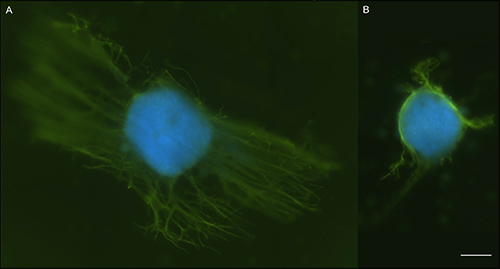
Figure 5. NGF Bioactivity Test. Dorsal Root Ganglia primary cells were harvested from 8 day old chick eggs and seeded on scaffolds with NGF filled microspheres (A) or empty microspheres (B). DRGs were stained with a neurofilament antibody, FITC secondary antibody and DAPI to visualize the cell nuclei. Images show increased neurite outgrowth in the presence of NGF loaded microspheres as indicated by FITC stained neurites (green). This shows released NGF is viable and can promote growth. Images were taken with a confocal microscope. Scale bar = 200 µm. Please click here to view a larger version of this figure.
