Here, we present a procedure to fluorescently functionalize the disulfides on Qβ VLP with dibromomaleimide. We describe Qβ expression and purification, the synthesis of dibromomaleimide-functionalized molecules, and the conjugation reaction between dibromomaleimide and Qβ. The resulting yellow fluorescent conjugated particle can be used as a fluorescence probe inside cells.
The recent rise in virus-like particles (VLPs) in biomedical and materials research can be attributed to their ease of biosynthesis, discrete size, genetic programmability, and biodegradability. While they're highly amenable to bioconjugation reactions for adding synthetic ligands onto their surface, the range in bioconjugation methodologies on these aqueous born capsids is relatively limited. To facilitate the direction of functional biomaterials research, non-traditional bioconjugation reactions must be considered. The reaction described in this protocol uses dibromomaleimides to introduce new functionality in the solvent exposed disulfide bonds of a VLP based upon Bacteriophage Qβ. Furthermore, the final product is fluorescent, which has the added benefit of generating a trackable in vitro probe using a commercially available filter set.
Using nano-sized viral capsids has emerged as an exciting field, which aims to broaden the scope of applications in biomedical research1,2,3. Recombinantly expressed virus-like particles (VLPs) are structurally derived from viruses, but they lack the original viral genetic material making them non-infectious proteinaceous nanoparticles. As the surface features are genetically programed and each capsid is expressed identically to the ones before and after it, it is possible to know the location and number of reactive side chains of the amino acids with atomistic precision. In many cases, both the exterior and interior surfaces possess many kinds of solvent exposed amino acid residues, which can feasibly be functionalized through bioconjugation reactions – reactions that form covalent bonds between a biomolecule and a synthetic molecule4,5.
Bioconjugation reactions help biomolecules of interest have more diverse functionalities in a relatively straightforward fashion. Molecules of interest, such as therapeutic drugs6, fluorescent tags7 and polymers8,9 can be pre-synthesized and characterized before they are attached on the surface of VLPs. A particularly common VLP in biomedical and biomaterials research has been the VLP based upon Bacteriophage Qβ, which, as recombinantly expressed, is a 28 nm icosahedral viral capsid10. The most common reaction sites on Qβ are lysines by a wide margin, though we have recently communicated the successful conjugation11 of dibromomaleimide compounds to the reduced disulfides that line the pores of Qβ via a Haddleton-Baker reaction. The reaction proceeds with good yield and, equally importantly, without losing the thermal stability of the particles. At the same time, this reaction generates conjugation-induced fluorescence, which can be used to track the uptake of these particles into cells. In this work, we demonstrate the conjugation of polyethylene glycol (PEG) onto the surface of Qβ through the Haddleton-Baker reaction, which results in a bright yellow fluorophore. These particles can then be tracked as they are taken in by cells. The protocol herein will help researchers generate new fluorescent PEGylated proteinaceous nanoparticles based upon Qβ, though its principles are applicable to one of the many other VLPs containing solvent exposed disulfides.
1. Preparation
2. Expression of Qβ
3. Purification of Qβ
4. Quantification and Confirmation of the Product
5. Conjugating DB Compounds on Qβ
The dibromomaleimide derivatives can be synthesized through the condensation reaction between dibromomaleimide anhydride and primary amines15. Alternatively, a mild synthetic method16 using N-methoxycarbonyl activated 3,4-dibromomaleimide was exploited here by reacting with methoxypolyethylene glycol (PEG) to yield DB-PEG (Figure 1). NMR was used to identify the compound structure (Figure 2). Qβ VLP is a 28 nm icosahedral proteinaceous nanoparticle, which is composed of 180 identical coat proteins. The coat proteins tend to form noncovalent interlocking dimers through their α-helical domains with the β-sheets from the adjacent coat proteins17. VLPs tend to be selected for their stability at high temperatures, extreme pHs, and in various solvent compositions18. In this case, Qβ VLP is more stable than other RNA phages in the Leviviridac family owing to 180 inter-strand disulfide bonds located at the five- and six- fold axes of symmetry on the capsid19 (Figure 3). These hexameric and pentameric structures are linked by disulfides, which can be visualized by non-reducing SDS-PAGE19. Ten equivalents of TCEP (tris(2-carboxyethyl)phosphine) (0.70 µmol), relative to the disulfides in 1 mg of Qβ (0.070 µmol coat protein, 0.070 µmol disulfides), were used to reduce all the disulfides to generate the reduced Qβ capsids (rQβ) at room temperature in one hour, and non-reducing SDS-PAGE shows that all the higher order structures were reduced to monomeric coat proteins (Figure 4 and Figure 5A). The reaction to rebridge them was done as a one-pot synthesis, as the crude rQβ admixture was added directly to 20 equivalents of a solution of DB-PEG (1.4 µmol) in sodium phosphate (10 mM, pH 5.00, 10% DMF) following the one-hour reduction reaction11. There was observable bright yellow fluorescence under 365 nm UV lamp (Figure 5D) immediately after addition of the DB-PEG. The mixture was then incubated at RT overnight on a rotisserie, followed by purification using centrifugal filter (MWCO = 10 kDa) rinsing with the desired buffer three times to remove excess amounts of small molecules. The Qβ-malemide conjugates were resuspended into 10 mM sodium phosphate solution (pH 5.00) to promote photostability. The conjugation was confirmed by non-reducing SDS-PAGE under UV and coomassie blue staining (Figure 5A). All the bands showed fluorescence (Figure 5A) under UV which colocalized with the coomassie blue staining, representing a successful conjugation. The integrity of Qβ-PEG conjugates were confirmed by native agarose gel electrophoresis and transmission electron microscopy (TEM) (Figure 5B, C).
The fluorescence spectroscopy showed the excitation and emission maxima of Qβ-maleimide (Qβ-Μ) and Qβ-PEG to be around 400 nm and 540–550 nm, respectively (Figure 6). This aligns with the commercially available GFP-uv filter set, whose excitation wavelength is 405 nm and emission wavelength is 500–540 nm. The convenient alignment of the photophysical properties of the conjugates with the commercially available filter sets permit using Qβ-PEG as an in vitro probe, which was done and imaged in Figure 7. Qβ-PEG (200 nM) was incubated with Mouse Raw-264.7 cells in serum-free DMEM medium, followed by nucleus staining. Colocalization images in Figure 7 shows that yellow fluorescent particles were uptaken by Raw-264.7 cells and can be tracked after four hours of incubation. The unfunctionalized Qβ VLPs show negligible fluorescence.
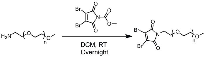
Figure 1: Synthetic scheme of DB-PEG. Please click here to view a larger version of this figure.
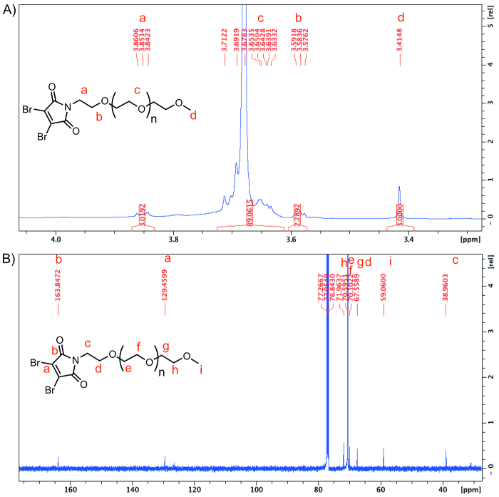
Figure 2: NMR characterization. (A) 1H NMR and (B) 13C of DB-PEG. Please click here to view a larger version of this figure.
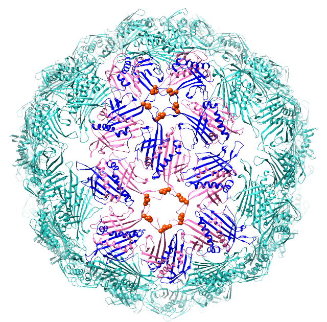
Figure 3: Crystallographic structure of Qβ VLP capsid as processed in Chimera (PDB ID: 1QBE). Two cysteine residues (Cys 74 and Cys 80) are shown in orange.
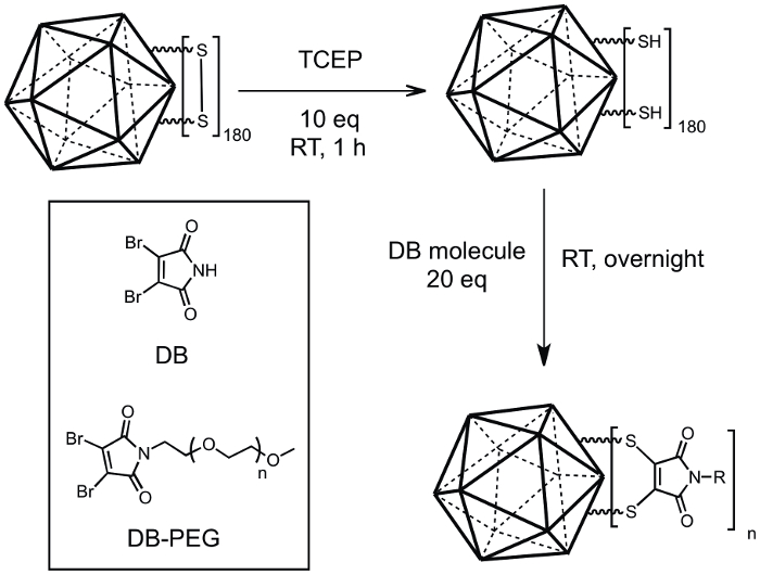
Figure 4: Conjugation scheme of Qβ-maleimide (Qβ-M) conjugates. Qβ (0.07 µmol of disulfides) was reduced using 10 equivalents of TCEP (0.7 µmol) at RT for one hour followed by addition of 20 equivalents of dibromomaleimide compounds (DB and DB-PEG) (1.4 µmol). Please click here to view a larger version of this figure.
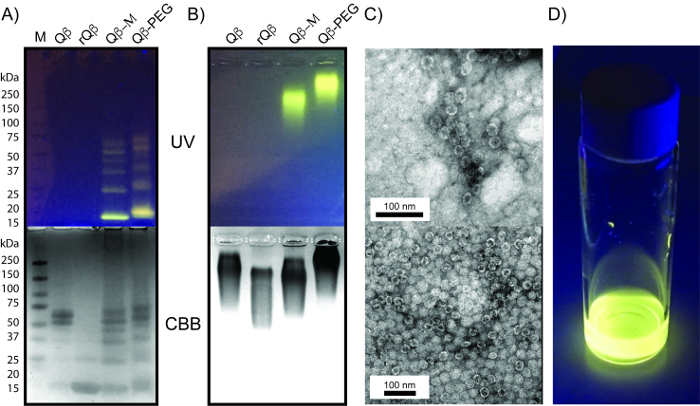
Figure 5: Characterization of Qβ, rQβ, Qβ-M and Qβ-PEG. (A) Non-reducing SDS-PAGE and (B) native agarose gel of Qβ, rQβ, Qβ-M and Qβ-PEG under UV (top) and coomassie blue staining (bottom). (C) TEM micrograph of Qβ-M (top) and Qβ-PEG (bottom). (D) Photograph of Qβ-PEG reaction mixture under 365 nm UV illumination. Please click here to view a larger version of this figure.
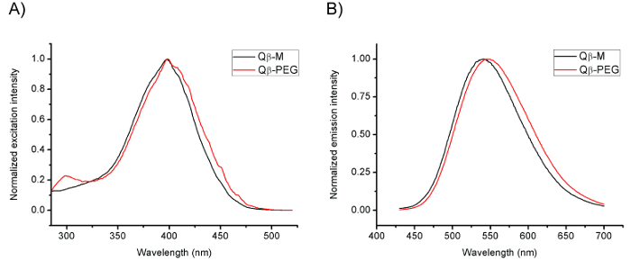
Figure 6: Fluorescence spectra. Fluorescence excitation (A) and emission (B) spectra of Qβ-M and Qβ-PEG in 0.1 M of potassium phosphate buffer (pH 7.00). The excitation maximum is around 400 nm and emission maximum is around 540–550 nm. Please click here to view a larger version of this figure.
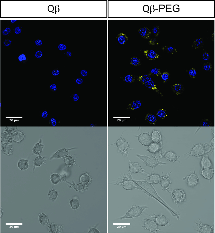
Figure 7: Confocal fluorescence images of Qβ-PEG conjugate in macrophage 264.7 cells. Blue: NucRed Live 647 ReadyProbes Reagents. Yellow: Qβ-PEG. Top images: merged blue and yellow channels. Bottom images: bright field images. Filter sets: uv-GFP (λex = 405 nm, λem = 500–540 nm), Cy5. Time of incubation was 4 h. Please click here to view a larger version of this figure.
Compared to smaller protein purification, a unique step in purifying bacteriophage Qβ is the sucrose gradient centrifugation. After the chloroform/n-butanol extraction step, Qβ is further purified using 5-40% sucrose gradients. During centrifugation, particles are separated based on their sizes. Larger particles travel to the higher density region, while smaller particles stay in the lower density region. Qβ travels to the lower third of the gradient and remains there while smaller protein impurities are trapped at the top of the centrifuge tube. The colloidal suspension of Qβ shows strong Tyndall scattering in the sucrose gradient, which can be seen as a blue band when an LED light is shined from underneath. This band is then easy to extract using a long syringe needle. Qβ in sucrose is then pelleted by ultracentrifugation, yielding a clear pellet. The pellet can be resuspended into the desired buffer or further purified by fast protein liquid chromatography (FPLC). The purity of the resulting particle can be confirmed by SDS-PAGE.
TCEP is not stable in phosphate buffer, so a 100x TCEP stock solution should be freshly prepared in water right before the reduction. In addition, TCEP neither contains free thiol nor reacts towards other amino acids, so it is not necessary to remove the excess of TCEP prior to performing the conjugation reaction. Dibromomaleimide compounds are dissolved in pH 5.00 sodium phosphate solution, followed by adding the reduced Qβ. At pH 5.00, cysteine (reduced disulfide) is protonated, which promotes the conjugation reaction with dibromomaleimide by preventing re-oxidation back to a disulfide. Under 365 nm UV illumination, the reaction mixture emits yellow fluorescence immediately after reduced Qβ was added into DB compounds. Differently from attaching pre-synthesized fluorophores on Qβ, this fluorescence is induced by the conjugation reaction. The fluorophore is formed as soon as the new bonds between sulfur and the maleimide ring are created. Fluorescence spectra shows that the excitation (400 nm) and emission (550 nm) maxima fit the uv-GFP filter set in a confocal microscope (λex = 405 nm, λem = 500−540 nm). Qβ-PEG was then incubated with macrophage Raw 264.7 in serum-free DMEM. After four hours of incubation, Qβ-PEG was uptaken and punctate yellow fluorescent compartments can be visualized inside the cells. One fact that needs to be mentioned is the fluorescence may be transferred for some extent to bovine serum albumin (BSA) in serum or other cysteine-rich substances inside the lysosomes or the late endosomes of cells. Over time, this will weaken the fluorescence inside the cell for cell tracking applications; however, it also provides an opportunity for a new design for drug delivery systems.
In conclusion, we have demonstrated conjugating DB-PEG on Qβ VLP through dibromomaleimide-thiol chemistry. The all-in-one conjugation reaction not only functionalizes the disulfides along the pores on VLP with PEG, but also makes it a fluorescently labeled proteinaceous nanoparticle.
The authors have nothing to disclose.
J.J.G. acknowledges the National Science foundation (DMR-1654405) and Cancer Prevention Research Institute of Texas (CPRIT) (RP170752s) for their support.
LB Broth (Miller) | EMD Millipore | 1.10285.0500 | |
Tryptone, Poweder | Research Products International | T60060-1000.0 | |
Yeast Extract, Poweder | Research Products International | Y20020-1000.0 | |
Anhydrous magnesium sulfate | P212121 | CI-06808-1KG | |
Sodium Chloride (Crystalline/Certified ACS) | Fisher Scientific | S271-10 | |
Potassium Chloride | Fisher Scientific | BP366-500 | |
Elga PURELAB Flex 3 Water Purification System | Fisher Scientific | 4474524 | |
Potassium Phosphate Monobasic | Fisher Scientific | BP362-1 | |
Potassium Phosphate Dibasic Anhydrous | Fisher Scientific | P288-500 | |
Sucrose | Fisher Scientific | S25590B | |
Ethanol | Fisher Scientific | BP2818500 | |
Isopropyl β-D-1-thiogalactopyranoside (IPTG) | Sigma Aldrich | I6758-1G | |
Fiberlite F10-4×1000 LEX rotor | Fisher Scientific | 096-041053 | |
Ammonium Sulfate | P212121 | KW-0066-5KG | |
Chloroform | Alfa Aesar | 32614-M6 | |
1-Butanol | Fisher Scientific | A399-4 | |
SW 28 Ti Rotor, Swinging Bucket, Aluminum | Beckman Coulter | 342204: SW 28 Ti Rotor/ 342217: Bucket Set | |
Type 70 Ti Rotor, Fixed Angle, Titanium, 8 x 39 mL, | Beckman Coulter | 337922 | |
Coomassie (Bradford) Protein Assay | Fisher Scientific | PI23200 | |
TRIS Hydrochloride | Research Products International | T60050-1000.0 | |
Tetramethylethylenediamine | Alfa Aesar | J63734-AC | |
Tris(2-carboxyethyl)phosphine hydrochloride | Sigma Aldrich | C4706-2G | |
2 3-Dibromomaleimide 97% | Sigma Aldrich | 553603-5G | |
Polythylene Glycol | Alfa Aesar | 41561-22 | |
Sodium Phosphate | Fisher Scientific | AC424375000 | |
Acrylamide/bis-Acrylamide | P212121 | RP-A11310-500.0 | |
Sodium dodecyl sulfate | Sigma Aldrich | L3771-100G | |
Ammonium Persulfate | Fisher Scientific | BP179-100 | |
FV3000 confocal laser scanning microscope | Olympus | FV3000 | |
Labnet Revolver Adjustable Rotator | Thomas Scientific | 1190P25 | |
1000 mL Sorvall High Performance Bottle, PC, with Aluminum Cap | Thermo Scientific | 010-1459 | |
Nalgene Centrifuge Bottles with Caps, Polypropylene Copolymer | Thermo Scientific | 3141-0250 | |
Nunc Round-bottom tubes; 38 mL; PC | Thermo Scientific | 3117-0380 | |
2 L Narrow Mouth Erlenmeyer Flasks with Heavy Duty Rim | Pyrex | 4980-2L | |
Amicon Ultra-4 Centrifugal Filter Units | Millipore Sigma | UFC801024 | |
M-110P Microfluidizer Materials Processor | Microfluidics | M-110P | |
Nalgene High-Speed Polycarbonate Round Bottom Centrifuge Tubes | Thermo Scientific | 3117-0380PK | |
Bottle, with Cap Assembly, Polycarbonate | Beckman Coulter | 41121703 | |
Cylinder, Graduated – Polypropylene 250 mL | PolyLab | 80005 | |
533LS-E Series Steam Sterilizers | Getinge | 533LS-E | |
TrueLine, Cell Culture Plate, Treated, PS, 96 Well, with Lid | LabSource | D36-313-CS | |
Falcon 15 mL Conical Centrifuge Tube | Fisher Scientific | 14-959-53A | |
Microcentifuge Tube: 1.5mL | Fisher Scientific | 05-408-129 | |
VWR Os-500 Orbital Shaker | VWR Scientifc Products | 14005-830 | |
Tetra Handcast systems | Bio-Rad | 1658000FC | |
Polypropylene, 250 mL | Beckman Coulter | 41121703 | |
Spectrofluorometer NanoDrop | Thermo Fisher Scientific | 3300 | |
Long Needle | Hamilton | 7693 | |
Exel International 5 to 6 cc Syringes Luer Lock | Fisher Scientific | 14-841-46 | |
P1000 Pipetman | Gilson | F123602 | |
P200 Pipetman | Gilson | F123601 | |
P100 Pipetman | Gilson | F123615 | |
P20 Pipetman | Gilson | F123600 | |
P10 Pipetman | Gilson | F144802 | |
Intel Weighing PM-100 Laboratory Classic High Precision Laboratory Balance | Intelligent Weighting Technology | IWT_PM100 | |
Falcon 50 mL Conical Centrifuge Tube | Fisher Scientific | 14-432-22 | |
4–15% Mini-PROTEAN TGX Gel, 10 well, 50 µl | Bio-Rad | 456-1084 |