小非编码RNA作为额外的球员基因表达的识别推出了新的复杂的基因组编程/基因调控。不同品种的非编码RNA在神经细胞,包括非编码小分子RNA 1-4功能的重要性。小分子RNA(MIR或miRNA)为例说明在开发大脑5个不同的和不断变化的表达谱。有针对性的鸡胚蛋内电穿孔提供了开发过程中的基因表达和沉默的时间和空间控制一个独特的机会。
本视频演示了不同的步骤,使用在OVO电6-10在鸡脑的特定区域进行miR的异位表达。为了确保在这些细胞中的小非编码RNA的效果持久,miR的DNA序列克隆到单或双顺反子载体。 在OVO电中,miR含v厄克托基是通过使一个小窗口,在蛋壳后露出的胚胎注入到中脑的神经管。转染的脑小加(阳极)和减号(阴极)的特定区域的铂电极被放置在特定的位置上。对于腹侧中脑的转染,所述阳极被置于左侧中脑腹侧和上面的中脑的右半边的阴极施加电流前下方。在蛋壳的开口被封闭,磁带和胚胎孵育只要所需的任何分析。该方法最初是由村松等 6所描述和特定区域的转染改善了由百濑等 8。
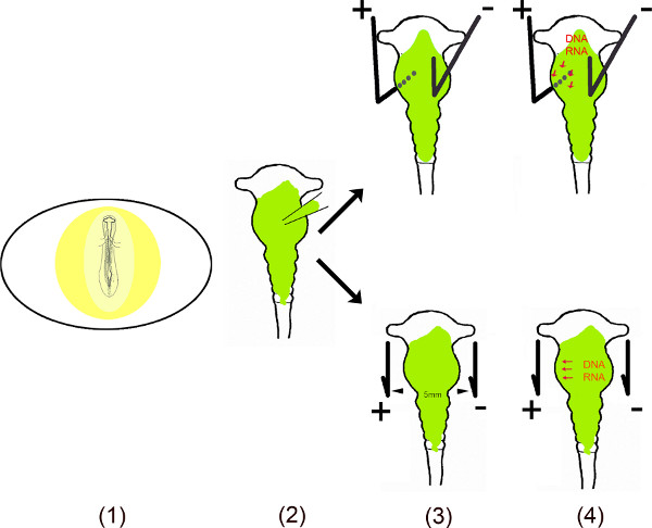
原理概述。
- 在鸡蛋胚胎是通过切割一个小窗口,进入次曝光Ë蛋壳。
- 溶解的载体(s)是注入到使用微毛细管中脑。
- 两个电极 – 平行放置,或根据与上面的胚胎 – 产生一个脉冲电场。
- 电场在时间上产生毛孔中的细胞膜,从而促进进入细胞由带负电荷的DNA(或RNA)被吸引到阳极11,12。
