Quantifying cell division and expansion is of crucial importance to the understanding of whole-plant growth. Here, we present a protocol to calculate cellular parameters determining maize leaf growth rates and highlight the use of these data for investigating molecular growth regulatory mechanisms by directing developmental stage-specific sampling strategies.
Growth analyses are often used in plant science to investigate contrasting genotypes and the effect of environmental conditions. The cellular aspect of these analyses is of crucial importance, because growth is driven by cell division and cell elongation. Kinematic analysis represents a methodology to quantify these two processes. Moreover, this technique is easy to use in non-specialized laboratories. Here, we present a protocol for performing a kinematic analysis in monocotyledonous maize (Zea mays) leaves. Two aspects are presented: (1) the quantification of cell division and expansion parameters, and (2) the determination of the location of the developmental zones. This could serve as a basis for sampling design and/or could be useful for data interpretation of biochemical and molecular measurements with high spatial resolution in the leaf growth zone. The growth zone of maize leaves is harvested during steady-state growth. Individual leaves are used for meristem length determination using a DAPI stain and cell-length profiles using DIC microscopy. The protocol is suited for emerged monocotyledonous leaves harvested during steady-state growth, with growth zones spanning at least several centimeters. To improve the understanding of plant growth regulation, data on growth and molecular studies must be combined. Therefore, an important advantage of kinematic analysis is the possibility to correlate changes at the molecular level to well-defined stages of cellular development. Furthermore, it allows for a more focused sampling of specified developmental stages, which is useful in case of limited budget or time.
Análise de crescimento depende de um conjunto de ferramentas que são comumente usados por cientistas de planta para descrever as diferenças de crescimento genótipo determinado e / ou respostas fenotípicas a fatores ambientais. Eles incluem tamanho e peso medições da planta inteira ou um órgão e cálculos de taxas de crescimento para explorar os mecanismos subjacentes de crescimento. Crescimento dos órgãos é determinada por divisão celular e de expansão ao nível celular. Portanto, incluindo a quantificação desses dois processos no crescimento análises é fundamental para compreender as diferenças no crescimento do órgão inteiro 1. Por conseguinte, é fundamental ter uma metodologia adequada para determinar os parâmetros de crescimento celular que é relativamente fácil de usar por laboratórios não especializados.
A análise cinemática já foi estabelecida como uma abordagem fornecendo uma estrutura poderosa para o desenvolvimento de modelos de crescimento de órgãos 2. A técnica foi optimizado para sistemas lineares,tais como raízes e folhas de Arabidopsis thaliana monocotiledóneas, mas também para os sistemas não lineares, tais como folhas dicotiledóneas 3. Hoje em dia, esta metodologia está cada vez mais sendo usado para estudar como genética, hormonal, de desenvolvimento e fatores ambientais influenciam a divisão celular e expansão em vários órgãos (Tabela 1). Além disso, também fornece uma estrutura para conectar processos celulares aos seus regulamentos bioquímicas, moleculares e fisiológicos subjacentes (Tabela 2), embora as limitações podem ser impostas por tamanho do órgão e da organização espacial para técnicas que exigem maiores quantidades de material vegetal (por exemplo, metabólitos medições, proteômica, etc.).
Folhas de monocotiledóneas, como o milho (Zea mays) da folha, representam sistemas lineares em que as células se movem a partir da base da folha em direcção a ponta, passando sequencialmente através da zona de meristema e alongamento para atingir a maturidadezona. Isto o torna um sistema modelo ideal para estudos quantitativos dos padrões espaciais de crescimento de 4. Além disso, folhas de milho têm zonas de grande crescimento (meristema e zona de alongamento abrangendo vários centímetros 5) e proporcionar possibilidades para estudos em outros níveis organizacionais. Isto permite a investigação dos mecanismos de regulação (putativos) que controlam a divisão celular e expansão, quantificada por análise cinemática através de uma série de técnicas moleculares, medições fisiológicas e abordagens de biologia celular (Tabela 2).
Aqui, nós fornecemos um protocolo para a realização de uma análise cinemática em folhas de monocotiledôneas. Em primeiro lugar, vamos explicar como conduzir uma análise adequada de ambos divisão celular e alongamento celular como uma função da posição ao longo do eixo da folha e como calcular parâmetros cinemáticos. Em segundo lugar, também mostrar como este pode ser usado como uma base para a concepção de amostragem. Aqui, discutimos dois casos: de alta resolução de uma amostragemd focado amostragem, permitindo uma melhor interpretação dos dados e economia de tempo / dinheiro, respectivamente.
Tabela 1. Visão Geral de cinemática analisa métodos de quantificação da divisão celular e expansão em vários órgãos.
órgão | referência |
folhas monocotiledôneas | 16, 20, 21, 22 |
dicas de raiz | 2, 23, 24, 25, 26, 27, 28, 29 |
folhas de dicotiledôneas | 21, 30, 31 |
atirar meristema apical | 32 |
Tabela 1. Visão Geral de cinemática analisa métodos de quantificação da divisão celular e expansão em vários órgãos.
<p class="jove_content" fo:keep-together.within-page = "1">
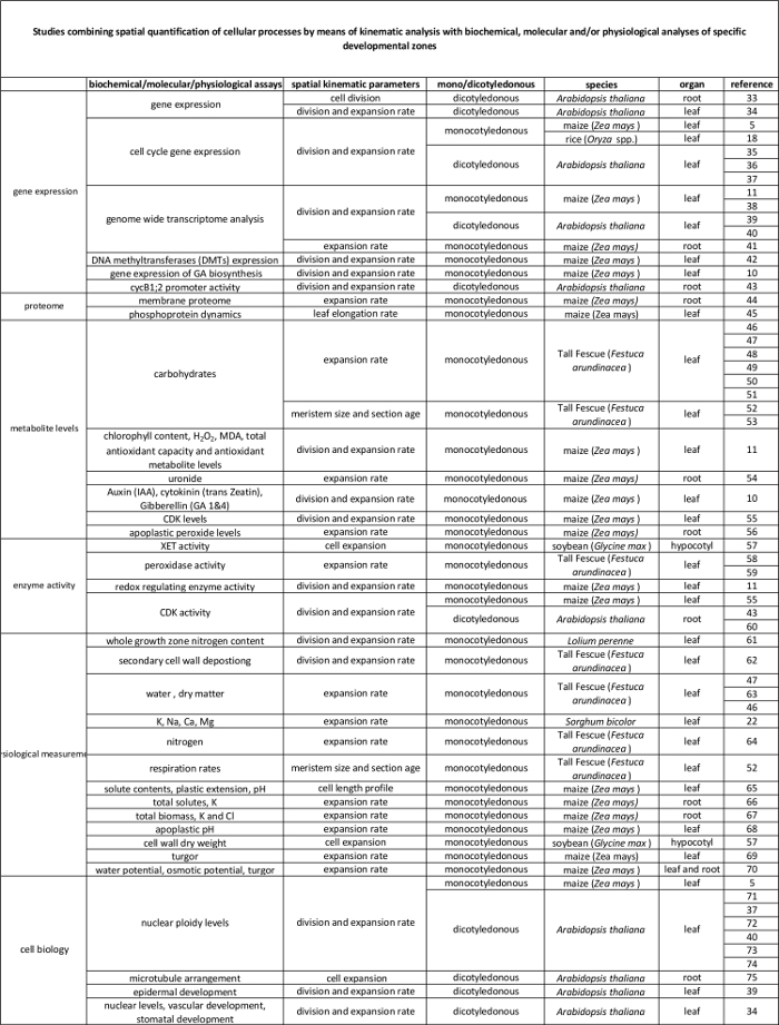
Tabela 2. A ligação entre os processos celulares quantificados pela análise cinemática a sua regulação ao nível molecular. As referências a diversos estudos de ligação a quantificação de processos celulares com os resultados de ensaios bioquímicos e moleculares em várias espécies e órgãos. Endotransglucosylase xiloglucano (XET), malondialdeído (MDA), quinases dependentes da ciclina (CDK). Por favor clique aqui para ver uma versão maior desta tabela.
Uma análise cinemática em folhas de milho integral permite a determinação da base celular de crescimento da folha e permite a concepção de estratégias de amostragem eficientes. Embora o protocolo é relativamente simples, alguma cautela é recomendado nas seguintes passos críticos: (1) É importante destacar as, folhas mais jovens fechados (passo 2.3), sem danificar o meristema, uma vez que a determinação comprimento meristema (passo 3) requer a completa meristema de estar presente. Alguma prática de antemão…
The authors have nothing to disclose.
Este trabalho foi apoiado por uma bolsa de doutoramento da Universidade de Antuérpia para VA; uma bolsa de doutoramento do Flamengo Science Foundation (FWO, 11ZI916N) para KS; subvenções de projectos do FWO (G0D0514N); uma bolsa de investigação actividades de investigação concertada (GOA), "A Biologia de Sistemas Abordagem de Folha Morfogênese" do conselho da Universidade de Antuérpia pesquisa; eo Interuniversitário de atração poloneses (IUAP VII / 29, MARS), "Milho e Arabidopsis raízes e parte aérea" do Escritório Belga Federal de Ciência Política (BELSPO) para GTSB Han Asard, Bulelani L. Sizani e Hamada AbdElgawad contribuíram para o vídeo .
Pots | Any | Any | We use pots with the following measueres, but can be different depending on the treatment/study : bottom diameter: 11cm, opening diameter: 15 cm, height: 12 cm. We grow one maize plant per pot. |
Planting substrate | Any | Any | We use potting medium (Jiffy, The Netherlands), but other substrates can be used, depending on treatment/study. |
Ruler | Any | Any | An extension ruler that covers at least 1,5 meters is needed to measure the final leaf length of the plants. |
Seeds | Any | NA | Seeds can be ordered from a breeder. |
Scalpel | Any | Any | The scalpel is used during leaf harvesting to detach the leaf of interest from its surrounding leaves and right after harvesting to cut a proper sample for cell length and meristem length measurements. |
15 ml falcon tubes | Any | Any | The 15 ml falcon tubes are used for storing samples used for cell length measurements during sample clearing with absolute ethanol and lactic acid. |
Eppendorf tubes | Any | Any | The eppendorf tubes are used for storing samples used for meristem length measurements in ethanol:acetic acid 3:1 (v:v) solution. |
Gloves | Any | Any | Latex gloves, which protect against corrosive reagents. |
Acetic acid | Any | Any | CAUTION: Corrosive to metals, category 1 Skin corrosion, categories 1A,1B,1C Serious eye damage, category 1; Flammable liquids, categories 1,2,3 |
Absolute ethanol | Any | Any | CAUTION: Hazardous in case of skin contact (irritant), of eye contact (irritant), of inhalation. Slightly hazardous in case of skin contact (permeator), of ingestion |
Lactic acid >98% | Any | Any | CAUTION: Corrosive to metals, category 1 Skin corrosion, categories 1A,1B,1C Serious eye damage, category 1 |
Sodium chloride (NaCl) | Any | Any | |
Ethylenediaminetetraacetic acid (EDTA) | Any | Any | CAUTION: Acute toxicity (oral, dermal, inhalation), category 4 Skin irritation, category 2 Eye irritation, category 2 Skin sensitisation, category 1 Specific Target Organ Toxicity – Single exposure, category 3 |
Tris(hydroxymethyl)aminomethane hydrochloride (Tris-HCl) | Any | Any | This material can be an irritant, contact with eyes and skin should be avoided. Inhalation of dust may be irritating to the respiratory tract. |
4′,6-Diamidine-2′-phenylindole dihydrochloride (DAPI) | Any | Any | Cell permeable fluorescent minor groove-binding probe for DNA. Causes skin irritation. May cause an allergic skin reaction. May cause respiratory irritation. |
Ice | Any | NA | The DAPI solution has to be kept on ice. |
Fluorescent microscope | AxioScope A1, Axiocam ICm1 from Zeiss or other | Any fluorescent microscope can be used for determining meristem length. | |
Microscopic slide | Any | Any | |
Cover glass | Any | Any | |
Tweezers | Any | Any | Tweezers are needed for unfolding the rolled maize leaf right after harvesting in order to cut a proper sample for cell length and meristem length measurements. |
Image-analysis software | Axiovision (Release 4.8) from Zeiss | NA | The software can be downloaded at: http://www.zeiss.com/microscopy/en_de/downloads/axiovision.html. Other softwares such as ImageJ (https://imagej.nih.gov/ij/) could be used as well. |
Microscope equipped with DIC | AxioScope A1, Axiocam ICm1 from Zeiss or other | Any microscope, equipped with differential interference contrast (DIC) can be used to measure cell lengths. | |
R statistical analysis software | R Foundation for Statistical Computing | NA | Open source; Could be downloaded at https://www.r-project.org/ |
R script | NA | NA | We use the kernel smoothing function locpoly of the Kern Smooth package (Wand MP, Jones MC. Kernel Smoothing: Chapman & Hall/CRC (1995)). The script is available for Mac and Windows upon inquire with the corresponding author. We have versions for Mac and Windows. |