1. A Matrigel-Based Tube Formation Assay to Assess the Vasculogenic Activity of Tumor Cells
- Preparation of tumor cells and microvascular endothelial cells: brain tumor cells such as U87 cells, melanoma cells B16F1, breast cancer MDA-MB-435, and colon cells HCT116 were grown in DMEM supplemented with 10% FBS and penicillin/streptomycin (all from Invitrogen). Endothelial cell line human microvascular endothelial cells (HMVECs) were cultured in EBM2 medium (Lonza) supplemented with 1 μg/ml hydrocortisone and 1 ng/ml epidermal growth factor, 10% FBS and penicillin/streptomycin. Cells were washed with PBS three times and detached with 0.05% trypsin/EDTA (Invitrogen). After centrifugation with 2,000 rpm for 5 min, cell pellets were washed again with PBS. Then, the pelleted cells were counted with a hemocytometer.
- Matrigel preparation: An aliquot of growth factor-reduced Matrigel (BD Bioscience) was warmed up at room temperature. Before completely thawed, it was transferred onto ice and its liquid was kept on ice for at least 10 min. Then 50 μl of Matrigel was plated to 96-well plates at a horizontal level that allows the Matrigel to distribute evenly, and incubated for 30 min at 37°C.
- Tube formation: Cells (1-2 x 10⁴) were re-suspended with serum-free DMEM for tumor cells or EBM-2 for endothelial cells, and loaded on the top of the Matrigel. If this assay was used for measuring effects of some agents on tubule development, these agents (stimulators or inhibitors) were added to the serum-free medium. Each conditional group contained 4-6 wells.
- Image and data analysis: Following incubation at 37°C overnight, each well was analyzed directly under a microscope. If tubules were needed for fixation, 100 μl of 10% formalin saline-based solution was gently added and ten min later, it was ready for analysis. Under a microscope with 10x phase contrast, tubules in each field were imaged and an average of tubules from 3-5 random fields in each well was counted.
2. Representative Results:
HMVECs were used as a positive control, as these tubules were developed from clear elongated cell bodies that connect to form polygon network. B16F1, U87, and MDA-MB-435 cells developed vascular tubules similar to those formed by HMVECs, but HCT116 cells did not (Figure 1). A cell dose-dependent tube formation was tested in U87 cells. As demonstrated in Figure 2, 10,000 cells formed discontinued tubules. Once the cells were doubled, a solid vascular network was comparable to those seen in HMVECs. In contrast, cells less than 5,000 failed to form a vascular phenotype.
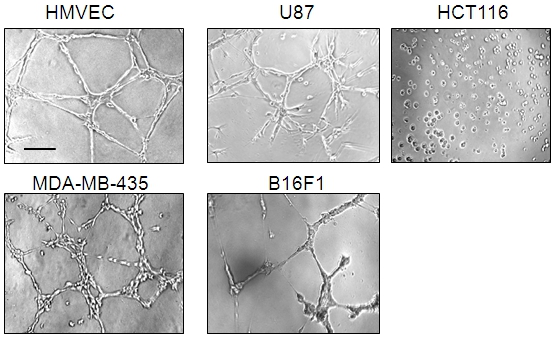
Figure 1. Tube formation induced by HMVECs, U87, MDA-MB-435, and B16F1 cells,
but not HCT116 cells. All the cells (2 x 10⁴) were loaded on Matrigel and incubated
overnight. Tubules were imaged using phase contrast. HMVECs were used as a positive
control. A representative of 3-5 fields was shown. Bar: 100 μm.
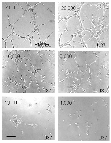
Figure 2. U87 cell-induced tubules in a cell number-dependent manner. Different
numbers of U87 cells as indicated in the corners were used for tube formation. HMVECs
were used for a positive control. Bar: 100 mm.
