We present a protocol on how to utilize high-throughput cryo-electron tomography to determine high resolution in situ structures of molecular machines. The protocol permits large amounts of data to be processed, avoids common bottlenecks and reduces resource downtime, allowing the user to focus on important biological questions.
Cryo-electron tomography (Cryo-ET) is a powerful three-dimensional (3-D) imaging technique for visualizing macromolecular complexes in their native context at a molecular level. The technique involves initially preserving the sample in its native state by rapidly freezing the specimen in vitreous ice, then collecting a series of micrographs from different angles at high magnification, and finally computationally reconstructing a 3-D density map. The frozen-hydrated specimen is extremely sensitive to the electron beam and so micrographs are collected at very low electron doses to limit the radiation damage. As a result, the raw cryo-tomogram has a very low signal to noise ratio characterized by an intrinsically noisy image. To better visualize subjects of interest, conventional imaging analysis and sub-tomogram averaging in which sub-tomograms of the subject are extracted from the initial tomogram and aligned and averaged are utilized to improve both contrast and resolution. Large datasets of tilt-series are essential to understanding and resolving the complexes at different states, conditions, or mutations as well as obtaining a large enough collection of sub-tomograms for averaging and classification. Collecting and processing this data can be a major obstacle preventing further analysis. Here we describe a high-throughput cryo-ET protocol based on a computer-controlled 300kV cryo-electron microscope, a direct detection device (DDD) camera and a highly effective, semi-automated image-processing pipeline software wrapper library tomoauto developed in-house. This protocol has been effectively utilized to visualize the intact type III secretion system (T3SS) in Shigella flexneri minicells. It can be applicable to any project suitable for cryo-ET.
Type III secretion systems (T3SS) are essential virulence determinants for many Gram-negative pathogens. The injectisome, also known as the needle complex, is the central T3SS machine required for direct translocation of effector proteins from the bacterium into eukaryotic host cells1, 2. The injectisome comprises an extracellular needle, a basal body, and a cytoplasmic complex also known as the sorting complex3. Previous studies have elucidated 3-D structures of purified injectisomes from Salmonella and Shigella, along with the atomic structures of major basal body proteins4, 5. Recent in situ structures of injectisomes from Salmonella, Shigella, and Yersinia were revealed by cryo-ET6, 7. However, the cytoplasmic complex, essential for effector selection and needle assembly, has not been visualized in those structures.
Cryo-ET is the most suitable technique for imaging molecular machinery at nanometer resolution within its native cellular context (in situ). Nevertheless, the achievable resolution by cryo-ET is limited by specimen thickness. To overcome the drawback, we imaged intact injectisomes in a virulent Shigella flexneri strain that was genetically modified to produce minicells thin enough for cryo-ET. Another limitation of cryo-ET is the sensitivity of the sample to the radiation induced by the electron beam, which very quickly destroys the high-resolution information in the sample. As a result, extremely low doses are used for individual tilt-images so that a suitable dose can be distributed amongst the full tilt-series. This greatly lowers the signal-to-noise ratio (SNR) in the final reconstruction, which makes it difficult to differentiate the structural features of the subject from the large amount of noise in the tomogram and limits the resolution that can be achieved by cryo-ET. Conventional image processing such as Fourier and real-space filters as well as down sampling can be used to increase contrast, but at the expense of filtering out much of the high-resolution information. Recently, sub-tomogram averaging has made it possible to greatly increase the SNR and subsequently the final resolution in some cases to sub-nanometer levels8, 9. A more detailed analysis of complexes is made possible by computationally extracting thousands of sub-tomograms containing the areas of interest from the original tomograms and then aligning and averaging the sub-tomograms to determine in situ complex structures with higher SNR and higher resolution. These methods can be integrated with genetic approaches to provide even greater insights into macromolecular assemblies and their dynamic conformations in the native cellular context.
In general, tens or even hundreds of thousand sub-tomograms need to be averaged in order to determine high-resolution structures in situ. The acquisition of a sufficient number of tilt-series needed to produce this large number of sub-tomograms quickly becomes a bottleneck. The resulting tilt-series are often affected by beam-induced shift, stage backlash, as well as magnification, rotation and skew defects, which must be solved to bring the tilt-series into alignment prior to reconstruction. The tilt series is typically aligned by tracking gold fiducial markers, which are traditionally selected manually through inspection of the tilt-series, causing yet another bottleneck. Many software packages have been developed for automated tilt-series acquisition through computer-controlled electron microscopes10, 11, 12, tilt-series alignment and reconstruction13, 14 and sub-tomogram averaging15-18. As these packages handle discrete operations in the workflow of cryo-ET, it becomes desirable to build a higher level of abstraction into the process to systematically streamline the entire scheme into a single pipeline. Therefore, we developed a software wrapper library "tomoauto" designed to organize a number of these packages into a single semi-automated unit, allowing for simple user operation while maintaining full configuration of each component in a centralized manner. The library is open-source, well documented, continually developed and freely available for use, tailored development or further integration by means of an online remote source code repository (http://github.com/DustinMorado/tomoauto).
This high-throughput cryo-ET pipeline has been utilized to visualize intact injectisomes in S. flexneri minicells. A total of 1,917 tomograms were generated using this method, revealing a high-resolution in situ structure of the intact machine including the cytoplasmic sorting platform determined by sub-tomogram averaging19. Together with molecular modeling of wild-type and mutant machines, our high-throughput pipeline provides a new avenue to understand the structure and function of the intact injectisome in the native cellular context.
1. Minicell Preparation
2. EM Grid Preparation
3. High-throughput Automated Tilt-series Collection
4. High-throughput Automated Tilt-series Processing and Reconstruction Using Tomoauto
5. Sub-tomogram Averaging
NOTE: We use the i3 package15 (http://www.electrontomography.org/) to process sub-tomogram averaging experiments, however the protocol described applies generally to most available sub-tomogram averaging software packages16to process sub-tomogram averaging experiments, however the protocol described applies generally to most available sub-tomogram averaging software packages16-18.
Samples of minicells S. flexneri were collected and processed as showed in the schematic Figure 1 using tomoauto following the pipeline detailed in Figure 2. Tilt-series were collected using SerialEM10, which allows for high-throughput tilt-series acquisition at points designated by the user on low-magnification montage maps (Figure 3). Micrographs were collected using dose-fractionation mode on a direct-detection device camera to reduce beam-induced motion22 (Figure 4). Tomoauto coordinates motion correction taking a collection of dose-fractionated micrographs processing each with MOTIONCORR22 and assembles the results into a tilt-series to be further processed (Movie 1).
The most general application of tomoauto is the automatic alignment of an initial tilt-series. Tomoauto composes sequential execution of the necessary commands in IMOD13 to coarsely align the tilt-series and generate an initial fiducial model tracking the colloidal gold particles in the sample, which are in turn used to generate the final alignment. The accuracy of this fiducial model is essential to the quality of the reconstructed tomogram, and so the user is able to visually inspect the automatically calculated fiducial model before proceeding with reconstruction or afterwards to identify tilt-series that should be processed manually. Figure 5 shows two tilt-series coarsely-aligned and the determined fiducial model as generated by tomoauto. Figures 5A, C show the untilted images and in both the fiducial model is correct with model points centered on fiducial markers. Figures 5B, D show the corresponding tilt-series at 50 degrees and while the model in Figure 5B is still tracking the gold particles correctly, several model points (red) in Figure 5D have strayed from their corresponding gold markers and the model is not suitable for fine alignment. This error can be measured quantitatively as the mean residual error between the center of the model point and the likely center of the gold marker, and tomoauto can be configured to alert the user when the measured error exceeds a user-defined threshold to expedite inspection. Tilt-series that are insufficiently aligned automatically can then be aligned manually. We find that tomoauto successfully aligns around 80%-90% of our collected tilt-series (Movie 2).
After a tilt-series has been successfully aligned, it must be reconstructed into the final tomogram. Tomoauto has been designed so that the user may use IMOD13 or tomo3d26 to generate the final reconstruction. We currently use tomo3d to take advantage of several features in modern multi-core computer processing units (CPUs) to greatly reduce the reconstruction time. The final tomogram as shown in Figure 6 and Movie 3 is a 3-D volume of the imaged sample that can then be used for cellular annotation by segmentation, or sub-tomogram averaging to obtain higher resolution information of the molecular machinery within the sample. Sub-tomogram averaging increases both the SNR and decreases the artifacts produced by the missing-wedge by averaging out the high-levels of noise in individual tomograms and utilizing the large number sub-tomograms in a well distributed set of random orientations with respect to the missing wedge to limit artifacts and improve the final resolution. The 2.7nm sub-tomogram average of the intact S. flexneri T3SS is shown in Figure 7 as deposited in the EMDB (EMD-2667), which shows the large improvement capable with this technique compared to the injectisome displayed in a single tomogram in Figure 6B.
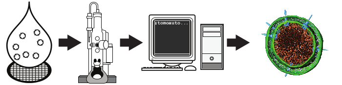
Figure 1. Schematic overview of high-throughput cryo-electron tomography. A liquid suspension is rapidly frozen on an EM grid and a set of tilt-series is collected by an automated computer-controlled electron microscope. The resulting micrographs are processed automatically using tomoauto to generate the tomogram. The final step here is a segmented S. flexneri minicell from a tomogram generated by this protocol from Hu et al. 201515. Please click here to view a larger version of this figure.
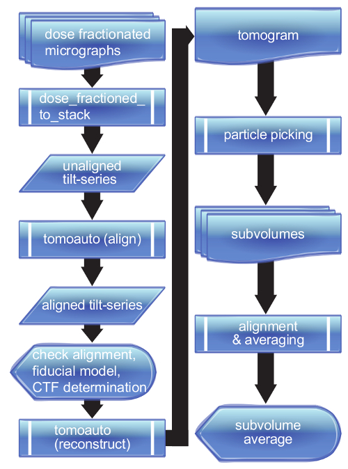
Figure 2. Flowchart of tomoauto process. A breakdown of the tomoauto workflow shows how data is processed from a collection of dose-fractionated micrographs all the way to a final sub-tomogram average. Sub-process symbols detail the tasks that tomoauto coordinates to process input by running the configured appropriate software. Data symbols show output generally not used by the user, while document and multi-document symbols show the output actually handled by the user. Finally display symbols show where user intervention occurs in the workflow. Please click here to view a larger version of this figure.
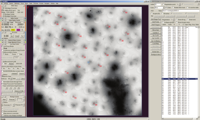
Figure 3. Batch tilt-series acquisition with SerialEM Navigator. Positions of montage maps are stored as stage positions (shown selection) in the Navigator window list shown on the left side of the screen, and the currently loaded map is displayed in the buffer window along with the selected points labeled numerically with a red cross added to the map for acquisition. Acquisition points are listed by label in the Navigator window and can be set to acquire using the "Tilt series" checkbox. Please click here to view a larger version of this figure.
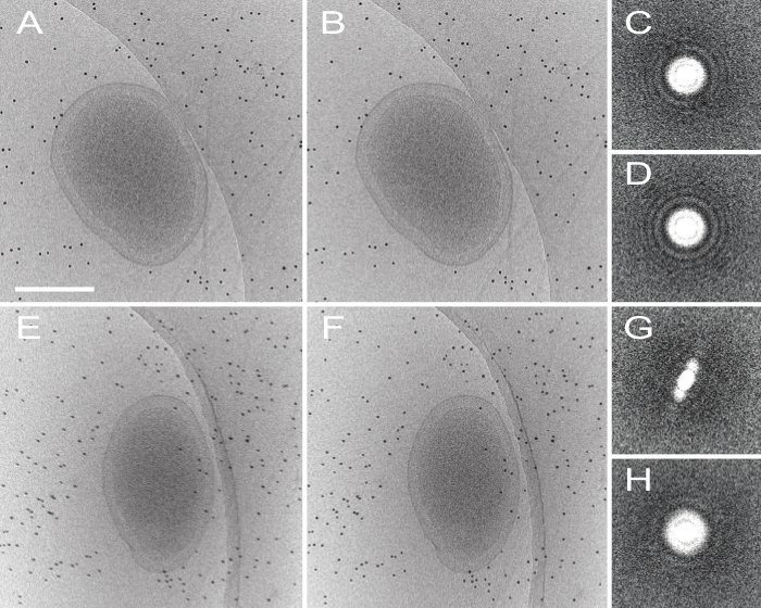
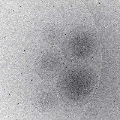
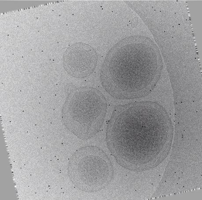
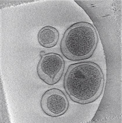
Figure 4. Effect of motion-correction on dose-fractionated data. (A) Shows an untilted and uncorrected micrograph, and the motion-corrected image (processed by MOTIONCORR) is shown in (B), the contrast is slightly improved after correction. Improvement can be seen more apparently by looking at the Fourier transform of the micrograph before (C) and after (D) motion correction. Images E-H show the same information but with a micrograph tilted at 60 degrees, where contrast is diminished and Thon rings visible in C and D are no longer visible at high tilt. Scale bar 250 nm. Please click here to view a larger version of this figure.
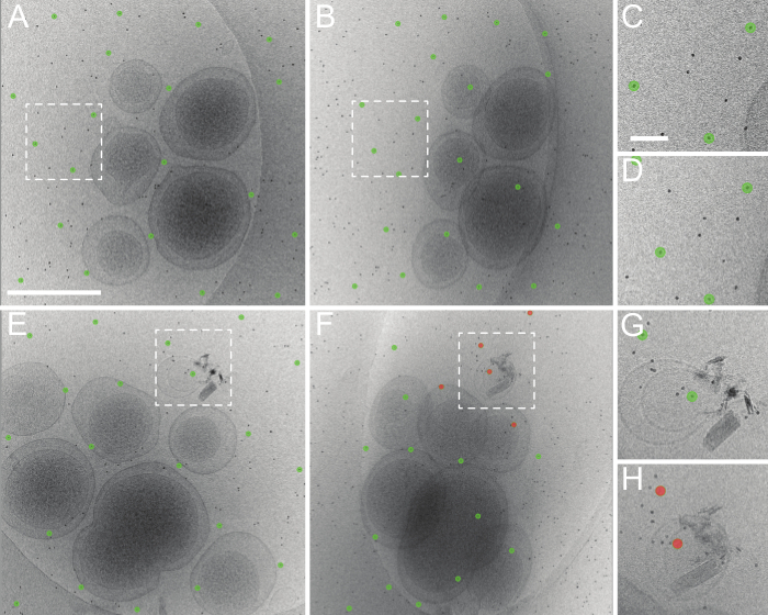
Figure 5. Good and bad results of tomoauto automated tilt-series alignment. (A) An untilted roughly aligned micrograph and the fiducial model produced automatically using tomoauto. (B) The determined fiducial model at 50 degrees tilt. The model still fits well and is centered on the appropriate fiducial markers. (C, D) Shows the model in (A, B) respectively, zoomed in at the boxed area. This tilt-series was aligned with a mean residual error of 1.06 pixels. (E) An untilted micrograph and fiducial model from another tilt-series and (F) the series at 50 degrees tilt. Here we see that model has lost track of several fiducial markers (shown in red) and this is representative of a bad automated tracking. (G, H) Shows the model in (E, F) respectively, zoomed in at the boxed area. This tilt-series was aligned with a mean residual error of 3.51 pixels and had to be processed by manual alignment of the series. (A) Scale bar 500 nm (C) Scale bar 50 nm. Please click here to view a larger version of this figure.
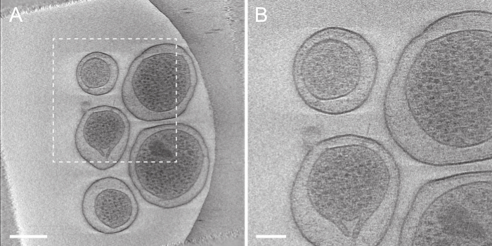
Figure 6. Tomogram generated automatically by tomoauto. (A) This displays a projection of seven slices from the center of the reconstruction of the tilt-series displayed in Figure 4A. Scale bar 250 nm. (B) A zoomed in view of the boxed area in (A) displaying an intact injectisome. Scale bar 100 nm. Please click here to view a larger version of this figure.
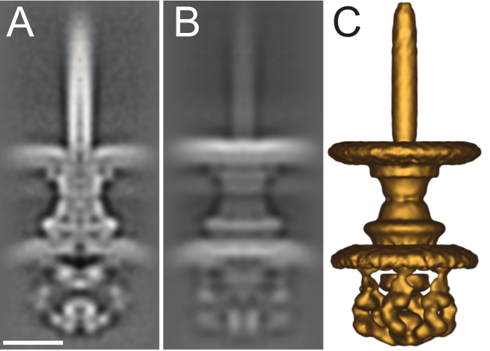
Figure 7. Sub-tomogram average of intact S. flexneri type III secretion system. (A) Central slice of the 2.7 nm sub-tomogram average of the intact S. flexneri T3SS from EMDB (EMD-2667). (B) Full projection along the X-axis of the volume. (C) Isosurface rendering of the volume viewed at a contour threshold of 130 in IMOD. Scale bar 5 nm. Please click here to view a larger version of this figure.
Movie 1: Animation of unaligned tilt-series (Right click to download). This animation runs through the tilt-series as initially collected by SerialEM. Translational shifts are easily identified by the erratic path of individual fiducial markers from image to image, and these shifts along with less noticeable defects must be corrected before the tilt-series can be reconstructed.
Movie 2: Animation of aligned tilt-series (Right click to download). This animation runs through the same micrographs displayed in Movie 1 after automated alignment by tomoauto. The erratic paths of fiducial markers now follow a smooth trajectory through the tilt-series, and the tilt-axis is aligned vertically with respect to the viewer.
Movie 3: Animation of reconstructed tilt-series (Right click to download). This animation runs through the tomogram shown in Figure 6 generated after automated reconstruction of the tilt-series displayed in Movie 2 by tomoauto.
The high-throughput method described here enabled us to process 1,917 cryo tilt-series and produce over 4,500 sub-tomograms of the intact S. flexneri injectisome19. The collected data led to the detailed characterization of in situ injectisome, including the cytoplasmic sorting complex. The method was also utilized to visualize several mutant cells with specific deletion of putative protein components, which helped elucidate the composition of the sorting platform of the injectisome. Our method provided new avenues to investigate the structure-function relationship of the injectisome. As a result, the new stage was set for further dissection of the mechanisms underlying T3SS-mediated secretion and pathogenesis.
The protocol presented here describes high-throughput cryo-ET of intact S. flexneri, but is applicable to any project suitable for cryo-ET. This method has been used in the structural characterization of the flagellar motor of Borrelia burgdorferi27, the infection of E. coli minicells by bacteriophage T728, and chemoreceptor arrays in E. coli29. By facilitating the collection of massive datasets, it is possible to screen multiple mutants as well as image a large number of conditions that permit inferences of dynamic processes such as machine assembly27 and the progress of phage infection28. By collecting multiple software packages and allowing for full control over processing execution, users are able to customize different combinations of packages for optimal results. Chen et al.21 previously published a similar protocol describing collecting data using the software package Leginon12 and automating tilt-series processing using IMOD13 and RAPTOR24. The current protocol complements this method detailing an alternative method to collecting and processing data while also showing how much the technology and procedure has advanced, driven by the increased focus on high-resolution through sub-tomogram averaging, with dose-fractionated data, automated CTF estimation and correction, and more robust automated alignment routines within IMOD13. While the previous protocol goes into visual detail with emphasis on data collection, this method focuses on the details of processing the collected data.
High-throughput methods allow for massive data collection that maximize usage of microscope and computer resources, while limiting the amount of tedious user manual interventions that slow down the project and act as a major bottleneck. The wrapper library tomoauto has been designed to allow full configuration of all parameters used in each software package in a simple and centralized manner. Once a suitable configuration has been determined, it is then easy to apply the settings to whole dataset. A majority of the tilt-series can be processed with acceptable results (Figure 4A), while a minor subset is required to be processed manually. These tilt-series are usually less ideal acquisitions plagued by excessive image shift, poor contrast, or a lack of sufficient fiducial markers which causes the automated fiducial tracking routine to fail (Figure 4B). To obtain the best possible tomograms, extensive care must be taken at every crucial step from sample preparation, image acquisition, to image processing.
Further developments in high-throughput cryo-ET such as automated sub-tomogram extraction by template matching and the integration of modern sub-tomogram averaging software packages such as Dynamo into existing workflow pipelines like tomoauto are now being investigated. The recent advent of new-generation direct detection device cameras has made major improvements in increasing tilt-series SNR and enabling more consistent CTF determination due to the higher efficiency of the detector. The use of new full gold grid-types may decrease tilt-series collection defects, improving the success rate for automated tilt-series processing and reconstruction with less need for manual intervention30. Finally with the now ubiquitous use of computer clusters and graphic processing units (GPUs) to parallelize and speed up large dataset processing, development of pipelines that can utilize these systems will soon hopefully be able to shorten execution time from days to hours, providing users with still even less downtime in between experiment design and meaningful data analysis while still increasing dataset size and achieving higher resolutions.
The authors have nothing to disclose.
We thank Dr. William Margolin for comments. We are grateful for the support on SerialEM from Drs. David Mastronarde and Chen Xu. D.M., B.H. and J.L. were supported by Grant R01AI087946 from the National Institute of Allergy and Infectious Diseases, Grants R01GM110243 and R01GM107629 from the National Institute of General Medical Sciences (NIGMS), and Grant AU-1714 from the Welch Foundation. The direct electron detector was funded by National Institutes of Health Award S10OD016279.
Glycerol | Sigma-Aldrich | G9012 | |
Tyrptic Soy Broth | Sigma-Aldrich | 22092 | |
Spectinomycin | Sigma-Aldrich | S0692 | |
Electroporation Apparatus | Bio-rad | 165-2100 | |
1 mm Cuvette | BTX | 45-0124 | |
1.5 mL Cryogenic Tube | Thermoscientific | 5000-1020 | |
1.5 mL Microcentrifuge Tube | Sigma-Aldrich | Z336769 | |
Holey Carbon Grids | Quantifoil (Electron Microscopy Sciences) |
Q2100CR2 | R2/2 200 Cu |
Glow Discharge Device | In-House | Commercial Alternative Available | |
Vacuum Desiccator | Sigma-Aldrich | Z119016 | Used in In-House Glow Discharge Device |
High-Frequency Generator | Electro-Technic Products | BD-10A | Used in In-House Glow Discharge Device. CAUTION: This device generates high voltages. |
Centrifuge | |||
Forceps | Dumont (Electron Microscopy Sciences) |
72705-D | Style 5 Anti-magnetic |
Colliodal Gold | Aurion | BSA 10nm | |
Filter Paper | Whatman | #2 | |
Ethane | Matheson Tri-Gas | UN1035 | |
Nitrogen | Matheson Tri-Gas | UN1977 | |
Plunger Device | In-House | Commercial Alternative Available | |
Cryogenic Grid Storage Box | Electron Microscopy Sciences | 71166-30 | |
Transmission Electron Microscope | FEI | Tecnai Polara F30 (300 KeV) |
|
Direct Detection Device Camera | Gatan | K2 Summit | |
Tomogram Acquisiton Software | SerialEM | http://bio3d.colorado.eud/SerialEM Alternatives: UCSF Tomography, Leginon, FEI Batch Tomography | |
Beam-induced Motion Correction Software | MOTIONCORR | http://cryoem.ucsf.edu/software/driftcorr.html Requires >2GB Nvidia GPU | |
Tilt-Series Alignment Software | IMOD | http://bio3d.colorado.edu/IMOD Alternatives: XMIPP, Protomo | |
Automatic Fiducial Marker Modelling Software | IMOD | Alternatives: RAPTOR (Included in IMOD0 (Usable in tomoauto) |
|
CTF Determination Software | IMOD | Alternatives: CTFFIND http://grigoriefflab.janelia.org/ctf (Usable in tomoauto) |
|
Tilt-Series Reconstruction Software | tomo3d | https://sites.google.com/site/3demimageprocessing/tomo3d Alternatives: IMOD, XMIPP http://xmipp.cnb.csic.es , Protomo | |
Tilt-Series Automated Processing Software | tomoauto | https://github.com/DustinMorado/tomoauto | |
Particle Picking Software | i3 | http://www.electrontomography.org Alternatives: IMOD | |
Subvolume Averaging Software | i3 | Alternatives: PEET http://bio3d.colorado.edu/PEET, Dynamo https://dynamo.bioz.unibas.ch , PyTom http://pytom.org |