Here, we present a protocol describing a streamlined method for the efficient generation of plasmids expressing both the CRISPR enzyme and associated single guide RNA (sgRNAs). Co-transfection of mammalian cells with this sgRNA/CRISPR vector and a dual luciferase reporter vector that examines double-strand break repair allows evaluation of knockout efficiency.
Although highly efficient, modification of a genomic site by the CRISPR enzyme requires the generation of a sgRNA unique to the target site(s) beforehand. This work describes the key steps leading to the construction of efficient sgRNA vectors using a strategy that allows the efficient detection of the positive colonies by PCR prior to DNA sequencing. Since efficient genome editing using the CRISPR system requires a highly efficient sgRNA, a preselection of candidate sgRNA targets is necessary to save time and effort. A dual luciferase reporter system has been developed to evaluate knockout efficiency by examining double-strand break repair via single strand annealing. Here, we use this reporter system to pick up the preferred xCas9/sgRNA target from candidate sgRNA vectors for specific gene editing. The protocol outlined will provide a preferred sgRNA/CRISPR enzyme vector in 10 days (starting with appropriately designed oligonucleotides).
The CRISPR sgRNAs comprise a 20-nucleotide sequence (the protospacer), which is complementary to the genomic target sequence1,2. Although highly efficient, the ability of the CRISPR/Cas system to modify a given genomic site requires the generation of a vector carrying an efficient sgRNA unique to the target site(s)2. This paper describes the key steps in the generation of that sgRNA vector.
For successful genome editing using the CRISPR/Cas system, the use of highly efficient sgRNAs is a crucial prerequisite3,4,5. Since engineered nucleases used in genome editing manifest diverse efficiencies at different targeted loci1, a pre-selection of candidate sgRNA targets is necessary in order to save time and effort6,7,8,9. A dual luciferase reporter system has been developed to evaluate knockout efficiency by examining double-strand break repair via single strand annealing3,10. Here we use this reporter system to choose a preferred CRISPR sgRNA target from different candidate sgRNA vectors designed for specific gene editing. The protocol stated here has been implemented in our group and collaborating laboratories for the last few years to generate and evaluate CRISPR sgRNAs.
The following protocol sums up how to design suitable sgRNA through network software. Once the suitable sgRNAs are selected, we describe the different steps to obtain the required oligonucleotides as well as the approach for inserting the paired oligonucleotides into the pX330-xCas9 expression vector. We also present a method for assembling sgRNA-expressing and dual luciferase reporter vectors based on the ligation of these sequences into a predigested expression vector (steps 2-10, Figure 1A). Finally we describe how to analyze the the DNA cutting efficiency for each of the sgRNAs (steps 11-12).
1. sgRNA oligonucleotide design
2. Oligonucleotide modification
3. Oligonucleotide annealing
4. sgRNA/CRISPR vector digestion
5. Ligation of the annealed sgRNA oligonucleotides to the expression vector
6. Competent cell transformation
7. Identification of the correct recombinant plasmids by PCR
8. Validate the sequence of sgRNA expression plasmid
9. Construction of dual luciferase reporter vector
10. Cell transfection
11. Dual-luciferase detection
The methods outlined in this protocol are for the construction of sgRNA and xCas9 expression vectors and then for the optimization screening of sgRNA oligos with relatively higher gene targeting efficiencies. Here we display a representative example of 3 sgRNA targets to sheep DKK2 exon 1. SgRNA and xCas9 expressing vectors can be built by predigesting the vector backbone (Figure 2) followed by ligating it in a series of short double-strand DNA fragments through annealing oligo pairs. The positive colonies could be detected through specific primer pairs guided PCR (Figure 3). An 440 bp DNA fragment from sheep DKK2 exon 1 was subcloned into pSSA-Dual plasmid15 by using double digestion with AscI and SalI, resulting in pSSA-Dual-DKK2. The gene targeting capacities of pX330-xCas9-T1, pX330-xCas9-T2 and pX330-xCas9-T3 were simutanusly detected (Figure 5), and then the last sgRNA vector was identified as the relatively better one, which we pick up for sheep gene editing research at the next step.
This detection method combines a single strand annealing (SSA) mechanism (Figure 1B and Figure 4) with a luciferase report gene in order to monitor DNA cutting efficiency. As illustrated in Figure 4, SSA is a process initiated when a double strand break is made between two repeated sequences oriented in the same direction. Single strand regions are created adjacent to the breaks that extend to the repeated sequences so the complementary strands can anneal to each other. This annealing intermediate can be processed by digesting away the single stranded tails and filling in the gaps. The Dual-Luciferase Reporter gene mainly includes the luciferase genes from the firefly Photinus pyralis and from Renilla reniformis (also known as sea pansy). The activities of firefly and renilla luciferases are measured sequentially from a single sample. When the recognition area that has the termination codon is not cut in its middle, the gene in SSA system is blocked and cannot be translated into a functional protein. When the processing takes place, the SSA system will merge the homologous sequence automatically, and overlapping sequences becomes a single sequence, gene gets recombination repaired and can then be read throughout producing a functional protein.
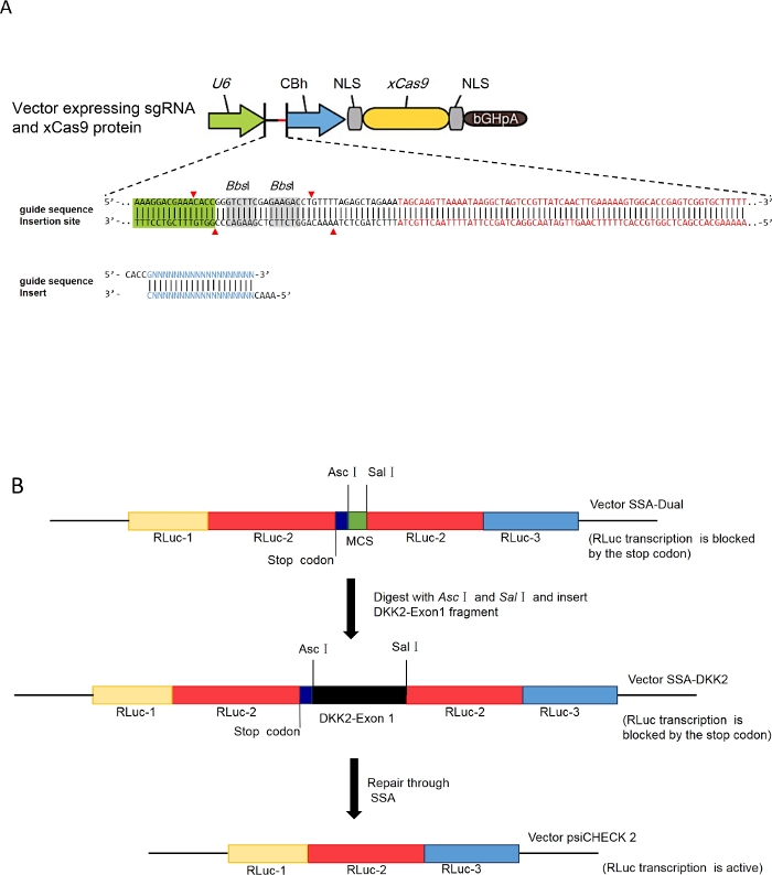
Figure 1: Schematic representation of the different steps of the cloning process.
(A) Schematic of sgRNA vector construction, adapted from the protocols of Feng Zhang Lab. (B) Schematic of dual luciferase reporter vector building (select vector pSSA-DKK2 as an example), DNA fragments Rluc-, Rluc-2 and Rluc-3 represent three parts of full-length coding sequence of Renilla luciferase. MCS represents “multiple cloning site”. Please click here to view a larger version of this figure.
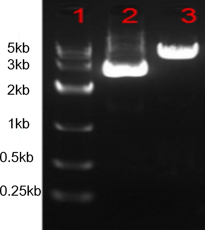
Figure 2: Predigesting the vector backbone pX330-xCas9 with BbsI.
1: DNA Marker, 2: pX330-xCas9 plasmid, 3: pX330-xCas9 plasmid digested with BbsI. Please click here to view a larger version of this figure.
Figure 3: Specific primer pairs guided PCR for DKK2-T1 sgRNA vector detection.
Lanes 1-7 indicate PCR bands for 7 different bacteria colonies separately. Please click here to view a larger version of this figure.
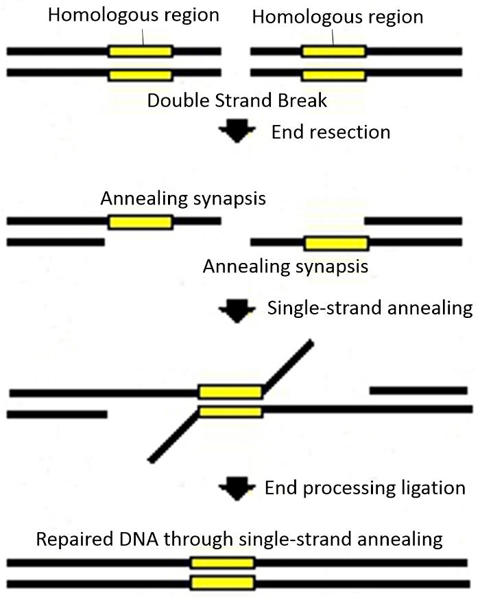
Figure 4: Schematics for single strand annealing (SSA). Please click here to view a larger version of this figure.
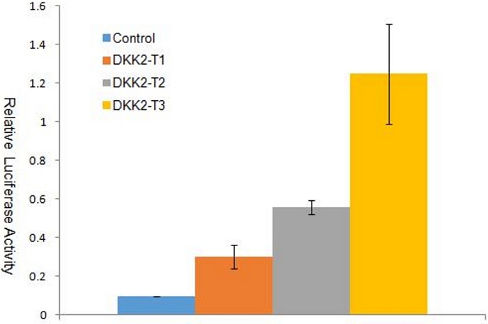
Figure 5: Dual luciferase assay with reporter vector pSSA-DKK2 in cell line PIEC.
The Ranilla luciferase luciferase activities are significantly induced (P<0.01, Student's t test) in PIEC by overexpresing of pX330-xCas9-T1, pX330-xCas9-T2 or pX330-xCas9-T3 with the fold change of 3.15, 5.84 or 13.10, respectively. The error bars indicate standard deviations (SD)(n=3) for each group. Please click here to view a larger version of this figure.
Name | Genomic DNA Targets (5’-3’) | sgRNA Oligos (5’-3’) | ||
T1 | TGCCTGCTCCTACTGGCCGCGG | T1-F: CACCGTGCCTGCTCCTACTGGCCGC | ||
T1-R: AAACGCGGCCAGTAGGAGCAGGCAC | ||||
T2 | ATCAAGTCCTCTCTGGGCGGGG | T2-F: CACCGATCAAGTCCTCTCTGGGCGG | ||
T2-R: AAACCCGCCCAGAGAGGACTTGATC | ||||
T3 | GCCCGCGAGCTGCCGAACTGTG | T3-F: CACCGCCCGCGAGCTGCCGAACTG | ||
T3-R: AAACCAGTTCGGCAGCTCGCGGGC |
Table 1: sgRNA targets and oligos designed for gene editing of sheep DKK2.
PCR Component | 25 µL reaction |
10 µM Forward Primer (e.g. T1-F) | 1 µL |
10 µM Reverse Primer BbsI-R | 1 µL |
10x PCR Buffer | 2.5 µL |
2.5 mM dNTPs | 1 µL |
bacterial fluid | 1 µL |
DNA Taq Polymerase | 2.5 units |
Nuclease-free water | Up to 25 µL |
Table 2: PCR mixture for positive bacterial colonies detection.
Reagent | Volume |
pSSA-Dual vector (synthesized DNA fragment or PCR product) | 1-2 μg |
CutSmart Buffer | 5 μL |
AscI | 1 μL |
SalI-HF | 1 μL |
Distilled water | up to 50 μL |
Total volume | 50 μL |
Table 3: Double digestion of pSSA-Dual vector and synthesized DNA fragment (or PCR product).
Reagent | Volume |
pSSA-Dual vector(predigested) | 0.5 μg |
DNA fragments containing the sgRNA targets (predigested) | 0.2 μg |
T4 DNA Ligase Buffer (10x) | 1 μL |
T4 DNA Ligase | 1 μL |
Distilled water | up to 10 μL |
Total volume | 10 μL |
Table 4: A ligation reaction of DNA fragments containing the sgRNA targets into a predigested pSSA-Dual vector.
Step 1: reagent preparation | |
Transfection reagent | 2 μL |
Table 5: Details of cell transfection in a 24 well plate.
The sgRNA vector cloning procedures we have described here facilitates efficient production of sgRNAs, with most of the costs derived from the oligonucleotide ordering and vector sequencing. While the outlined method is designed to allow users to generate sgRNAs for use with CRISPR/Cas9, the protocol can easily be adapted for use with Cas9 orthologues or other RNA-guided endonucleases such as Cpf1, introducing minor modifications to the vector backbone and the oligonucleotide overhanging sequences.
The protocol outlined above will provide a preferred sgRNA target in 10 days when starting with appropriately designed oligonucleotides. This includes the sgRNA design (1 h, steps 1 – 2), dilution, aliquot and annealing of the oligonucleotides (30 min, step 3), digestion and purification of the sgRNA expression vector (3 h, step 4), the cloning of the sgRNA oligonucleotides into a predigested empty vector (overnight, steps 5 – 6), the PCR detection of the colonies (4-5 h, step 7) and the validation of the sequence of the sgRNA expression plasmid (24 h, step 8). Meanwhile, the construction and sequence validation of dual luciferase reporter vector takes 5 – 7 d (step 9). The preparation of the cell line, plasmid transfection and dual luciferase detection take about 48 h (steps 10 – 11). The failure rates for generating each plasmid are nearly 0.
The most critical step of the protocol is the restrictive enzyme digestion of sgRNA vector by enzymes such as BbsI. If the digestion is not sufficient, then the positive rate of bactera colonies might be very low. While the PCR-based detection approach could sensitively monitor the digestion efficiency and the positive rate. The forward primers, formerly used as forward oligos of the inserted sgRNA fragment, ensured highly effective distinction of right insertions and empty vectors. One of the advantages of this strategy is precisely the efficient detection of the positive colonies by PCR before DNA sequencing. As stated above, sequence verification is still relatively more expensive compared to PCR, especially when incomplete digestion of BbsI occurs. In terms of resource saving, we paired the forward oligos for sgRNA fragment annealing (such as T1-F, T2-F or T3-F) with the sequencing primer BbsI-R for PCR amplification. More than 500 sgRNA vectors have been successfully constructed in the lab through this strategy for colony identification.
Another merit of the protocol is the effective pre-selection of candidate sgRNA targets using dual luciferase reporter system. Huge variance of cutting efficiency indeed exists at different sgRNA targets4. To identify a highly efficient sgRNA target by T7E1 assay or Surveyor nuclease assay1 in a difficult-to-transfect cell line is time and labor consuming. In addition, for PCR amplification in the T7E1 assay or Surveyor nuclease assay, it is sometimes difficult to specifically amplify the target sequence in the genome DNA because of the lack of suitable primer pairs1. In contrast, the dual luciferase reporter assay is independent of genome amplification procedures. Previously we have used this method to get highly active sgRNAs16,19.
Despite the clear advantages of the method described in this work, there are also some limitations that must be pointed out. Although it ensures a high rate of success, the luciferase-based method for sgRNA selection proposed could be more expensive than other approaches (surveyor/high-resolution melt analysis, etc.) due to the need to synthesize the target sequence. In addition, this approach is not faster than either of these alternative methods due to the need to clone the reporter plasmid if an easy-to-transfect cell line will be conducted for gene targeting. It is also necessary to clarify that the method is suitable for making knockouts but not to edit specific nucleotides. Additionally, only double-stranded breaks and not single-stranded breaks can be evaluated.
The authors have nothing to disclose.
This project was funded by First Class Grassland Science Discipline Program of Shandong Province (China), National Natural Science Foundation of China (31301936, 31572383), the Special Fund for Agro-scientific Research in the Public Interest (201403071), National risk assessment major special project of milk product quality and safety (GJFP201800804) and Projects of Qingdao People's Livelihood Science and Technology (19-6-1-68-nsh, 14-2-3-45-nsh, 13-1-3-88-nsh).
A new generation of full touch screen gradient PCR instrument | LongGene | A200 | Target gene amplification |
AscI restriction enzymes | New England Biolabs | R0558V | Cutting target vectors |
BbsI restriction enzyme | New England Biolabs | R0539S | Cutting target vectors |
Clean workbench | AIRTECH | SW-CJ-2FD/VS-1300L-U | A partial purification device in the form of a vertical laminar flow, which creates a local high clean air environment |
DH5α Competent Cells | TaKaRa | K613 | Plasmid vector transformation |
Dual-Luciferas Reporter Assay System | Promega | E1910 | Dual-luciferas reporter assay |
Electric thermostatic water bath | Sanfa Scientific Instruments | DK-S24 | Heating reagent by constant temperature in water bath |
Electrophoresis | Beijing Liuyi Biotechnology Co., Ltd. | DYY-6C | Control voltage, current, etc. |
Eppendorf Reference 2 | Eppendorf China Ltd. | Reference 2 | Accurately draw and transfer traces of liquid |
Gel imaging analyzer | Beijing Liuyi Biotechnology Co., Ltd. | WD-9413B | For the analysis of electrophoresis gel images |
GloMax 20/20 Luminometer | Promega | E5311 | Detect dual luciferase activity |
High speed refrigerated centrifuge | BMH | sigma 3K15 | Nucleic acid extraction and purification |
Intelligent biochemical incubator | Sanfa Scientific Instruments | SHP-160 | Provide a suitable temperature environment for the enzyme digestion experiment |
LB Broth Agar | Sangon Biotech | A507003-0250 | For the cultivation of E.coli |
Lipofectamine 3000 Transfection Reagent Kit | Thermo Fisher | L3000015 | DNA Transfection |
SalI restriction enzymes | New England Biolabs | R3138V | Cutting target vectors |
SanPrep Column DNA Gel Extraction Kit | Sangon Biotech | B518131-0050 | Recycling DNA fragments |
SanPrep Column Plasmid Mini-Preps Kit | Sangon Biotech | B518191-0100 | Extraction of plasmid DNA |
T4 DNA Ligase | New England Biolabs | M0202V | Link DNA fragment |
TaKaRa MiniBEST DNA Fragment Purification Kit Ver.4.0 | TaKaRa | 9761 | DNA purification |
Vertical pressure steam sterilizer | JIBIMED | LS-50LD | High temperature and autoclave to kill bacteria, fungi and other microorganisms in laboratory equipment |
Water bath thermostat | Changzhou Guoyu Instrument Manufacturing Co., Ltd. | SHZ-82 | Let the bacteria keep shaking, which is good for contact with air. |