1. Optical Calibration: Measuring Light Output
- To prepare the light delivery apparatus, connect a broadband light source (e.g., Xenon or tungsten lamp) to an appropriate power supply. Position a collimating lens in front of the light source to produce a collimated beam of light. Pass the light through a liquid heat filter to remove most of the heat from the light beam. Depending on the application, focus the collimated beam on to the entrance aperture of a liquid light guide, which provides for more flexible delivery of the light (e.g., into an incubator).
- At the other end of the liquid light guide, position a second collimating lens and then a holder for the interference and neutral density filters. Check that this arrangement produces an evenly illuminated spot of light of the desired waveband and intensity.
Note: The distance between the end of the light guide and the specimen plane may need to vary depending on the area that must be illuminated, but remember that as the distance from the end of the light guide increases, light intensity will decrease. In the present study, this distance is 14cm and produces a beam cross section that evenly illuminates a 3×3 well area on a 96-well plate. - Ensure that stray light from the lamp and associated optical components is unable to reach the specimen.
- Turn on the water cooler for the liquid heat filter and ensure there is exchange of water through the filter jacket. Turn on the lamp power source and wait for at least 5 min for the broadband light source to stabilize. Note: The use of the water cooler is required to prevent over-heating of the device.
- Select a narrowband interference filter (usually described by their center wavelength, peak transmittance and full width at half maximum (FWHM) bandwidth) to generate the desired waveband of illumination. Note: The narrowband interference filters used in the current study were 442 nm, 550 nm, 671 nm and 810 nm.
- Measure the light produced by the lamp at the plane where the specimen is to be positioned during treatment. Measure light using a calibrated irradiance probe (cosine collector) connected to a suitable spectroradiometer, using propriety software according to manufacturer’s instructions.
- Use neutral density filters to adjust the intensity of light until the desired output is obtained. Note: In the present study, the intensity of light passed by each interference filter is adjusted to give an equal quantal output at each of the four treatment wavebands. It is important to check the calibration regularly, as the lamp output is subject to change.
Note: Following the set up of the apparatus, the cells are ready to be illuminated. Figure 1 is a representation of the light delivery apparatus used in the current study.
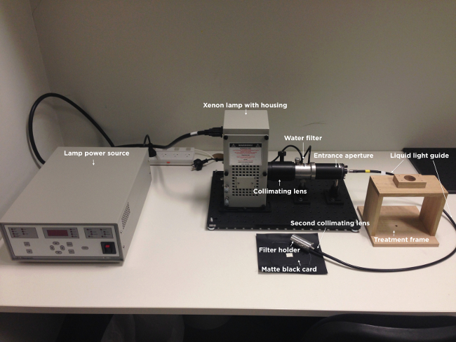
Figure 1. Image of the light delivery apparatus. Illustrated are the light power source, xenon lamp with housing, collimating lens, water filter, entrance aperture, liquid light guide, second collimating lens, filter holder, treatment frame and matte black card. Note that the narrowband wavelength and intensity filters are not shown.
2. Cell Preparation
- The described R/NIR-LT delivery method can be applied to any cell type or in vitro model system; as such the following descriptions of cell culture are general, using well-established techniques to culture pheochromocytoma (PC12), Müller (rMC1) and primary mixed retinal cells.
- Prior to cell seeding for light treatment and oxidative stress assay, culture immortalized cells in their respective growth media containing appropriate supplements (e.g., FBS, antibiotics) in T75 flasks until 70-80% confluent.
- Detach immortalised cells from T75 flasks, using the method appropriate for the cell type to be tested e.g., trypsin. If necessary, before proceeding with the seeding of cells in 96-well assay plates, coat assay plate wells with 10µg/ml poly-L-lysine for 1h (e.g., for PC12 cells) or 10µg/ml poly-L-lysine for 1h followed by an O/N incubation with 10µg/ml laminin (e.g., for mixed retinal cells).
- Centrifuge cells to collect as appropriate (e.g., 3 min at 405 x g for PC12 cells or 10 min at 218 x g for rMC1 cells), remove the supernatant and resuspend in 8 ml of appropriate cell culture media.
- If mixed retinal cells are to be used, prepare from P0-5 neonatal rat pups by enzymatic digestion (papain) according to established procedures 19
- Count the number of viable cells excluding 0.4% (w/v) trypan blue dye, using a haemocytometer.
- Adjust cell density such that cells will be approximately 70-80% confluent after 24 or 48h in culture. (for PC12 cells the seeding density is 4.0 x 10⁵ viable cells/ml, for rMC1 cells, it is 2.5 x 10⁵ viable cells/ml and for mixed retinal cells it is 8 x 10⁵ viable cells/ml). After adding cells to either clear or black 96-well plates (used for the H2O2 or DCFH-DA assay respectively), in 100 µl of appropriate growth media, allow plate to sit on a flat surface at 37°C, 5% CO2 (or whatever conditions are appropriate for the specific cell type) for at least 24 hr to allow cell adherence.
- Culture cells in growth media in the appropriate 96 well trays until they are 70-80% confluent (24h for PC12 and rMC1 cells, 48h for mixed retinal cells).
3. Adding Glutamate Stressor to Cells
- Prepare L-glutamic acid monosodium salt hydrate stressor concentrations of 0-10mM in appropriate full growth culture media.
- Remove media from the cells and gently wash cells 3 times with PBS (PC12) or HBSS (rMC1 and mixed retinal cells). After washes, add glutamate-containing media to a total volume of 100 µl/well.
4. First Dosages of Light Treatment
- Immediately upon addition of glutamate or the stressor of choice, expose cells to light treatment at the desired wavelengths and intensities by placing under the prepared light delivery apparatus. Be sure to alter the quantal output of the light beam using the established combinations of neutral density filters depending on the wavelength being administered.
Note: In the current experiments, expose cells to light treatment for 3 min maintain the temperature by resting the 96 well plates on a 37oC heat pad. Treatment time may be varied as required.- Place a matte black card underneath the cells to prevent light reflecting into adjacent wells in the 96-well tray designated for other treatment parameters.
- If treatment is desired for longer lengths of time, add 25mM Hepes buffer to maintain pH of the cell culture media or ideally, place the apparatus and treatment plates within an incubator with CO2 concentration regulated to 5% CO2, or conditions that are optimal for the specific cell type.
- Following completion of the light treatment, place the cells on a level surface and incubate at 37°C and 5% CO2 (or whatever conditions are optimal for the cell type) for 24h.
Note: Additional dosages of R/NIR-LT can be administered as described above, as desired.
5. Final Dosage of Light Treatment and Detection of ROS
- Before performing the final round of light treatment, prepare the reagents to be used for ROS detection. Prepare 50mM citrate buffer (pH6.0), Triton X100, and H2O2 detection reagent working solution (1:2:97 ratio of 10mM H2O2 detection reagent stock solution; 10 U/ml horseradish peroxidase; 50mM sodium citrate (pH 6.0)). Prepare DCFH-DA at a final concentration of 100µM in appropriate media.
Note: DCFH-DA reagent for PC12 cells is in RPMI media, DCFH-DA reagent for rMC1 cells is in DMEM media. Note that commercially available detection reagents have differential sensitivities to specific ROS and reagents should be chosen carefully to provide the information desired for an individual application. - Administer light treatment to the cells, as described in step 4.1-4.1.2. Ensure the cells are being treated with the desired fluences / wavelengths of light.
- Immediately following light treatment, remove the glutamate-containing media and wash twice with appropriate buffer solution (for PC12 cells, PBS is used and for rMC1 cells, HBSS is used). Perform the ROS assays on the cells as follows:
- H2O2 assay: add 45 µl of 50mM citrate buffer (pH 6.0) and 5 µl Triton X100 to each of the wells. Gently shake the cells on an orbital shaker for 30 sec and incubate at 37°C and 5% CO2 for 15 min. Add 50 µl of H2O2 detection working solution and incubate for 30 min at RT. Measure fluorescence using a plate reader with an excitation wavelength of 530nm and an emission wavelength of 480nm.
- DCF assay: add 100 µl of the 100 µM solution of DCFH-DA to each of the wells and incubate for 30min at 37°C and 5% CO2. Remove the media containing 100 µM DCFH-DA and wash with appropriate buffer solution (as described above) twice. Add 90 µl of buffer solution and 10 µl Triton X100 to each of the wells and gently shake on an orbital shaker for 30 sec before 15 min incubation at 37°C. Measure DCF derived fluorescence using a plate reader with an excitation wavelength of 480nm and an emission wavelength of 530nm.
- Express ROS values relative to the protein concentration of cells remaining in the wells, using a colorimetric kit to quantify protein concentration according to manufacturer’s instructions, with reference to a standard curve to calculate mg protein.
The output of light delivered at a wavelength of 670nm was calibrated using neutral density filters in order to irradiate cells with a range of fluences encompassing a dose of 670nm light previously shown to be beneficial in vivo (0.3 J/cm2) 20. As the number of neutral density filters in front of the light source increased, the intensity (W/m2) decreased, allowing less light to pass to the target area. Table 1 presents the calibration data of 670nm light generated from the light source fitted with a wavelength filter and includes the number of ND filters used and the intensity of light generated as a result, at described distances from the light output. Fluence, or dose of 670nm light (J/cm2), was calculated from the equation: [Dose (J/cm2) = (Light intensity (W/m2) / 10,000) x time (s)], where the time of treatment was 180s.
| Number of ND filters | Distance from light output (cm) | Intensity (W/m^2) | Dose (J/cm^2) |
| 0 | 10.5 | 20.11 | 0.38 |
| 0 | 14 | 10.55 | 0.19 |
| 1 | 14 | 4.91 | 0.075 |
| 2 | 14 | 2.28 | 0.041 |
| 3 | 14 | 1.03 | 0.018 |
| 4 | 14 | 0.47 | 0.0085 |
| 5 | 14 | 0.21 | 0.0038 |
| 6 | 14 | 0.094 | 0.00169 |
| 7 | 14 | 0.045 | 0.00081 |
| 8 | 14 | 0.021 | 0.000378 |
| 9 | 14 | 0.013 | 0.000234 |
Table 1: Output of the light delivery apparatus fitted with the 670nm wavelength filter.Number of ND filters refers to the number of neutral density filters fitted to the front of the light source output. Intensity (W/m2) refers to the intensity of the light as reported by the propriety software. Fluence, or dose was calculated by the equation[Dose (J/cm2) = (Light intensity (W/m2) / 10,000) x time (s)], where the time of exposure was 180s and distance from the light output was 10.5 or 14 cm.
Two clinically relevant quantal fluences of light were chosen to investigate differential effects of R/NIR-LT wavelength on production of ROS. A dose that can reach CNS tracts following transmission through overlying tissue using LED devices(i.e., 1.78 W/m2 at 670nm) 20 equated to 0.03 J/cm2 for a 3 min treatment, or 4.9 x 10¹⁴ photons/cm2/s. The light source equipped with filters to result in emission of 442, 550, 670 or 830nm was then calibrated using combinations of neutral density filters to emit equal quantal outputs (photons) for each wavelength as opposed to energy outputs (J/cm2), and the dosages (J/cm2) and intensities in W/m2 calculated (Table 2a). An additional higher dose that was within the recommended guidelines to stimulate cellular activity 21 was also used (1.29 x 10¹⁵ photons/cm2/s), and calibration conducted for each wavelength (Table 2b).
| Wavelength (λ) | Dose (J/cm^2) | Intensity (W/m^2) | Emission (Photons/cm^2/s) |
| 442 | 0.057 | 3.21 | 4.8 x 10^14 |
| 550 | 0.051 | 2.87 | 5.0 x 10^14 |
| 670 | 0.032 | 1.78 | 4.9 x 10^14 |
| 830 | 0.018 | 1.01 | 4.9 x 10^14 |
Table 2: Calibration of intensity of dosage delivered by equal numbers of photons of light at varying wavelengths. Intensity (W/m2), dosage for a 3 min treatment (J/cm2) and emission (photons/cm2/s) when output of xenon light emitting 442, 550, 670 and 830nm are calibrated to emit A) 4.9 x 10¹⁴ photons/cm2/s or B) 1.3 x 10¹⁵ photons/cm2/s at a distance of 14 cm from the light output.
In order to assess whether the light delivered by the apparatus was toxic to cells at the dosages used, we assessed the protein content remaining in PC12 cell culture wells following the ROS assays, using a colorimetric protein assay. There was no significant loss of protein at any of the higher output dosages of the light (P > 0.05), indicating that the dosage of light delivered was not causing cell death (Figure 2) and is appropriate to use for assessments of oxidative metabolism.
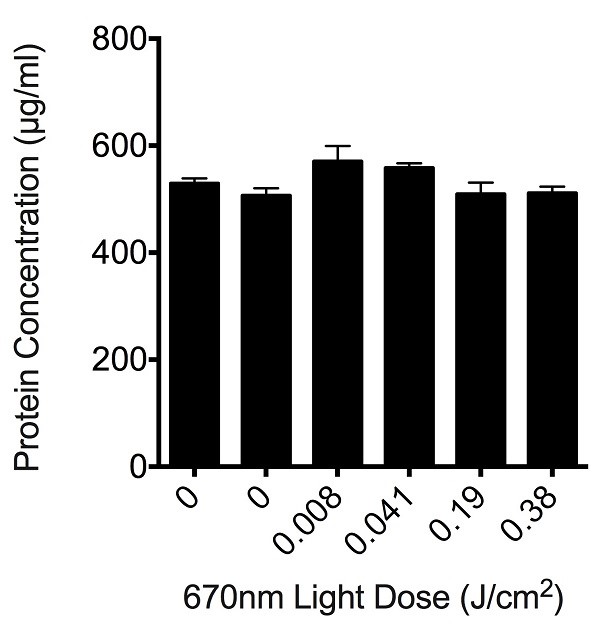
Figure 2: Effect of varying doses of light on the total protein concentration remaining in culture wells following R/NIR-LT and ROS assay. Histogram bars are the mean ± S.E.M protein concentrations in PC12 cell culture wells, 6 replicates / concentration, experiments were repeated 3 times. There were no statistically significant differences between control and any of the treatment groups as determined by analysis of variance (ANOVA), p > 0.05.
We initially assessed the effects of 670nm light, delivered at fluences ranging from 0.0085 to 0.38 J/cm2, as an example wavelength to assess suitability of the light delivery apparatus. No significant effect of 670nm light was observed at any of the fluences tested when assessing either H2O2 or DCF fluorescence in PC12, rMC1 or mixed retinal cells stressed with glutamate (Figure 3, P > 0.05). Similarly, there were no significant effects of varying wavelengths of R/NIR-LT delivered at 4.9 x 10¹⁴ photons/cm2/s or 1.3 x 10¹⁵ photons/cm2/s on ROS production, when assessing H2O2 or DCF fluorescence (P > 0.05, data not shown). Our ability to detect changes in reactive species is confirmed by an increase in fluorescence of the DCFH-DA reactive dye at 13.44 ± 0.67 mM glutamate to 22.10 ±2.10 at 10mM glutamate. While our data do not reveal positive effects of R/NIR-LT delivered using our light delivery apparatus on ROS production in the selected model system, neither were there negative effects as cells were not compromised, indicated by sustained protein content in culture wells following light therapy (Figure 2). As such, the described method provides a protocol for treating cells or mitochondria with a defined dosage of photons at a range of wavelengths and may be used to assess higher dosages and alternative outcome measures, which may enable optimization of R/NIR-LT parameters.
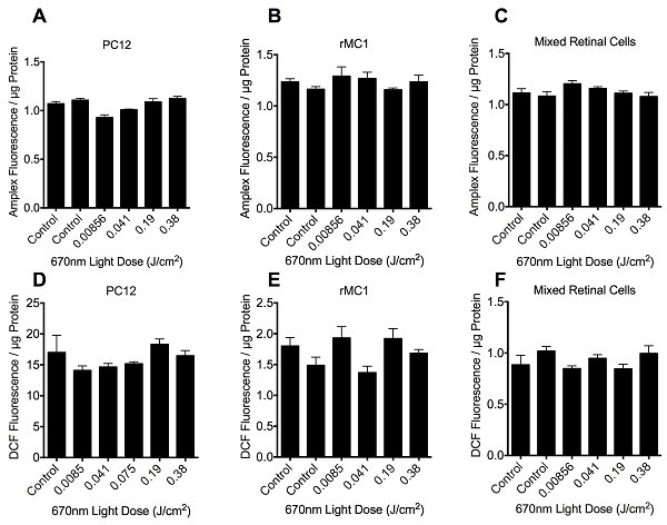
Figure 3: Quantification of H2O2 (A-C) and DCF (D-F) fluorescence in PC12 (A, D), rMC1 (B, E) or mixed retinal cell (C, F) cultures in the presence of 10mM glutamate stressor, following 670nm irradiation therapy at fluence doses ranging from 0 – 0.38 J/cm2. Histogram bars represent the mean arbitrary fluorescence units / µg protein ± SEM. There were no statistically significant differences between control and any of the treatment groups as determined by ANOVA, p > 0.05, 6 replicates per group, experiments were repeated 3 times.
